
Article Figures & Data
Figures
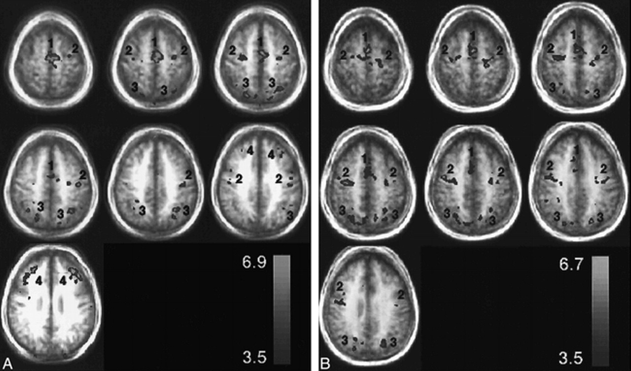
- fig 1.
Representative group axial activation maps (t > 3.5) for the visually guided saccade paradigm through the SEF (1), FEF (2), IPS (3), and PFC (4). The activation is displayed as a hot iron color scale (bottom right) where red is the lowest t threshold and yellow is the peak t value. The activation is displayed over group structural images to show the degree of blurring from biological anatomic variation. A relates to patients with pAD and B to control volunteers.
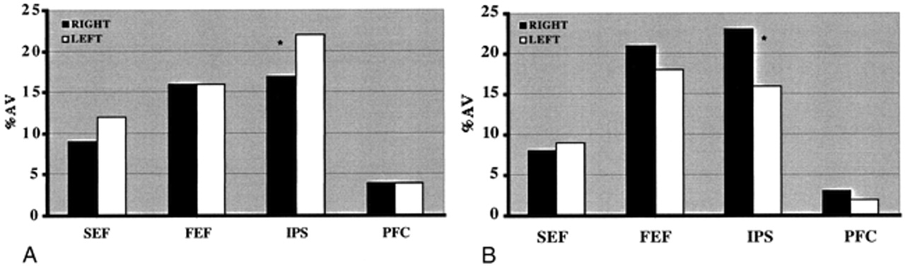
- fig 2.
Percent activation volume in each of four areas of activation in right and left cerebral hemispheres for the visually guided saccade paradigm. The regions are SEF, FEF, IPS, and PFC. The voxel numbers for each region were pooled from the activation maps of each participant. Table 3 shows that there is a corresponding significant difference (P < .02, unpaired two-tail t test) in LR for IPS between the normal group and the pAD group.
A, pAD group (n = 18). *, statistically significant difference (P < .10, paired two-tailed t test) between the volume of activation for the right and left hemispheres.
B, Control group (n = 10). *, statistically significant difference (P < .001, paired two-tailed t test) between the volume of activation for the right and left hemispheres.
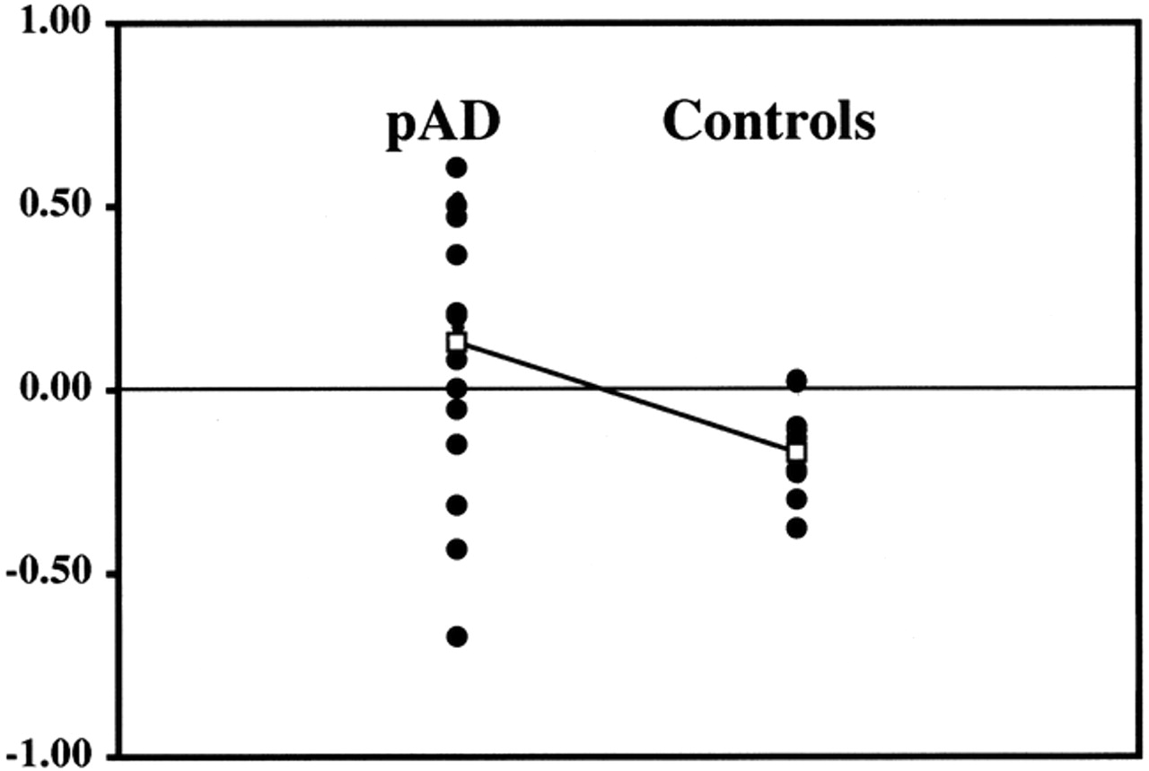
- fig 3.
LR for IPS of individual participants • of the pAD and control groups. The mean LR □ for each group was significantly different (P < .02, unpaired two-tailed t test). The other regions of interest (PFC, SEF, FEF) did not show significant separations (not shown)
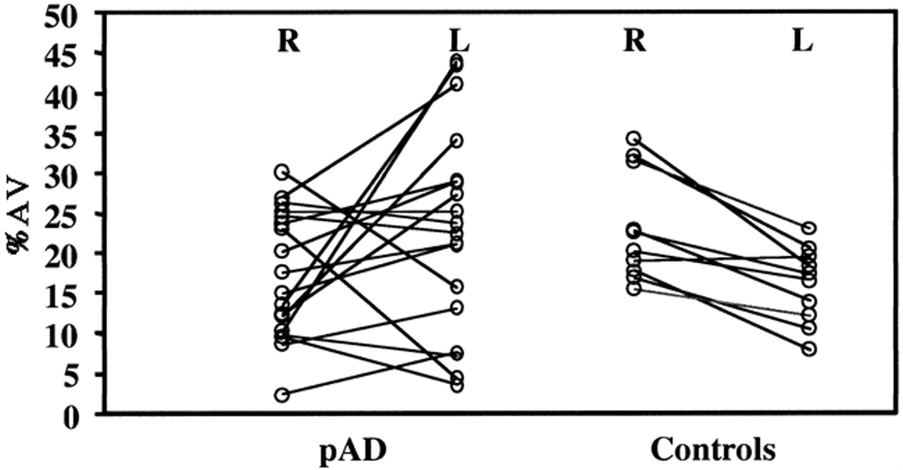
- fig 4.
Percent activation volume for the IPS regions of the right (R) and left (L) hemispheres of individual participants in the pAD and control groups, showing the group mean values. Right and left values are connected for each participant to show both individual trends within each group


Tables
 TABLE 1:
Activation data for individual control subjects (n = 10, mean and SD) tabulated as Talairach coordinates, Brodmann areas, and % signal change for activation peaks in regions of supplementary eye fields (SEF), frontal eye fields (FEF), prefrontal cortex (PFC), and intraparietal sulcus (IPS) for the left and right hemispheres for the visually guided saccade paradigm
- TABLE 2:
Activation data for individual subjects with probable Alzheimer's disease (n = 15, mean and SD) tabulated as Talairach coordinates, Brodmann areas, and % signal change for activation peaks in regions of SEF, FEF, PFC, and IPS as in Table 1
- TABLE 3:
Mean LRs for the four regions of interest defined in Table 1 for the pAD and elderly control groups


