Advertisement

Main menu

  • Home
  • Content
    • Current Issue
    • Accepted Manuscripts
    • Article Preview
    • Past Issue Archive
    • Video Articles
    • AJNR Case Collection
    • Case of the Week Archive
    • Case of the Month Archive
    • Classic Case Archive
  • Special Collections
    • AJNR Awards
    • Low-Field MRI
    • Alzheimer Disease
    • ASNR Foundation Special Collection
    • Photon-Counting CT
    • View All
  • Multimedia
    • AJNR Podcasts
    • AJNR SCANtastic
    • Trainee Corner
    • MRI Safety Corner
    • Imaging Protocols
  • For Authors
    • Submit a Manuscript
    • Submit a Video Article
    • Submit an eLetter to the Editor/Response
    • Manuscript Submission Guidelines
    • Statistical Tips
    • Fast Publishing of Accepted Manuscripts
    • Graphical Abstract Preparation
    • Imaging Protocol Submission
    • Author Policies
  • About Us
    • About AJNR
    • Editorial Board
    • Editorial Board Alumni
  • More
    • Become a Reviewer/Academy of Reviewers
    • Subscribers
    • Permissions
    • Alerts
    • Feedback
    • Advertisers
    • ASNR Home

User menu

  • Alerts
  • Log in

Search

  • Advanced search
American Journal of Neuroradiology

American Journal of Neuroradiology

ASHNR American Society of Functional Neuroradiology ASHNR American Society of Pediatric Neuroradiology ASSR
  • Alerts
  • Log in

Advanced Search

  • Home
  • Content
    • Current Issue
    • Accepted Manuscripts
    • Article Preview
    • Past Issue Archive
    • Video Articles
    • AJNR Case Collection
    • Case of the Week Archive
    • Case of the Month Archive
    • Classic Case Archive
  • Special Collections
    • AJNR Awards
    • Low-Field MRI
    • Alzheimer Disease
    • ASNR Foundation Special Collection
    • Photon-Counting CT
    • View All
  • Multimedia
    • AJNR Podcasts
    • AJNR SCANtastic
    • Trainee Corner
    • MRI Safety Corner
    • Imaging Protocols
  • For Authors
    • Submit a Manuscript
    • Submit a Video Article
    • Submit an eLetter to the Editor/Response
    • Manuscript Submission Guidelines
    • Statistical Tips
    • Fast Publishing of Accepted Manuscripts
    • Graphical Abstract Preparation
    • Imaging Protocol Submission
    • Author Policies
  • About Us
    • About AJNR
    • Editorial Board
    • Editorial Board Alumni
  • More
    • Become a Reviewer/Academy of Reviewers
    • Subscribers
    • Permissions
    • Alerts
    • Feedback
    • Advertisers
    • ASNR Home
  • Follow AJNR on Twitter
  • Visit AJNR on Facebook
  • Follow AJNR on Instagram
  • Join AJNR on LinkedIn
  • RSS Feeds

AJNR Awards, New Junior Editors, and more. Read the latest AJNR updates

Research ArticleBRAIN

CSF Flow Studies of Intracranial Cysts and Cyst-like Lesions Achieved Using Reversed Fast Imaging with Steady-State Precession MR Sequences

Karl T. Hoffmann, Norbert Hosten, Bernd U. Meyer, Simone Röricht, Christian Sprung, Johann Oellinger, Matthius Gutberlet and Roland Felix
American Journal of Neuroradiology March 2000, 21 (3) 493-502;
Karl T. Hoffmann
aFrom the Departments of Radiology (K.T.H., N.H., J.O., M.G., R.F.), Neurology (B.U.M., S.R.), and Neurosurgery (C.S.) Charité, Campus Virchow-Klinikum, Humboldt-University, Berlin, Germany.
  • Find this author on Google Scholar
  • Find this author on PubMed
  • Search for this author on this site
Norbert Hosten
aFrom the Departments of Radiology (K.T.H., N.H., J.O., M.G., R.F.), Neurology (B.U.M., S.R.), and Neurosurgery (C.S.) Charité, Campus Virchow-Klinikum, Humboldt-University, Berlin, Germany.
  • Find this author on Google Scholar
  • Find this author on PubMed
  • Search for this author on this site
Bernd U. Meyer
aFrom the Departments of Radiology (K.T.H., N.H., J.O., M.G., R.F.), Neurology (B.U.M., S.R.), and Neurosurgery (C.S.) Charité, Campus Virchow-Klinikum, Humboldt-University, Berlin, Germany.
  • Find this author on Google Scholar
  • Find this author on PubMed
  • Search for this author on this site
Simone Röricht
aFrom the Departments of Radiology (K.T.H., N.H., J.O., M.G., R.F.), Neurology (B.U.M., S.R.), and Neurosurgery (C.S.) Charité, Campus Virchow-Klinikum, Humboldt-University, Berlin, Germany.
  • Find this author on Google Scholar
  • Find this author on PubMed
  • Search for this author on this site
Christian Sprung
aFrom the Departments of Radiology (K.T.H., N.H., J.O., M.G., R.F.), Neurology (B.U.M., S.R.), and Neurosurgery (C.S.) Charité, Campus Virchow-Klinikum, Humboldt-University, Berlin, Germany.
  • Find this author on Google Scholar
  • Find this author on PubMed
  • Search for this author on this site
Johann Oellinger
aFrom the Departments of Radiology (K.T.H., N.H., J.O., M.G., R.F.), Neurology (B.U.M., S.R.), and Neurosurgery (C.S.) Charité, Campus Virchow-Klinikum, Humboldt-University, Berlin, Germany.
  • Find this author on Google Scholar
  • Find this author on PubMed
  • Search for this author on this site
Matthius Gutberlet
aFrom the Departments of Radiology (K.T.H., N.H., J.O., M.G., R.F.), Neurology (B.U.M., S.R.), and Neurosurgery (C.S.) Charité, Campus Virchow-Klinikum, Humboldt-University, Berlin, Germany.
  • Find this author on Google Scholar
  • Find this author on PubMed
  • Search for this author on this site
Roland Felix
aFrom the Departments of Radiology (K.T.H., N.H., J.O., M.G., R.F.), Neurology (B.U.M., S.R.), and Neurosurgery (C.S.) Charité, Campus Virchow-Klinikum, Humboldt-University, Berlin, Germany.
  • Find this author on Google Scholar
  • Find this author on PubMed
  • Search for this author on this site
  • Article
  • Figures & Data
  • Info & Metrics
  • Responses
  • References
  • PDF
Loading

Article Figures & Data

Figures

  • Tables
  • fig 1.
    • Download figure
    • Open in new tab
    • Download powerpoint
    fig 1.

    A, Diagram of the PSIF pulse sequence. pn (the PSIF echo) is a spin echo that results from refocusing the echo fn−1 by the RF pulse, n. fn−1 (the FISP echo) is a gradient echo of the RF pulse, n−1, refocused by the readout gradient, Gr. Only the PSIF echoes are sampled with this sequence. This results in TEeff > TR, with TE(p) = 2TR − TE(f) in the symmetric arrangement of RF pulses and gradients shown. The phase-encoding gradients are applied before and after the echo (including a compensating pulse) for a zero net phase-encoding gradient between RF pulses to avoid affecting the established steady state. Data are sampled during the interpulse interval to avoid interference with RF transmission (6).

    B, Alternating RF pulses and oscillating transverse magnetization in relation to the free induction decay (FID) and spin echo (SE) in SSFP imaging with the PSIF sequence.

  • fig 2.
    • Download figure
    • Open in new tab
    • Download powerpoint
    fig 2.

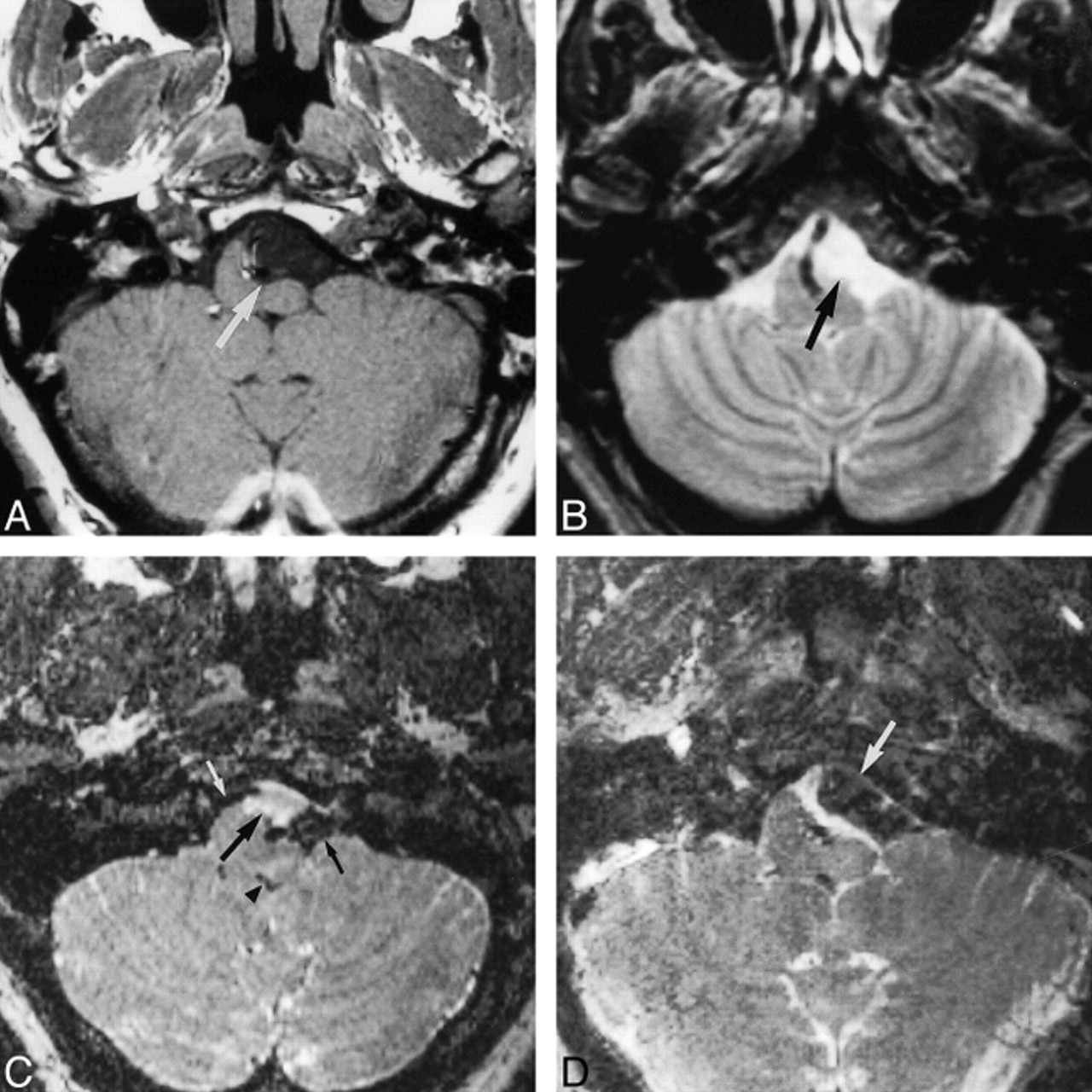
    19-year-old woman with incidentally found arachnoid cysts in the cerebellopontine angle.

    A, Transverse T2-weighted SE image (2000/80/1) shows high signal intensity within the enlarged cerebellopontine angles (arrows) and in the prepontine cistern. The shape of the enlarged CSF space suggests arachnoid cysts.

    B, On the corresponding SSFP image (20/25/1), signal attenuation due to CSF flow is found in the prepontine cistern and fourth ventricle (curved arrows). Persistently high signal intensity is found in the cerebellopontine cysts (straight arrows) due to stationary CSF, confirming the diagnosis of arachnoid cysts.

    C, Sagittal T1-weighted SE image (500/12/2) with suspected thin membrane (long arrow) between the isointense subpeduncular space (curved arrow) and the left cerebellopontine mass (short straight arrow).

    D, Corresponding sagittal SSFP image (500/12/2) shows functional separation of the hypointense pulsating CSF between the temporal bone and the cerebellar peduncles (curved arrow) and the hyperintense stationary CSF within the cyst (straight arrow).

  • fig 3.
    • Download figure
    • Open in new tab
    • Download powerpoint
    fig 3.

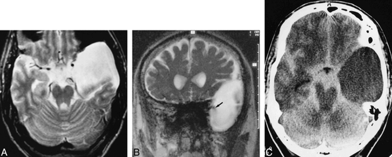
    44-year-old man with left-sided tinnitus and hearing loss, paraesthesia, and respiratory distress.

    A, T1-weighted transverse image (500/12/2) of the posterior fossa shows a paramedullary, space-occupying lesion with signal intensity similar to CSF, compressing the left ventrolateral portion of the medulla oblongata (arrow).

    B, T2-weighted SE image (2000/80/1) of the same section as in A. Different compartments are not visible; the lesion has the same signal intensity as the surrounding CSF spaces, and margins of the cyst are not reliably detectable, making the diagnosis of an arachnoidal cyst highly probable.

    C, Corresponding preoperative SSFP image (20/25/1) shows a circumscribed, hyperintense, premedullary lesion (large arrow), indicating stationary CSF of a noncommunicating arachnoid cyst in contrast to the significantly reduced signal of moving CSF in neighboring compartments (small arrows) and in the fourth ventricle (arrowhead). This finding strongly supports the decision for neurosurgical intervention.

    D, SSFP image (20/25/1) after membranectomy shows signal attenuation in most parts of the formerly hyperintense cyst (arrow), indicating communication with neighboring CSF spaces. The patient experienced relief of his symptoms.

  • fig 4.
    • Download figure
    • Open in new tab
    • Download powerpoint
    fig 4.

    31-year-old woman after membranectomy of left temporal and right temporofrontal arachnoid cysts.

    A, Transverse T2-weighted SE image (2000/80/1) shows slightly inhomogeneous, hyperintense temporal cysts (straight arrows). The frontal part of the large right-sided cyst is depicted (curved arrow).

    B, Corresponding SSFP image (20/25/1) of the same section clearly shows a jet phenomenon with distinct signal reduction arising from the right prepontine and left chiasmatic cisterns, indicating communication between the cysts and cisterns (black arrows). Marked signal reduction results from moving CSF in the prepontine cistern (white arrow). The contents of the arachnoid cysts appear, with local flow attenuation, more inhomogeneous in the SSFP image (20/25/1) owing to higher sensitivity to turbulent flow.

    C and D, Transverse (C) and coronal (D) CT cisternograms show homogeneous contrast enhancement within the basal cisterns and both the left temporal and right temporofrontal cysts. The regions of bilateral membranectomy are indicated by arrows. The coronal image also shows a frontal part of the right cyst (small arrow).

  • fig 5.
    • Download figure
    • Open in new tab
    • Download powerpoint
    fig 5.

    23-year-old man with a left-sided temporal arachnoid cyst, suffering from occasional headaches.

    A, Transverse T2-weighted SE image (2000/80/1) shows a hyperintense left temporal arachnoid cyst.

    B, Coronal PSIF image (20/25/1) depicts distinct signal reduction (arrow) arising from the basal cisterns, indicating probable communication.

    C, CT cisternogram (3-hour delay) shows slight contrast enhancement within the cyst, confirming slow communication. The contrast enhancement was confirmed by measuring a clear increase of intracystic HU. The site of communication was not detectable on the CT cisternogram owing to proposed valve mechanism with slow communication.

  • fig 6.
    • Download figure
    • Open in new tab
    • Download powerpoint
    fig 6.

    6-year-old girl with a system of temporal, supra- and parasellar, and paracerebellar arachnoid cysts.

    A, Transverse T2-weighted SE image (2000/80/1) shows a right and small left temporal, suprasellar, and paracerebellar cysts. Note hamartoma of the tuber cinereum (arrow) anterior to the rotated brain stem.

    B, Parasagittal PSIF image (20/25/1) shows some evidence of communication between the parasellar and paracerebellar cysts in the form of a continuous flow void (arrow).

    C, CT cisternogram shows contrast enhancement with layering in the parasellar and paracerebellar cysts (arrows), providing further evidence of communication. Layering was caused by the patient's prolonged supine position.

  • fig 7.
    • Download figure
    • Open in new tab
    • Download powerpoint
    fig 7.

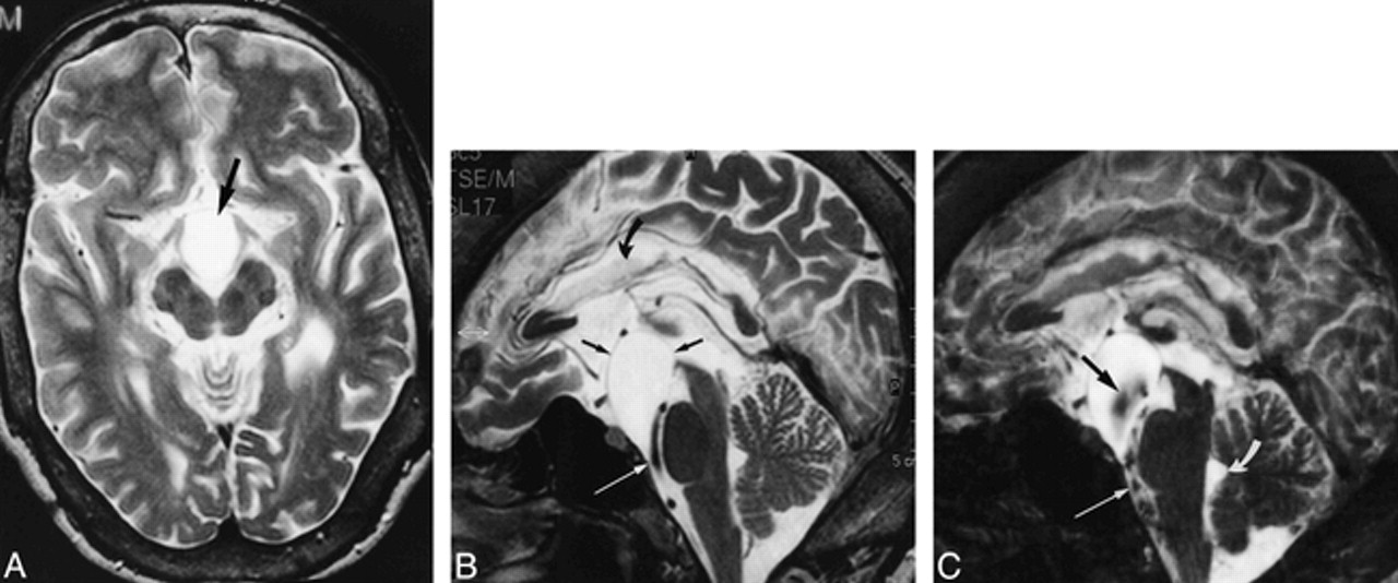
    28-year-old man with a prepontine arachnoid cyst, corpus callosum anomaly, hydrocephalus, ventriculoperitoneal shunt, and headaches.

    A, T2-weighted axial image (2000/80/1) shows a hyperintense cyst (arrow) within the prepontine cistern, pushing the crura cerebri and the optic chiasm apart.

    B, Midsagittal T2-weighted section (2000/80/1) shows defined margins of the cyst (straight black arrows), except for the narrowing between the basilar artery and skull base (white arrow). Note callosal dysplasia (curved arrow). The bottom of the third ventricle is elevated.

    C, Midsagittal SSFP image (20/25/1) reveals broad-based signal void within the cyst (black arrow) probably due to transmission of pulsations from the basilar artery. A continuous jet phenomenon between the prepontine cistern with pronounced signal void (straight white arrow) on the one hand and the cyst on the other hand is not demonstrable, making communication unlikely. Flow void within the fourth ventricle was absent (curved arrow) owing to occlusion of the aqueduct. Membranectomy was not performed, because the patient had no evidence of visual impairment.

Tables

  • Figures
  • TABLE 1:
    • View popup
    • Download powerpoint
    TABLE 1:

    Summary of cases

  • TABLE 2:
    • View popup
    • Download powerpoint
    TABLE 2:

    Summary of lesions in which additional information was obtained from reversed fast imaging with steady-state precession (PSIF) images as compared with spin-echo images

PreviousNext
Back to top

In this issue

American Journal of Neuroradiology
Vol. 21, Issue 3
1 Mar 2000
  • Table of Contents
  • Index by author
Advertisement
Print
Download PDF
Email Article

Thank you for your interest in spreading the word on American Journal of Neuroradiology.

NOTE: We only request your email address so that the person you are recommending the page to knows that you wanted them to see it, and that it is not junk mail. We do not capture any email address.

Enter multiple addresses on separate lines or separate them with commas.
CSF Flow Studies of Intracranial Cysts and Cyst-like Lesions Achieved Using Reversed Fast Imaging with Steady-State Precession MR Sequences
(Your Name) has sent you a message from American Journal of Neuroradiology
(Your Name) thought you would like to see the American Journal of Neuroradiology web site.
CAPTCHA
This question is for testing whether or not you are a human visitor and to prevent automated spam submissions.
Cite this article
Karl T. Hoffmann, Norbert Hosten, Bernd U. Meyer, Simone Röricht, Christian Sprung, Johann Oellinger, Matthius Gutberlet, Roland Felix
CSF Flow Studies of Intracranial Cysts and Cyst-like Lesions Achieved Using Reversed Fast Imaging with Steady-State Precession MR Sequences
American Journal of Neuroradiology Mar 2000, 21 (3) 493-502;

Citation Manager Formats

  • BibTeX
  • Bookends
  • EasyBib
  • EndNote (tagged)
  • EndNote 8 (xml)
  • Medlars
  • Mendeley
  • Papers
  • RefWorks Tagged
  • Ref Manager
  • RIS
  • Zotero
0 Responses
Respond to this article
Share
Bookmark this article
CSF Flow Studies of Intracranial Cysts and Cyst-like Lesions Achieved Using Reversed Fast Imaging with Steady-State Precession MR Sequences
Karl T. Hoffmann, Norbert Hosten, Bernd U. Meyer, Simone Röricht, Christian Sprung, Johann Oellinger, Matthius Gutberlet, Roland Felix
American Journal of Neuroradiology Mar 2000, 21 (3) 493-502;
del.icio.us logo Twitter logo Facebook logo Mendeley logo
  • Tweet Widget
  • Facebook Like
  • Google Plus One

Jump to section

  • Article
    • Abstract
    • Methods
    • Results
    • Discussion
    • Conclusion
    • Acknowledgments
    • Footnotes
    • References
  • Figures & Data
  • Info & Metrics
  • Responses
  • References
  • PDF

Related Articles

  • No related articles found.
  • PubMed
  • Google Scholar

Cited By...

  • No citing articles found.
  • Crossref
  • Google Scholar

This article has not yet been cited by articles in journals that are participating in Crossref Cited-by Linking.

More in this TOC Section

  • Usefulness of Quantitative Susceptibility Mapping for the Diagnosis of Parkinson Disease
  • White Matter Alterations in the Brains of Patients with Active, Remitted, and Cured Cushing Syndrome: A DTI Study
  • Qualitative and Quantitative Analysis of MR Imaging Findings in Patients with Middle Cerebral Artery Stroke Implanted with Mesenchymal Stem Cells
Show more BRAIN

Similar Articles

Advertisement

Indexed Content

  • Current Issue
  • Accepted Manuscripts
  • Article Preview
  • Past Issues
  • Editorials
  • Editor's Choice
  • Fellows' Journal Club
  • Letters to the Editor
  • Video Articles

Cases

  • Case Collection
  • Archive - Case of the Week
  • Archive - Case of the Month
  • Archive - Classic Case

More from AJNR

  • Trainee Corner
  • Imaging Protocols
  • MRI Safety Corner
  • Book Reviews

Multimedia

  • AJNR Podcasts
  • AJNR Scantastics

Resources

  • Turnaround Time
  • Submit a Manuscript
  • Submit a Video Article
  • Submit an eLetter to the Editor/Response
  • Manuscript Submission Guidelines
  • Statistical Tips
  • Fast Publishing of Accepted Manuscripts
  • Graphical Abstract Preparation
  • Imaging Protocol Submission
  • Evidence-Based Medicine Level Guide
  • Publishing Checklists
  • Author Policies
  • Become a Reviewer/Academy of Reviewers
  • News and Updates

About Us

  • About AJNR
  • Editorial Board
  • Editorial Board Alumni
  • Alerts
  • Permissions
  • Not an AJNR Subscriber? Join Now
  • Advertise with Us
  • Librarian Resources
  • Feedback
  • Terms and Conditions
  • AJNR Editorial Board Alumni

American Society of Neuroradiology

  • Not an ASNR Member? Join Now

© 2025 by the American Society of Neuroradiology All rights, including for text and data mining, AI training, and similar technologies, are reserved.
Print ISSN: 0195-6108 Online ISSN: 1936-959X

Powered by HighWire