
Article Figures & Data
Figures
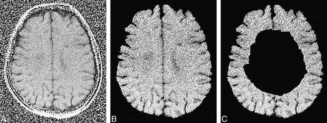
- fig 1.
Axial quantitative MTR image of the brain in a patient with MS, obtained at level of the roof of the lateral ventricles. A and B, The slice is shown before (A) and after (B) manual image segmentation, which precedes any MTR histographic analysis to exclude all extracranial tissues.
C, The same slice is shown after postprocessing (see Methods); only the cortical/subcortical brain tissue is left to enter the MTR histographic analysis.
- fig 2.
MTR histograms of cortical/subcortical brain regions from MS patients without (continuous line) and with (dotted line) cognitive impairment. Vertical lines represent the MTR values at which the integral of the histogram is 25%, 50%, and 75% of the total (ie, MTR25, MTR50 and MTR75). The histogram of MTR values from the cortical/subcortical brain tissue of impaired patients has a lower peak height and location than that obtained from unimpaired patients. For further details and statistical analysis, see text


Tables
 TABLE 1:
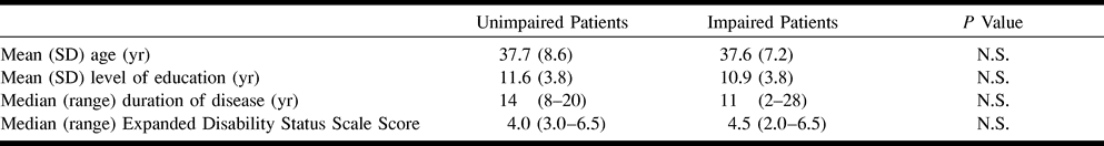
Main demographic and clinical data from MS patients without and with cognitive impairment
- TABLE 2:
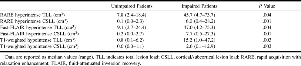
Total and cortical/subcortical brain MR lesion loads on RARE, fast-FLAIR, and T1-weighted images in MS patients without and with cognitive impairment
- TABLE 3:
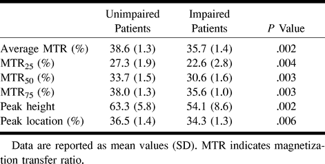
MT histogram-derived measures of the cortical/subcortical regions in MS patients without and with cognitive impairment


