Advertisement

Main menu

  • Home
  • Content
    • Current Issue
    • Accepted Manuscripts
    • Article Preview
    • Past Issue Archive
    • Video Articles
    • AJNR Case Collection
    • Case of the Week Archive
    • Case of the Month Archive
    • Classic Case Archive
  • Special Collections
    • AJNR Awards
    • Low-Field MRI
    • Alzheimer Disease
    • ASNR Foundation Special Collection
    • Photon-Counting CT
    • View All
  • Multimedia
    • AJNR Podcasts
    • AJNR SCANtastic
    • Trainee Corner
    • MRI Safety Corner
    • Imaging Protocols
  • For Authors
    • Submit a Manuscript
    • Submit a Video Article
    • Submit an eLetter to the Editor/Response
    • Manuscript Submission Guidelines
    • Statistical Tips
    • Fast Publishing of Accepted Manuscripts
    • Graphical Abstract Preparation
    • Imaging Protocol Submission
    • Author Policies
  • About Us
    • About AJNR
    • Editorial Board
    • Editorial Board Alumni
  • More
    • Become a Reviewer/Academy of Reviewers
    • Subscribers
    • Permissions
    • Alerts
    • Feedback
    • Advertisers
    • ASNR Home

User menu

  • Alerts
  • Log in

Search

  • Advanced search
American Journal of Neuroradiology

American Journal of Neuroradiology

ASHNR American Society of Functional Neuroradiology ASHNR American Society of Pediatric Neuroradiology ASSR
  • Alerts
  • Log in

Advanced Search

  • Home
  • Content
    • Current Issue
    • Accepted Manuscripts
    • Article Preview
    • Past Issue Archive
    • Video Articles
    • AJNR Case Collection
    • Case of the Week Archive
    • Case of the Month Archive
    • Classic Case Archive
  • Special Collections
    • AJNR Awards
    • Low-Field MRI
    • Alzheimer Disease
    • ASNR Foundation Special Collection
    • Photon-Counting CT
    • View All
  • Multimedia
    • AJNR Podcasts
    • AJNR SCANtastic
    • Trainee Corner
    • MRI Safety Corner
    • Imaging Protocols
  • For Authors
    • Submit a Manuscript
    • Submit a Video Article
    • Submit an eLetter to the Editor/Response
    • Manuscript Submission Guidelines
    • Statistical Tips
    • Fast Publishing of Accepted Manuscripts
    • Graphical Abstract Preparation
    • Imaging Protocol Submission
    • Author Policies
  • About Us
    • About AJNR
    • Editorial Board
    • Editorial Board Alumni
  • More
    • Become a Reviewer/Academy of Reviewers
    • Subscribers
    • Permissions
    • Alerts
    • Feedback
    • Advertisers
    • ASNR Home
  • Follow AJNR on Twitter
  • Visit AJNR on Facebook
  • Follow AJNR on Instagram
  • Join AJNR on LinkedIn
  • RSS Feeds

AJNR Awards, New Junior Editors, and more. Read the latest AJNR updates

Research ArticleBRAIN

Contrast-enhanced MR Angiography of Intracranial Giant Aneurysms

H. Rolf Jäger, Habib Ellamushi, Elizabeth A. Moore, Joan P. Grieve, Neil D. Kitchen and Wendy J. Taylor
American Journal of Neuroradiology November 2000, 21 (10) 1900-1907;
H. Rolf Jäger
  • Find this author on Google Scholar
  • Find this author on PubMed
  • Search for this author on this site
Habib Ellamushi
  • Find this author on Google Scholar
  • Find this author on PubMed
  • Search for this author on this site
Elizabeth A. Moore
  • Find this author on Google Scholar
  • Find this author on PubMed
  • Search for this author on this site
Joan P. Grieve
  • Find this author on Google Scholar
  • Find this author on PubMed
  • Search for this author on this site
Neil D. Kitchen
  • Find this author on Google Scholar
  • Find this author on PubMed
  • Search for this author on this site
Wendy J. Taylor
  • Find this author on Google Scholar
  • Find this author on PubMed
  • Search for this author on this site
  • Article
  • Figures & Data
  • Info & Metrics
  • Responses
  • References
  • PDF
Loading

Article Figures & Data

Figures

  • Tables
  • fig 1.
    • Download figure
    • Open in new tab
    • Download powerpoint
    fig 1.

    78-year-woman with bilateral cavernous carotid aneurysms. A–D, T2-weighted fast spin-echo (FSE) image (A) (4200/99/2 [TR/TEeff/excitations]) shows bilateral enlargement of cavernous sinus by giant aneurysms of internal carotid arteries. These are of mixed signal intensity with high signal in areas of turbulent flow. 3D-TOF (B) (40/6.9/1; flip angle, 20°) shows only the entering vessels, but not aneurysm sacs nor exiting vessels, which are seen on contrast-enhanced 3D-TOF (C) (40/6.9/1/45°) and dynamic contrast-enhanced MRA sequences (D) (6.4/1.4/1/30°, slice thickness = 1.5 mm). These also show extension of aneurysmal dilatation into both middle cerebral arteries (MCAs). On the contrast-enhanced 3D-TOF sequence (C), the signal intensity of aneurysm sacs and distal vessels is attenuated compared with that of contrast-enhanced dynamic MRA (D), which provides excellent contrast between vessels and background
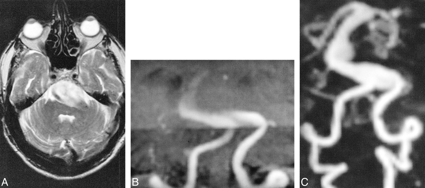
  • fig 2.
    • Download figure
    • Open in new tab
    • Download powerpoint
    fig 2.

    77-year-old man with giant basilar artery aneurysm. A–C, On T2-weighted FSE image (A) (5182/99/1), a giant basilar artery aneurysm is mainly of high signal. Patency of its lumen can therefore not be assumed. Targeted MIP projection of the posterior circulation from the 3D-TOF (B) (35/6/1/20°) sequences, acquired with overlapping slabs, shows the entering vessels, only a small part of the aneurysm sac, and no exiting vessels. Aneurysm and exiting vessels are well seen on dynamic contrast-enhanced MRA sequence (C) (5/2/1/25°)

  • fig 3.
    • Download figure
    • Open in new tab
    • Download powerpoint
    fig 3.

    62-year-old woman with giant aneurysm of left cavernous carotid artery (CCA). A–F, early (A) and late (B) frames of DSA run after left CCA injection. Early frame (A) shows entering vessel and high-velocity inflow jet. Full extent of aneurysm sac and exiting vessels are only shown on late frame of DSA run (B) where visualization of distal vessels is faint owing to turbulent flow and contrast dilution in the aneurysm sac. MIP of 3D-TOF (C) (40/6.9/1/20°) and contrast-enhanced 3D-TOF (D) (40/6.9/1/45°). Precontrast 3D-TOF (C) shows only entering vessels and high-velocity inflow jet, similar to early frame of the DSA (A), accounting for different projection angle, whereas contrast-enhanced 3D-TOF (D) also shows aneurysm sac and distal vessels. Axial source images of 3D-TOF (E) and contrast-enhanced 3D-TOF (F) show intraluminal clot. Low signal clot adherent to posterolateral wall of aneurysm is visible on 3D-TOF axial source images (E), but dropout of signal from nonlaminar flow anteriorly does not allow exclusion of additional clot. Interface between patent aneurysm lumen and low signal clot is much clearer on axial source images of contrast-enhanced 3D-TOF (F)

  • fig 4.
    • Download figure
    • Open in new tab
    • Download powerpoint
    fig 4.

    31-year-old man with ruptured giant aneurysm of the right MCA. A–E, T1-weighted SE image (A) (540/11/2) shows hematoma in right temporal lobe and insula with high signal from methemoglobin. It was shown to be due to rupture of fusiform giant aneurysm of right MCA by DSA (B). On 3D-TOF image (C) (40/6.9/1/20°), aneurysm lumen is indistinguishable from surrounding T1 contamination artifact. Contrast-enhanced 3D-TOF (D) (40/6.9/1/45°) faintly delineates aneurysm but remains severely degraded by T1 contamination artifact. Note that exiting vessel and M2 branches of right MCA, not seen by use of 3D-TOF (C), are, however, visualized. The dynamic contrast-enhanced MRA findings (E) (8.7/1.8/1/30°/3 mm), which is of low spatial resolution in this case, correlate well with those of DSA (B), showing only the aneurysm lumen, and not the hematoma

Tables

  • Figures
  • TABLE 1:
    • View popup
    • Download powerpoint
    TABLE 1:

    Demographic patient data and anatomical location of giant aneurysms

  • TABLE 2:
    • View popup
    • Download powerpoint
    TABLE 2:

    Visualization of entering vessels and aneurysm sac

PreviousNext
Back to top

In this issue

American Journal of Neuroradiology
Vol. 21, Issue 10
1 Nov 2000
  • Table of Contents
  • Index by author
Advertisement
Print
Download PDF
Email Article

Thank you for your interest in spreading the word on American Journal of Neuroradiology.

NOTE: We only request your email address so that the person you are recommending the page to knows that you wanted them to see it, and that it is not junk mail. We do not capture any email address.

Enter multiple addresses on separate lines or separate them with commas.
Contrast-enhanced MR Angiography of Intracranial Giant Aneurysms
(Your Name) has sent you a message from American Journal of Neuroradiology
(Your Name) thought you would like to see the American Journal of Neuroradiology web site.
CAPTCHA
This question is for testing whether or not you are a human visitor and to prevent automated spam submissions.
Cite this article
H. Rolf Jäger, Habib Ellamushi, Elizabeth A. Moore, Joan P. Grieve, Neil D. Kitchen, Wendy J. Taylor
Contrast-enhanced MR Angiography of Intracranial Giant Aneurysms
American Journal of Neuroradiology Nov 2000, 21 (10) 1900-1907;

Citation Manager Formats

  • BibTeX
  • Bookends
  • EasyBib
  • EndNote (tagged)
  • EndNote 8 (xml)
  • Medlars
  • Mendeley
  • Papers
  • RefWorks Tagged
  • Ref Manager
  • RIS
  • Zotero
0 Responses
Respond to this article
Share
Bookmark this article
Contrast-enhanced MR Angiography of Intracranial Giant Aneurysms
H. Rolf Jäger, Habib Ellamushi, Elizabeth A. Moore, Joan P. Grieve, Neil D. Kitchen, Wendy J. Taylor
American Journal of Neuroradiology Nov 2000, 21 (10) 1900-1907;
del.icio.us logo Twitter logo Facebook logo Mendeley logo
  • Tweet Widget
  • Facebook Like
  • Google Plus One

Jump to section

  • Article
    • Abstract
    • Methods
    • Results
    • Discussion
    • Conclusion
    • References
  • Figures & Data
  • Info & Metrics
  • Responses
  • References
  • PDF

Related Articles

  • No related articles found.
  • PubMed
  • Google Scholar

Cited By...

  • Surveillance of Unruptured Intracranial Saccular Aneurysms Using Noncontrast 3D-Black-Blood MRI: Comparison of 3D-TOF and Contrast-Enhanced MRA with 3D-DSA
  • Quality-Evaluation Scheme for Cerebral Time-Resolved 3D Contrast-Enhanced MR Angiography Techniques
  • Diagnostic Accuracy of Magnetic Resonance Angiography for Cerebral Aneurysms in Correlation With 3D-Digital Subtraction Angiographic Images: A Study of 133 Aneurysms
  • Crossref
  • Google Scholar

This article has not yet been cited by articles in journals that are participating in Crossref Cited-by Linking.

More in this TOC Section

  • Predictors of Reperfusion in Patients with Acute Ischemic Stroke
  • Enhanced Axonal Metabolism during Early Natalizumab Treatment in Relapsing-Remitting Multiple Sclerosis
  • Progression of Microstructural Damage in Spinocerebellar Ataxia Type 2: A Longitudinal DTI Study
Show more BRAIN

Similar Articles

Advertisement

Indexed Content

  • Current Issue
  • Accepted Manuscripts
  • Article Preview
  • Past Issues
  • Editorials
  • Editor's Choice
  • Fellows' Journal Club
  • Letters to the Editor
  • Video Articles

Cases

  • Case Collection
  • Archive - Case of the Week
  • Archive - Case of the Month
  • Archive - Classic Case

More from AJNR

  • Trainee Corner
  • Imaging Protocols
  • MRI Safety Corner
  • Book Reviews

Multimedia

  • AJNR Podcasts
  • AJNR Scantastics

Resources

  • Turnaround Time
  • Submit a Manuscript
  • Submit a Video Article
  • Submit an eLetter to the Editor/Response
  • Manuscript Submission Guidelines
  • Statistical Tips
  • Fast Publishing of Accepted Manuscripts
  • Graphical Abstract Preparation
  • Imaging Protocol Submission
  • Evidence-Based Medicine Level Guide
  • Publishing Checklists
  • Author Policies
  • Become a Reviewer/Academy of Reviewers
  • News and Updates

About Us

  • About AJNR
  • Editorial Board
  • Editorial Board Alumni
  • Alerts
  • Permissions
  • Not an AJNR Subscriber? Join Now
  • Advertise with Us
  • Librarian Resources
  • Feedback
  • Terms and Conditions
  • AJNR Editorial Board Alumni

American Society of Neuroradiology

  • Not an ASNR Member? Join Now

© 2025 by the American Society of Neuroradiology All rights, including for text and data mining, AI training, and similar technologies, are reserved.
Print ISSN: 0195-6108 Online ISSN: 1936-959X

Powered by HighWire