
Article Figures & Data
Figures
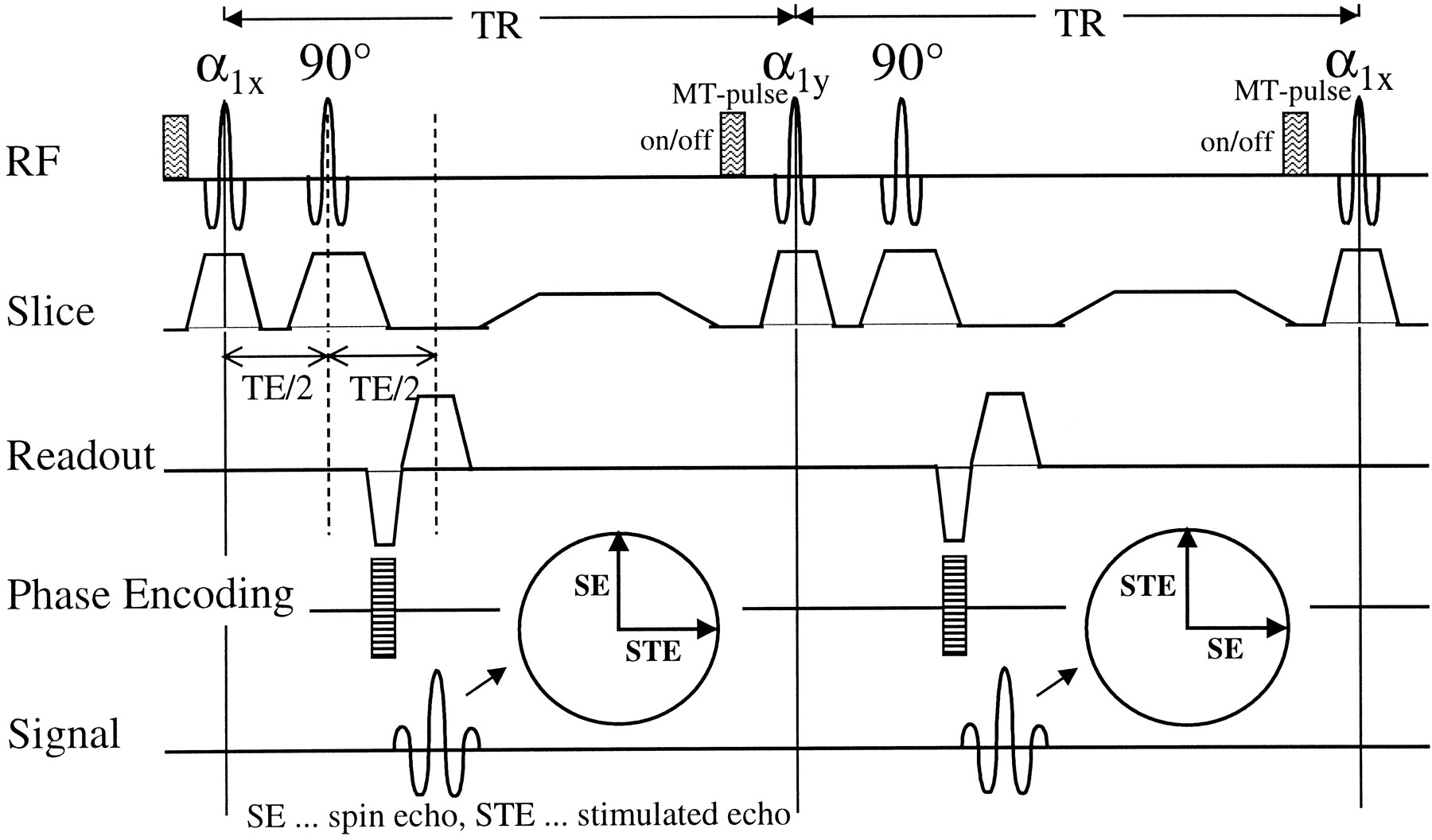
- fig 1.
The FastPACE sequence, as used for quantitative magnetization transfer imaging. Two phase-alternated acquisitions for a single phase-encoding step are shown. These two acquisitions are necessary to remove the background phase, because T1 is obtained from the phase of the composite echo. Each TR interval can be exploited for excitation of additional slices. MT saturation is achieved by performing a short binomial pulse before α1. Owing to multislice acquisition, a high saturation level can be achieved
- fig 2.
NAWM (circle) and “dirty white matter” (square) in MS on spin-echo proton density–weighted (2060/20/1) image (A). No corresponding changes are seen on the postcontrast T1-weighted (510/14/2) image (B). In the calculated image showing the transfer rate kfor, the area of dirty white matter is clearly visible (C)
- fig 3.
MTR, kfor, and T1free of different lesion types. The whiskers represent the range, the box represents the SD, and the line indicates the mean value. The mean value for NWM is marked by the solid line across the graph
- fig 4.
Ring-enhancing lesion surrounded by edema (arrows) and multiple non-active lesions. Proton density–weighted (2060/20/1) (A), postcontrast T1-weighted (510/14/2) (B), kfor (C), and T1free (D) images. The quantitative analysis in the acute lesions yielded the following results: ring-enhancing lesion: MTR = 16.4 %, kfor = 0.13 s−1, T1free = 1.5 s; edema: MTR = 41.1 %, kfor = 0.56 s−1, T1free = 1.2 s. Proton density–weighted (E) and postcontrast T1-weighted (F) images acquired 6 weeks later. Disappearance of perifocal signal abnormality supports the assumption of edema
- fig 5.
MTR versus kfor for different lesion types. The open squares show NWM. The filled circles show areas with diffuse changes. Non- active lesions are represented by the open circles, and active lesions are represented by the filled squares. The clustered groups show a good correlation between MTR and kfor; however, the overall relationship is non-linear


Tables

Mean values and SD of MTR, kfor, and Tfree for NWM, NAWM, and different lesion types

In this issue


Jump to section
Related Articles
Cited By...
- Quantitative magnetisation transfer imaging in relapsing-remitting multiple sclerosis: a systematic review and meta-analysis
- Dirty-Appearing White Matter: A Disregarded Entity in Multiple Sclerosis
- Diffusely Abnormal White Matter in Progressive Multiple Sclerosis: In Vivo Quantitative MR Imaging Characterization and Comparison between Disease Types