
Article Figures & Data
Figures
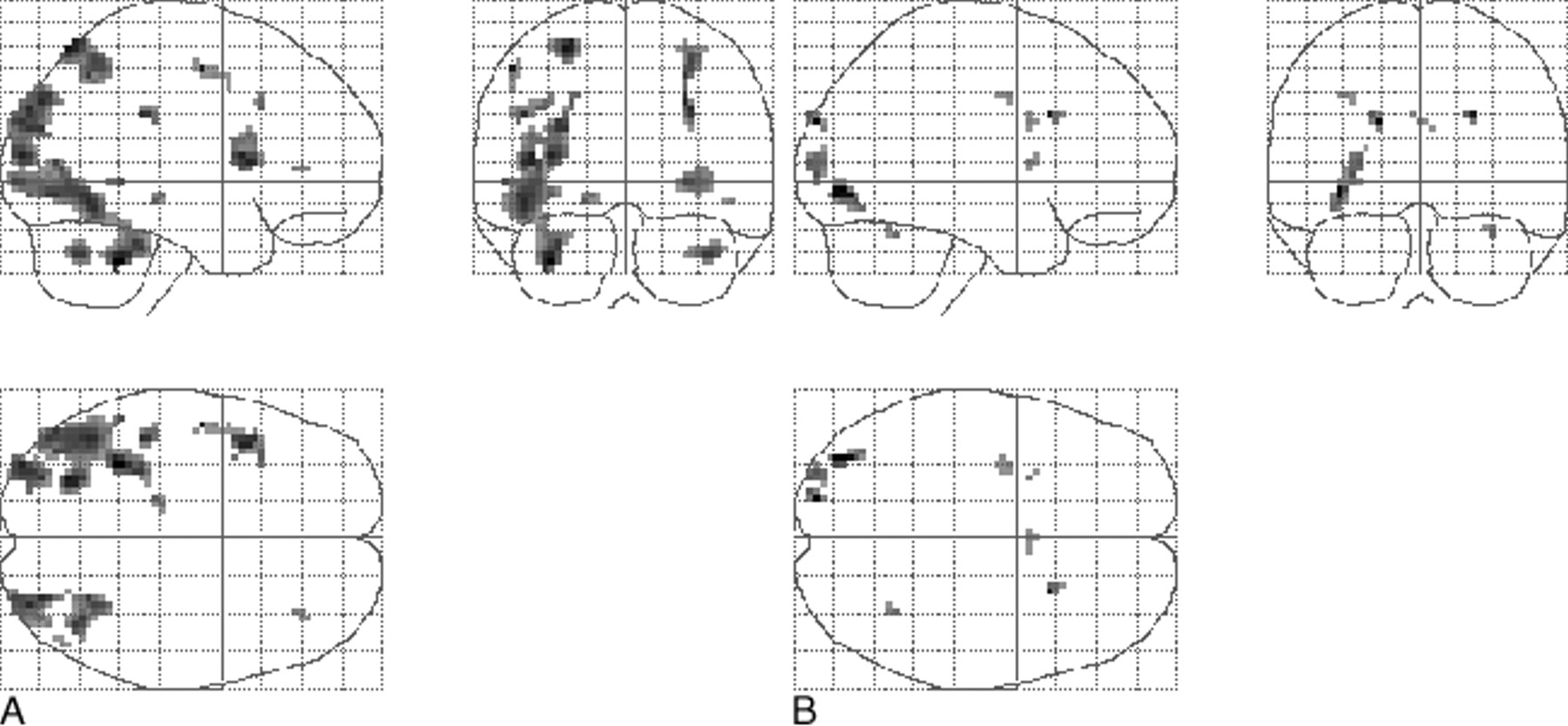
- fig 1.
Projection of average brain activation during encoding of color pictures in sagittal, coronal, and transverse directions in the control group and in the patients.
A, Control group (n = 10, left in picture is left in brain). A one-sample t test random effects analysis (P < .001; uncorrected; minimal cluster size, 108 mm3) was applied. Significant activation is observed in the occipital cortex, fusiform gyri, left parahippocampal gyrus, parietal lobe, and left inferior frontal gyrus.
B, Patients (n = 11). The main effect of signal increase is seen in the occipital cortex, right parahippocampal gyrus, fusiform gyrus, and cerebellum.
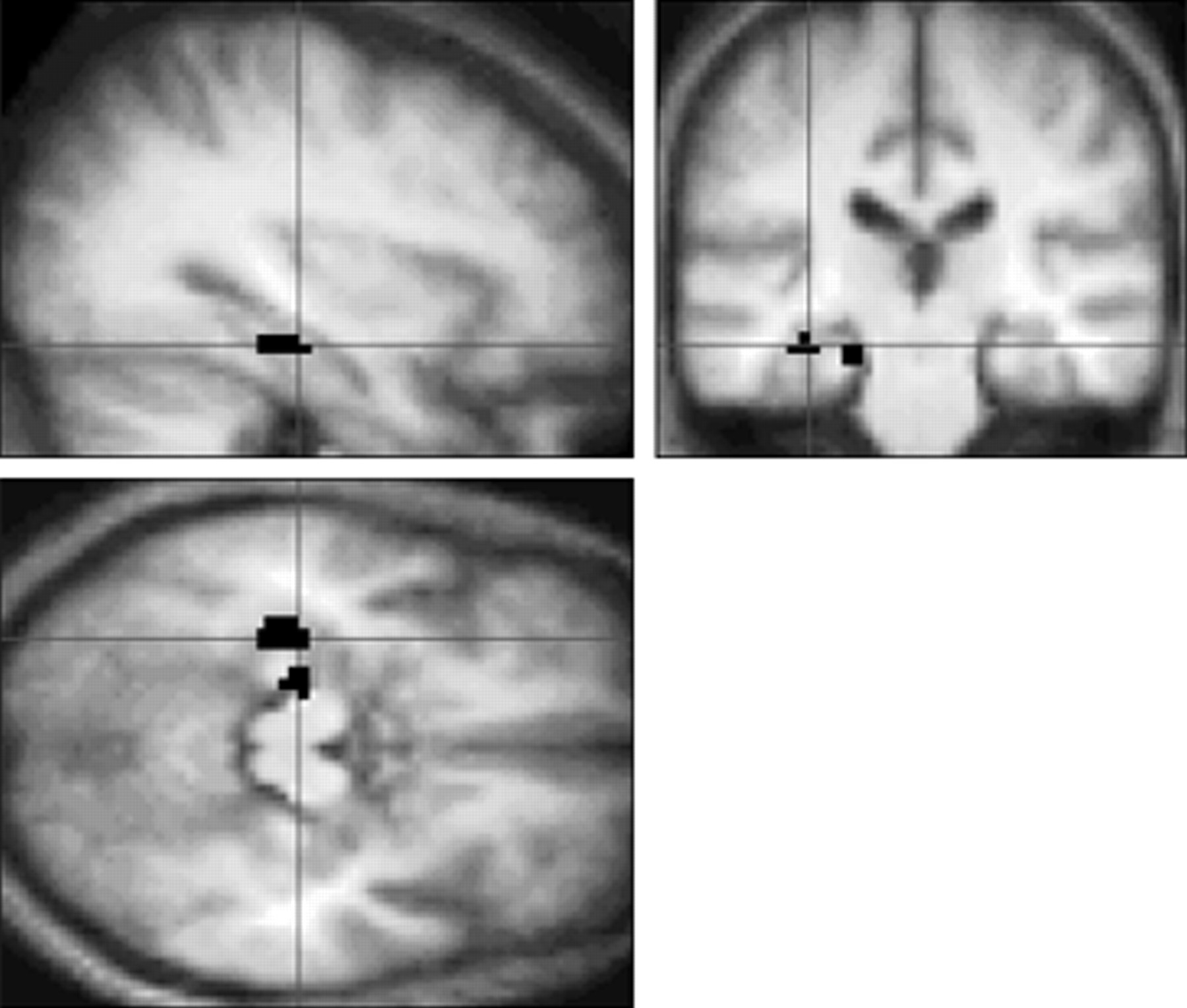
- fig 2.
Sagittal, coronal, and transverse sections showing a significant increase in brain activation in control volunteers compared with patients in the left hippocampus and parahippocampal gyrus (black areas) during the first task, after application of the region of interest analysis (P < .05, uncorrected). The same effect is seen in the right parahippocampal gyrus (not shown). Activation is projected on the average brain of the 10 control volunteers (3D gradient-echo, T1-weighted sequence with parameters 15/7/1). Left in the figure is left in the brain
- fig 3.
Projection of average brain activation during encoding of line drawings in sagittal, coronal, and transverse directions in the control group and in patients.
A, Control group (n = 10, left in the figure is left in the brain). A one-sample t test random effects analysis (P < .001; uncorrected; minimal cluster size, 108 mm3) was applied. Significant activation is observed in the cerebellum, occipital cortex, parietal cortex, inferior frontal gyrus, and left parahippocampal gyrus.
B, Patients (n = 8). The main effect of signal increase is seen in the occipital and parietal cortex, right precentral gyrus, left insula, left middle frontal gyrus, and cingulate sulcus.


Tables

Subject characteristics
In this issue


Jump to section
Related Articles
Cited By...
- mTOR Attenuation with Rapamycin Reverses Neurovascular Uncoupling and Memory Deficits in Mice Modeling Alzheimer's Disease
- Neurophysiological and brain structural markers of cognitive frailty differs from Alzheimers disease
- Brain imaging in dementia
- Functional and Structural MR Imaging in Neuropsychiatric Disorders, Part 1: Imaging Techniques and Their Application in Mild Cognitive Impairment and Alzheimer Disease
- Retinal pathology as biomarker for cognitive impairment and Alzheimer's disease
- Brain Imaging in Alzheimer Disease
- An inverse association of cardiovascular risk and frontal lobe glucose metabolism
- Relationship of fMRI activation to clinical trial memory measures in Alzheimer disease
- Impairment of nonverbal recognition in Alzheimer disease: A PET O-15 study
- Increased hippocampal activation in mild cognitive impairment compared to normal aging and AD
- Comparison of memory fMRI response among normal, MCI, and Alzheimer's patients
- Loss of frontal fMRI activation in early frontotemporal dementia compared to early AD
- fMRI studies of associative encoding in young and elderly controls and mild Alzheimer's disease
- Correlations between Visual Recognition Memory and Neocortical and Hippocampal Glucose Metabolism after Bilateral Rhinal Cortex Lesions in the Baboon: Implications for Alzheimer's Disease