
Article Figures & Data
Figures
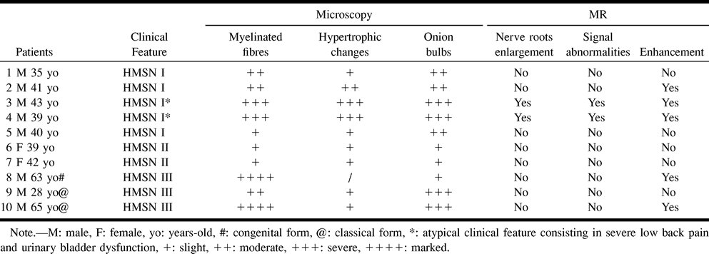
- fig 1.
A–E, MR images of the lumbosacral spine in a patient with CMT I and atypical clinical features (case 3).
Marked thickening of spinal nerve roots, completely filling the spinal canal, is seen on the sagittal (A) and axial (B) FSE T2-weighted (4000/120 [TR/TE]) images. Enhancement of hypertrophic spinal nerve roots and ganglia (arrows) is depicted in the postcontrast SE T1-weighted (TR/TE) sagittal (C) and axial (D) images. A coronal SPIR (E) postcontrast SE T1-weighted scan better depicts spinal ganglia hypertrophy and enhancement by supppressing signal from the paravertebral and foraminal fat.
- fig 2.
A–D, MR images of the lumbosacral spine in a patient with the congenital hypomyelinating form of DSD (case 8).
Marked diffuse enhancement of the cauda equina nerve roots in the absence of root enlargement is seen on pre- (A) and postcontrast (B) SE T1-weighted (500/15) sagittal images. Fat-suppressed (SPIR) coronal postcontrast T1-weighted image (C) enables better contrast between enhanced spinal ganglia (arrow) and surrounding fat-suppressed fat tissue signal compared with corresponding non-SPIR image (D).
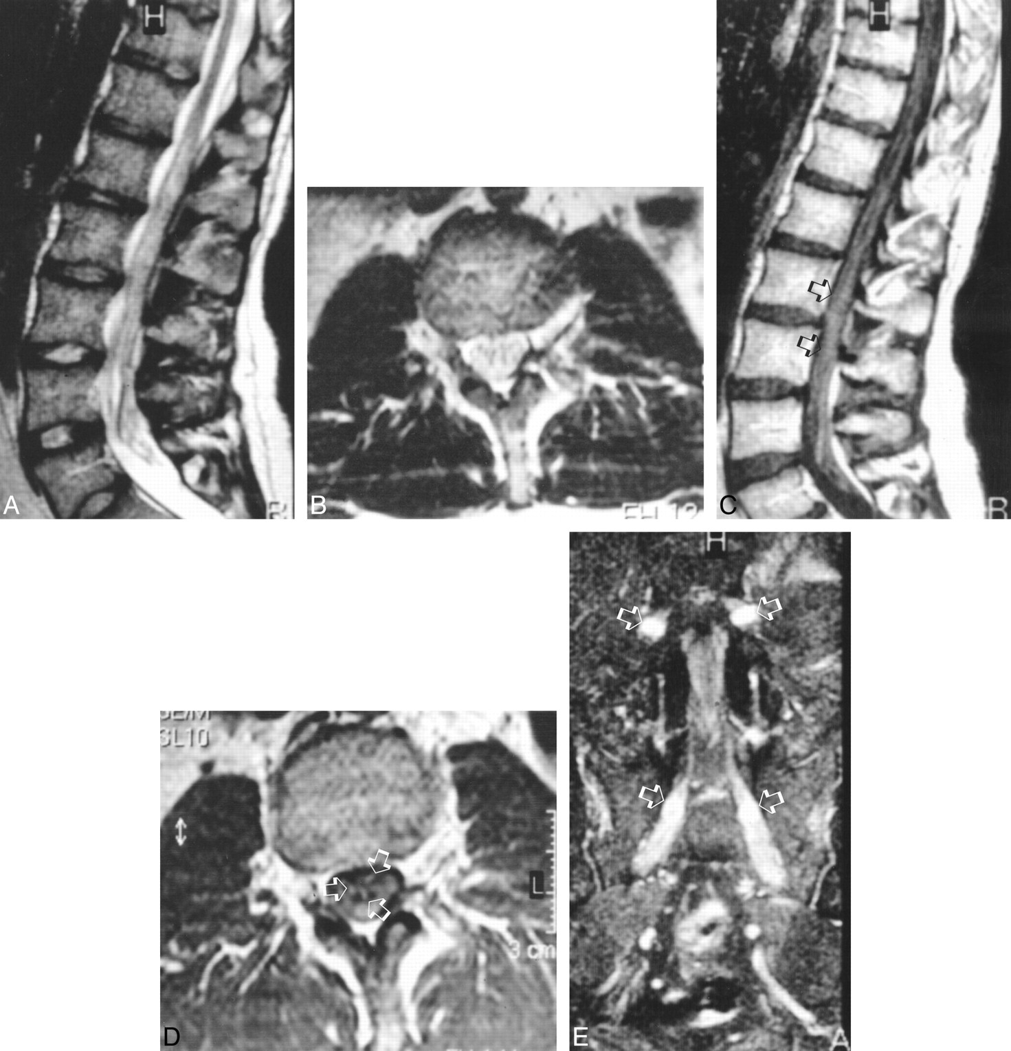
- fig 3.
Light microscopy of the sural nerve specimen (Toluidin blue stain, semi-thin section) in a patient with CMT I and atypical clinical symptoms (case 3).
Almost all fibers show “onion bulbs” (black arrowhead), with an absence of myelinated fibers, indicating active demyelination. Clusters of regenerating fibers are indicated by thin black arrow and collagen hypertrophy by large black arrow.
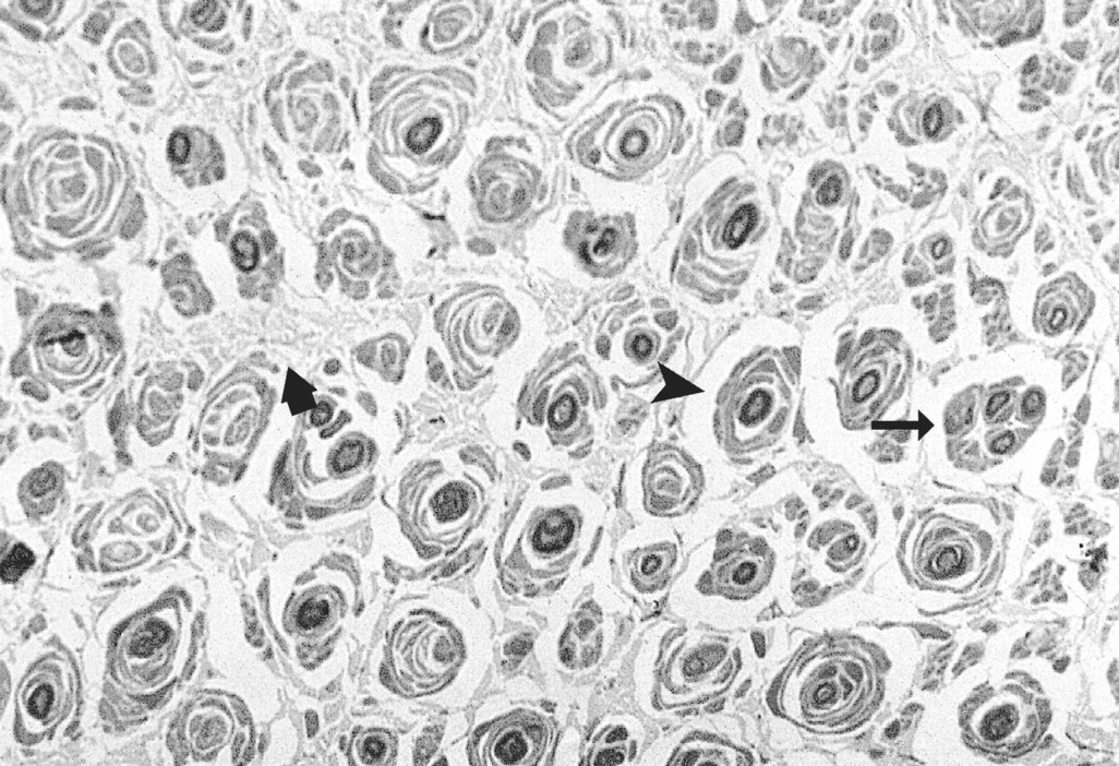
- fig 4.
A and B, LM and EM of the sural nerve specimen (Toluidin blue stain, semi-thin section) in a patient with the congenital hypomyelinating form of DSD (case 8).
On LM (A), a decreased number of myelinated fibers with peri- and epineural connective tissue hypertrophy is noted (arrow). Very few residual thinly myelinated fibers (open black arrow) are observed. Note the absence of onion bulbs and connective tissue hypertrophy (thick black arrows) with fibroblasts (black arrowhead). On EM (B), complete absence of myelin sheaths around axons is seen. A Schwann cell nucleus is indicated by the arrow.


Tables

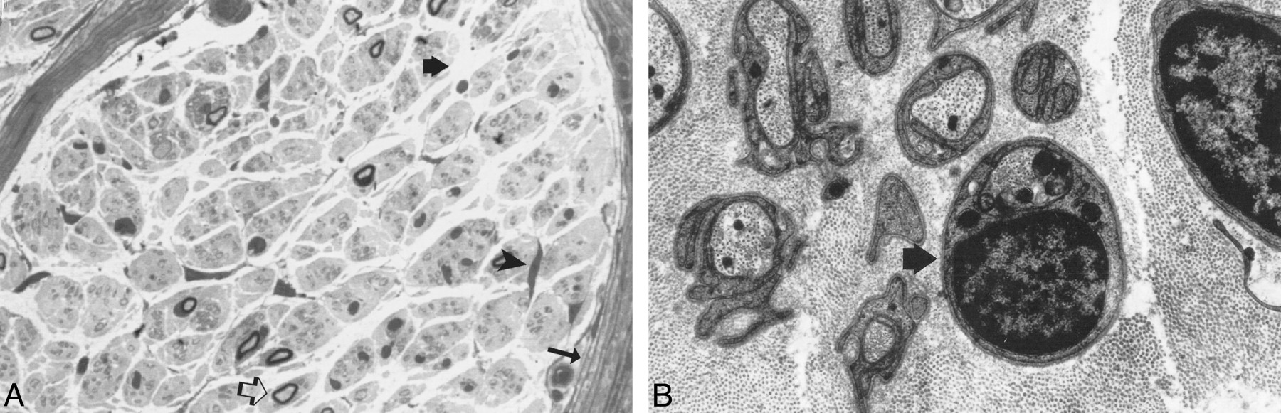
Correlations between pathologic features from sural nerve biopsy and lumbosacral MR examination


