
Article Figures & Data
Figures
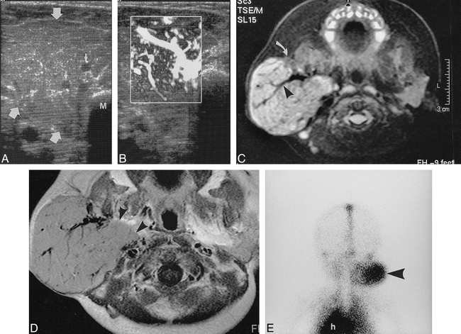
- fig 1.
Two-month-old female infant with right parotid hemangioendothelioma.
A, Transverse sonographic image shows a mildly lobulated tumor of intermediate echogenicity (large arrows) that enlarges and almost completely replaces the normal parotid gland. There are fine echogenic septations and large intratumoral blood vessels (small arrows). Note that involvement of the deep lobe of the gland is incompletely assessed because of the intervening mandibular angle (M).
B, Power Doppler imaging shows numerous large vessels within the tumor.
C, Fat-suppressed T2-weighted transverse image shows the mass to be hyperintense to the small amount of residual normal parotid (curved arrow, see also contralateral side). Large flow voids (arrowhead) represent intratumoral vessels.
D, Contrast-enhanced T1-weighted transverse image shows enhancement of the solid portion of the mass, which extends to involve the entire deep lobe (arrowheads).
E, Delayed planar image from 99mTc-labelled red cell scintigraphy (posterior view) shows uniform, well-circumscribed uptake (arrowhead), similar to that in the heart (h). Early dynamic images (not shown) revealed greater activity in the lesion than the heart in the first minute after injection.
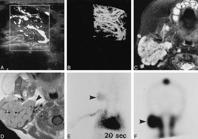
- fig 2.
Three-month-old female patient with right parotid hemangioendothelioma.
A, Power Doppler imaging shows numerous large intratumoral vessels, demonstrated to be branches of the external carotid artery and tributaries of the retromandibular vein.
B, Three-dimensional power Doppler imaging confirms the extreme vascularity of the parotid mass.
C, Fat-suppressed T2-weighted transverse image shows a hyperintense mass (small solid arrows) with numerous intratumoral vessels. Extension into the deep lobe (open arrow) is well shown. Long arrows identify the external carotid artery (medial) and retromandibular vein (lateral).
D, Contrast-enhanced T1-weighted transverse image shows enhancement of the solid portion of the mass, which involves the entire deep lobe (large arrowhead), and contains numerous large vessels (small arrowheads). The masseter (m) and medial pterygoid (p) are not involved.
E, Dynamic image from 99mTc-labelled red cell scintigraphy (anterior view, acquired at 20 seconds) shows uptake in the lesion (arrowhead), less than that in the heart.
F, Delayed coronal SPECT image shows intense uptake in a sharply defined lesion (arrowhead).


In this issue


Jump to section
Related Articles
Cited By...
- No citing articles found.