
Abstract
Summary: We present the MR imaging findings in a 43-year-old male patient with bilateral idiopathic sclerosing inflammation of the orbit. Bilateral enhancing retrobulbar masses, with concentric compression of the retrobulbar segment of the left optic nerve, were seen. MR imaging proved to be the only means to distinguish between the different intraorbital structures and to determine the exact site of optic nerve compression. To our knowledge, this is the first documented case of MR imaging findings of this entity.
Idiopathic sclerotic inflammation of the orbit (ISIO) is a distinct clinicopathologic entity characterized by progressive sclerotic inflammation that damages orbital structures primarily through cicatricial entrapment (1). Bilateral intraorbital fibrosis, appearing consistently and early, is the characteristic histologic finding that distinquishes idiopathic sclerotic inflammation from idiopathic orbital inflammation syndrome (IOIS [also known as orbital pseudotumor]) (1). Immunohistologic studies have confirmed that ISIO is more closely related to idiopathic benign retroperitoneal fibrosis, sclerotic mediastinitis, fibrotic thyroiditis, and sclerotic cholangitis than to IOIS (1, 2). Regarding these similarities, Comings et al have proposed that these different disorders are all manifestations of a single fibrosclerotic process that may be localized or multifocal (2). Disseminated manifestations of this fibrosclerotic process have been referred to as “multifocal fibrosclerosis” (1, 2). The present report describes the MR imaging appearence of bilateral idiopathic sclerotic inflammation of the orbit, with compression of the left optic nerve, in a rare case of multifocal fibrosclerosis.
Case Report
43-year-old white man had a 9-month history of histologically confirmed idiopathic retroperitoneal fibrosis, also known as Morbus Ormond, and bilateral ISIO. At his first admittance in a peripheral hospital, the patient presented with mild exophthalmus, xanthelasmas at the surface of both upper and lower lids, and mild limitation of the elevation and abduction of both globes. Increasing pain of the right and left flank alerted the ophthalmologist to recommend further investigation. CT of the abdomen revealed distinct increased attenuation of the retroperitoneal fat and the mesenteric root (30 × 18 × 8 cm [transverse × craniocaudal × sagittal diameter]), with bilateral cicatriceal entrappment of the kidneys, leading to extensive bilateral hydroureter and hydronephrosis. Subsequently, the patient underwent surgical removal of the retroperitoneal fibrosis to decompress both ureters. Histologic examination confirmed the diagnosis of idiopathic retroperitoneal fibrosis. CT of the orbits revealed diffuse, bilateral, mainly intraconal, masses infiltrating the orbital fat. After good postoperative recovery of bilateral hydroureter and hydronephrosis, and absent signs of visual loss, the patient left hospital on corticosteroid and immunsuppressive therapy (prednisolone, 20 mg; Imurek (Wellcome Foundation; London, UK), 50 mg [three times daily]). In spite of continued therapy, progressive gradual concentric vision loss on the left side occurred within the last 6 weeks. The patient was admitted to our ophthalmology department. Clinical data revealed mild hypothyroidism (no signs of Riedel's thyroiditis) and mild increased C-reactive protein. Slit-lamp examination showed dilated conjunctival and episcleral vessels of the left eye. Examination of the left fundus showed marked congestion of the subretinal veins and an indistinct border of the papilla of the optic nerve. The intraoccular pressure, measured by applanation tonometry, was 24 mm Hg on the left side and 20 mm Hg on the right side. Computer-aided perimetry revealed concentric impairment of the visual field on the left side and a full visual field on the right side. A- and b-mode sonography and unenhanced (owing to known adverse reaction to iodine contrast agent) CT (Plus 4, Siemens; Erlangen, Germany) of the orbits showed an increased density of the intra- and extraconal orbital fat and homogeneous soft-tissue masses in the frontal and sphenoid sinuses. Further differentiation of the intraconal structures was not possible on CT scans. Owing to the progressive left visual loss, surgical decompression of the left optic nerve was considered. MR imaging (Gyroscan ASC S15, Phillips; Best, Netherlands) of the orbits and the skull base, with and without gadolinium administration, was performed. MR scans revealed bilateral retrobulbar, mainly intraconal, homogeneous hypointense masses on T1- (Fig 1) and T2-weighted images. On contrast-enhanced T1-weighted images (0.2 mmol Gd/kgKG), these intraorbital masses showed homogeneous enhancement (Fig 2). The differentiation between the retrobulbar masses—the laterally displaced conal muscles, the ophthalmic artery, and the optic nerve—was possible on transverse (Fig 3) and coronal (Fig 4) turbo spin-echo T2-weighted fat-suppressed images. The optic nerve and its subarachnoid space were nicely depicted on both sides within these homogeneous hypointense intraconal masses. Just behind the globe, the left optic nerve appeared thinned over a distance of about 1.5 cm, with concentric compression of its subarachnoid space, which was clearly delineated at this location (Fig 4). The frontal and the sphenoid sinuses were filled by similar enhancing tissue with low signal on T1- and T2-weighted MR images. The skull base, especially the osseous frame of the orbit and the visible intracranial structures, revealed no abnormality. Trans-sphenoethmoidal endoscopic subtotal resection of the medial wall of the left orbit was performed to decompress the left optic nerve. The biopsies of the intraconal masses were fixed in formaldehyde solution and routinely processed for histologic evaluation. Tissue resection revealed predominantly fibrous connective tissue, varying from dense mature collagen bands to looser myxoid areas with foci of plump active fibroblasts. A mixed lymphocyte population was present, diffusely infiltrating the tissue. Scattered small blood vessels showed hyalinization of their walls.

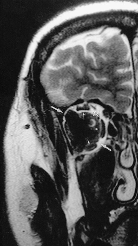
Axial unenhanced T1-weighted scan (507/15/4) [TR/TE/excitations] of the anterior skull base. Extensive homogeneous, bilateral, intraconal masses with low signal intensity are visible. The conal muscles are displaced laterally (white open arrow). The ophthalmic artery is apparent at the apex of the orbit (small white arrow).fig 2. Axial enhanced T1-weighted scan (507/15/4) of the anterior skull base. Marked homogeneous enhancement of the intraconal masses (black arrow) is visible. The left optic nerve is encircled and compressed by the retrobulbar enhancing masses and appears thinned at its distal segment (curved black arrow). There is no pathologic enhancement of the intracranial structures.fig 3. Axial turbo spin-echo T2-weighted image (2000/120/6) of the anterior skull base. Bilateral, marked hypointense, intraconal, retrobulbar masses are visible. The laterally displaced conal muscles (white open arrows) and the posterior segment of the left optic nerve and its subarachnoid space (small white arrows) are clearly apparent. The more distal segment of the subarachnoid space of the left optic nerve is compressed (curved white arrow)

Coronal turbo spin-echo T2-weighted image (2027/120/6) of the anterior skull base. An expanding retrobulbar mass with low signal intensity is visible in the right orbit (white arrow). The conal muscles are displaced without signs of infiltration (small white arrows). In the medial and upper quadrant of this mass, the optic nerve and its subarachnoid space are clearly visible (open white arrow)
Discussion
Histologically proved and radiographically documented ISIO associated with idiopathic retroperitoneal fibrosis is a rare entity. Reviewing the literature, we found only eight reported cases (3–9). On previous CT studies (6, 8, 9), ISIO was described as homogeneously enhancing intraorbital masses. Similiar to these reports, the CT images of our patient revealed diffuse increased attenuation of the intraorbital fat; however, further differentiation between the intraorbital muscles, the retrobulbar optic nerve, the retrobulbar ophthalmic artery, and the ISIO was not possible.
To our knowledge, this case report is the first description of the MR imaging features in a case of ISIO. The desmoplastic fibrous stroma of ISIO is presumably responsible for the low signal on T1- and T2-weighted images of the intraconal masses relative to the intraorbital fat and muscles. Distinct homogeneous enhancement of the intraorbital masses can be explained by histologically confirmed vascularity of the resected intraorbital fragments. MR imaging clearly delineates the extension into the paranasal sinuses—the bilateral, expansile, and compressive character of ISIO—which was confirmed by the following surgical intervention. In contrast to sonography and CT, MR imaging was the only diagnostic technique that revealed the necessary preoperative information concerning the delineation and the localization of the concentric left optic nerve compression.
The differential diagnosis of intraconal masses with low signal on T1- and T2-weighted images include orbital pseudotumor, fungal disease, orbital lymphoma, and ISIO.
Orbital pseudotumor, or IOIS, is the most common cause of proptosis in adults. Owing to fibroinflammatory stroma, diffuse IOIS may appear similar to ISIO on T1-weighted and T2-weighted images. For better differentiation between IOIS and ISIO, it is helpful to remember the diagnostic features that were cited by Mombaerts et al. These authors defined the “orbital pseudotumor” as a unilateral orbital mass lesion, clinically presenting with signs of mass effect, inflammation, and infiltration, showing either a focal or diffuse involvement of the intraorbital structures. Clinical investigations eliminate identifiable local and systemic causes. Orbital pseudotumor is not a lymphoid tumor. Histopathologic examination shows an orbital pseudotumor as a fibroinflammatory lesion. An atypical histopathologic pattern, such as extensive sclerosis, true vasculitis, granulomatous inflammation, and tissue eosinophilia, can be used for subclassification of the orbital pseudotumor. In absence of systemic fibroinflammatory, granulomatous, and vasculitic disease, these lesions can still be considered to belong to the spectrum of orbital pseudotumor (10). The pathogenesis of orbital pseudotumor remains elusive and the role of infection, autoimmune disorders, and abberant wound healing is still under investigation (10).
In addition to this definition of orbital pseudotumor, Rootman et al (1) and McCarthy et al (11) also investigated the histologic and immunohistologic pattern of patients who presented with simultaneous retroperitoneal fibrosis and a fibrosclerotic process in the orbit. Both authors concluded that the immunohistologic findings in these patients suggest that the dominant pathologic process is one of immune-mediated fibrosis rather than recurrent inflammation. Rootman et al introduced the term “idiopathic sclerotic inflammation of the orbit” describing the fibroinflammatory intraorbital mass as a distinct clinicopathologic entity and to document the closer relationship of this entity to other fibrosclerotic processes than is the case in the nonspecific orbital inflammatory syndrome (1). Simultaneous systemic disseminated fibrosclerotic processes are described as “multifocal fibrosclerosis” to document the suspected common dominant pathologic process of fibrosis (8). Idiopathic retroperitoneal fibrosis in multifocal fibrosclerosis is hypointense on T1- and T2-weighted images in relation to retroperitoneal fat, with diffuse homogeneous enhancement after intravenous administration of contrast agent (12). Similar signal intensities on T1- and T2-weighted images and similar enhancement support the theory that these are all manifestations of one single fibrosclerotic process that may be localized or multifocal (12).
As mentioned above, another diagnostic feature of IOIS is unilaterality, whereas, in our patient, extensive bilateral involvement of the intraorbital structures and extension into the paranasal sinuses were evident. Paranasal involvement is definitely not a diagnostic feature of IOIS, as described in the recent literature (10). Early extensive fibrosis is the characteristic diagnostic feature that will help to distinguish ISIO from an orbital pseudotumor on a histologic specimen (10).
Fungal masses are typically hypointense on T1- and T2-weighted images and mainly occur in immunocompromised patients. Invasive facial fungal disease originates mainly within the paranasal sinuses, subsequently invading the orbit and the skull base. Clouding of the sinuses, calcification within the masses, and the destruction of the anterior skull base in invasive fungal disease may be helpful in differentiating this condition from ISIO (13). Diffuse infiltration of the retrobulbar intraconal fat is documented in orbital lymphomas. Cytryn et al reported that lymphomas and inflammatory infiltrates appeared to be similar in signal intensity to extraocular muscles on T1-weighted images, but most of the lymphomas became brighter and inflammatory infiltrates became slightly darker on T2-weighted images (14). These results show that fibroinflammatory infiltrates can generally be distinguished from lymphomas by their signal intensities. Nevertheless, a few lymphomas may also be hypointense on T2-weighted images. In these patients, a blood cell count and biopsy will help to differentiate between these two entities. Additional information that helps in differentiating orbital lymphomas from ISIO is that the orbital lymphomas may appear either localized or diffuse but do not present such a cicatriceal entrapment of the optic nerve as presented in our patient.
Clinical symptoms of ISIO are dominated by cicatricial infiltration, mass effect, and chronic inflammation, which frequently lead to complete vision loss. The most common signs are pain, proptosis, lid swelling, restriction of extraocular movement, and ptosis. All of these symptoms can be explained by the mass effect of the intraorbital fibrosclerotic process. In about one third of the patients, blindness commonly occurs several months after onset of the inflammatory fibrosis. The absence of acute inflammatory signs, the recurrent progredient proptosis, the progredient vision loss, pain, and the impairment of extraocular movement during our patient's 9-month history of clinical manifestation of this disease support the theory that the underlying pathogenesis of ISIO is a chronic progredient immune-mediated fibrosclerotic process (1, 11). Xanthelasmas, as described above, seems to be an incidental finding, because the patient showed no signs of disturbance of lipid metabolism.
The striking features of bilateral intraorbital masses that suggest the diagnosis of ISIO rather than orbital pseudotumor, orbital mycosis, or orbital lymphoma are extension into the adjacent sinuses without bony erosion, low signal intensities on T1- and T2-weighted images with distinct enhancement, bilateral proptosis, progredient pain, cicatriceal entrapment of the optic nerve, impaired extraocular movement, ptosis, and additional uni- or bilateral hydronephrosis caused by histologically proved idiopathic retroperitoneal fibrosis.
Because the current mainstays of the treatment, corticosteroids and radiotherapy, are nonspecific, are frequently incomplete or instituted too late in the course, and are not directed at the immunopathologic process responsible for fibrosis, poor outcome is not surprising (1). The most disturbing long-term results of this entity are vision loss, proptosis, and restriction of extraocular movement. New therapeutic concepts recommend early introduction of immunosuppressive agents; nevertheless, reliable results have not been published yet (1).
Conclusion
A case of bilateral ISIO, with concentric left optic nerve compression, was present in a patient with multifocal fibrosclerosis. Bilateral intraorbital enhancing masses, with low signal intensities on T1- and T2-weighted images, were seen. Possible differential diagnoses of enhancing masses with low signal intensities on T1- and T2-weighted images were discussed, and specific diagnostic features of ISIO were concluded. A thorough knowledge of the radiologic features of ISIO enables the radiologist to diagnose this rare entity and to localize optic nerve compression in the case of vision loss. MR imaging of the orbit is a valuable diagnostic tool that provides the ophthalmologist with the necessary information to decide on a therapeutic course.
Footnotes
↵1 Address reprint requests to Gottfried J. Schaffler, Department of Radiology, University Hospital Graz, Auenbruggerplatz 9, A-8036 Graz Austria.
References
- Received August 21, 1998.
- Accepted after revision July 13, 1999.
- Copyright © American Society of Neuroradiology
In this issue


Jump to section
Related Articles
Cited By...
- No citing articles found.