
Article Figures & Data
Figures
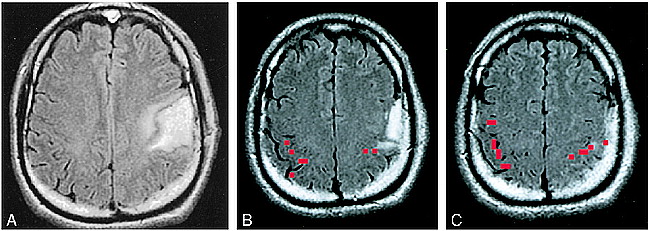
- fig 1.
A–C, Assessing the feasibility of surgery in a tumor patient, case 9 (Table 2): 38-year-old man with a biopsy-proved grade 2 oligodendroglioma in the left perirolandic region (A). Functional MR imaging was performed to assess the anatomic relationship between the tumor and the functional hand area. The thresholded activation map (red pixels) was superimposed on a FLAIR image. B represents the most inferior section on which functional activation was identified and C represents a more cephalic section. The apex of the tumor was in contact with the most inferior portion of the functional MR imaging activation (B). After considering the risk versus benefit of a proposed gross total resection, the patient opted for treatment with external beam radiation and no further surgery
- fig 2.
A and B, Assessing the feasibility of surgery in an epilepsy patient, case 12 (Table 1): 31-year-old man with life-long medically intractable seizures. An anatomic MR study (A) revealed bifrontal cortical developmental anomalies. Prolonged video-EEG recording showed unequivocally that the patient's typical seizures arose from the right frontal area. The characteristic landmarks used to define the anatomic central sulcus were absent. Functional MR imaging was performed (B) to ascertain the topographic relationship between the right frontal developmental anomaly and the hand sensorimotor functional area. This study showed that the functional hand area was posterior and superior to the anatomic developmental anomaly (arrow). The anatomic abnormality was resected, and the patient has been seizure-free for over 2 years
- fig 3.
A–D, Assessing feasibility of surgery in an epilepsy patient, case 23 (Table 1): 9-year-old boy with medically intractable partial-onset seizures. Anatomic MR image shows a pial-based abnormality in the left perirolandic region (A), which enhanced with contrast (B). This was interpreted to represent a pial angioma of Sturge-Weber. Functional MR imaging was performed to determine the topographic relationship between the lesion and the functional hand area (C and D). This study, particularly the section illustrated in C, which is inferior to the section in D, showed functional activation located directly beneath the pial angioma. Invasive extraoperative stimulation mapping confirmed the location of hand function identified by the functional MR imaging study. Surgery was deferred. The scalp marker over the right parietal region in A and C is a fiducial for frameless stereotactic surgery
- fig 4.
A–C, Surgical planning, case 9 (Table 1): 18-year-old boy with life-long medically intractable seizure disorder due to a perinatal hemorrhage (A). The patient had a spastic right hemiparesis but retained some right-hand function. Owing to the perceived proximity of the left frontal encephalomalacic cyst to the anatomic central sulcus, and the fact that the injury occurred in the perinatal period, the possibility of functional cortical reorganization was considered. Scalp EEG recordings revealed the site of seizure onset to be near the posterior margin of the cyst. Functional MR imaging was performed (B) to clarify the relationship between the functional sensorimotor area and the posterior margin of the cyst. Results of this study were used to guide surgical placement of EEG strips to ensure the functional hand area was covered by the subdural strips for purposes of extraoperative cortical stimulation mapping. The collage in C represents a 3D rendering of the brain with the functional MR imaging activation (red) embedded (on the reader's left). The orientation of the 3D rendering is identical to that of the exposed surgical field (on the reader's right), with the patient's nose toward the top of the page, the back of the head toward the bottom of the page, superior to the reader's right and inferior to the reader's left. Extraoperative ictal video-EEG recording and cortical stimulation mapping revealed that the functional hand area was located beneath the three electrode contacts to the left of the “T” of the more posterior strip, which coincided with the functional MR imaging study. The area of ictal onset was located beneath the three most cephalic electrodes to the right of the “T” of the more anteriorly positioned strip. The epileptogenic zone was resected and the patient has been seizure-free for over 2½ years
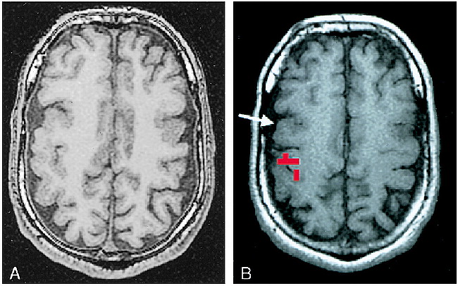
- fig 5.
A and B, Anatomic distortion due to tumor mass effect, case 2 (Table 2): 33-year-old man with a biopsy-proved recurrent high-grade astrocytoma. A, which shows enhancing areas within the tumor, was acquired with the patient in a rigid stereotactic frame, which accounts for the distortion of the scalp. Tumor mass effect produced substantial distortion of normal cortical landmarks, and functional MR imaging was performed to determine the topographic relationship between the tumor and functional hand area in the right hemisphere (B). Functional MR imaging showed that the tumor had displaced the functional hand area posteriorly. While the functional hand area was involved with tumor edema, the majority of the tumor volume was anterior to the motor strip. The patient underwent a tumor debulking procedure with intraoperative cortical stimulation mapping


