
Article Figures & Data
Figures
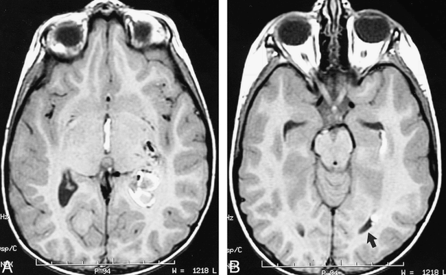
Fig 1. CT of previously healthy 5-year-old boy presenting with headache, disorientation, and somnolence is shown.
A and B, T1-weighted MR images of brain performed on the day of ictus show left periventricular hematoma with extension into atrium of adjacent left lateral ventricle. Note absence of blood within left occipital horn (arrow). Flow voids representing AVM feeding pedicles are present anteriorly within left basal ganglia.
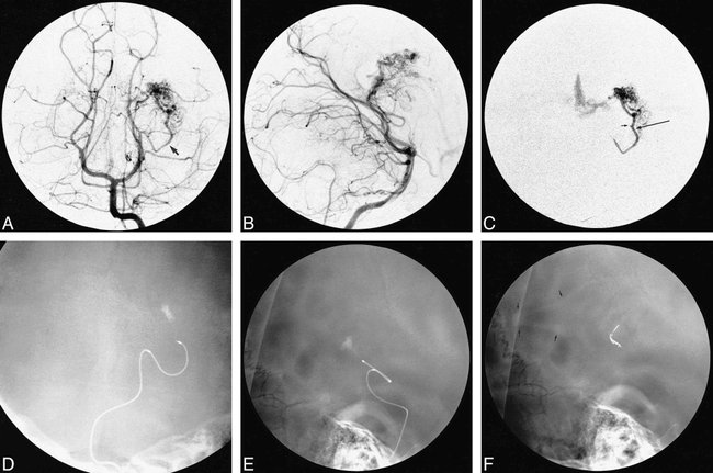
Fig 2. Embolization procedure conducted under digital roadmap and fluoroscopic guidance is shown.
A and B, Anteroposterior (AP) Towne's and lateral views of left vertebral injection show an enlarged left lateral posterior choroidal artery supplying AVM nidus (arrow).
C, AP view of superselective injection into left lateral posterior choroidal artery supplying AVM is shown. Position of microcatheter tip from which ethanol embolization was performed (long arrow). Note unopacified inflow (flow reversal) from small medial branch supplying normal choroid plexus of glomus (small arrow).
D and E, AP and lateral unsubtracted views (immediately after test injection) show persistent contrast filling of small choroidal branch indicating flow arrest and contrast stain in the region of atrial choroid plexus.
F, Unsubtracted lateral view showing contrast material within dependent portion of left lateral ventricle (arrows). Contrast medium appears to outline wall of ventricle. This appearance is most likely caused by preexistent intraventricular hematoma acting as radiolucent “filling defect.” Radioopaque fiber-coated coils within occluded left lateral posterior choroidal artery are visible anteriorly.
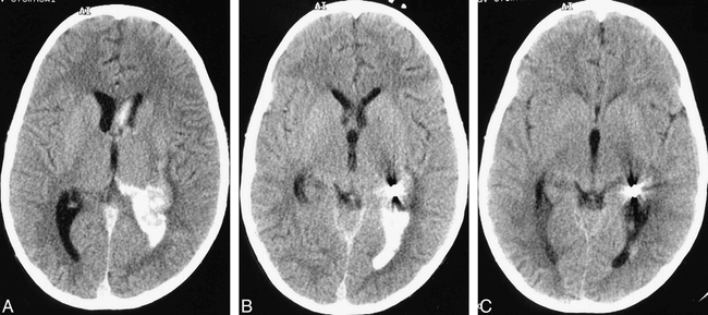
Fig 3. Postprocedural CT of brain is shown.
A and B, Immediate postembolization nonenhanced brain CT scan showing high-attenuation material consistent with iodinated contrast medium within dependent portion of left lateral ventricle. Size of the intraventricular hematoma has not increased.
C, Postembolization (at 20 hr), nonenhanced brain CT scan demonstrating almost complete resorption of high-attenuation iodinated contrast medium.


