
Article Figures & Data
Figures
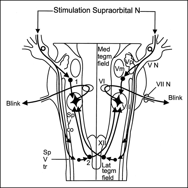
- fig 1.
Blink reflex. Diagram shows presumed location of the bulbar interneurons subserving the two components of the blink reflex: (1) interneurons subserving the ipsilateral early components; (2) interneurons subserving the bilateral late component. (Vm indicates trigeminal motor nucleus; Sp V co, spinal trigeminal complex; Sp V tr, spinal trigeminal tract; VI, abducens nucleus; VII, facial nucleus; VII, facial nerve; VN, trigeminal sensory root; XII, hypoglossal nucleus; Lat tegm field, lateral tegmental field; Med tegm field, medial tegmental field.) Modified from (5) and used with permission
- fig 2.
Masseter inhibitory reflex. Diagram shows presumed location of the bulbar interneurons subserving (1) the early (SP1) phase and (2) the late (SP2) phase of the masseter inhibitory reflex. (VN sens indicates trigeminal sensory root; VN mot, trigeminal motor root; for other abbreviations, see legend to fig 1.) Modified from (16) and used with permission
- fig 3.
Jaw-jerk reflex. Diagram shows the reflex arc subserving the jaw-jerk reflex. (Vmes indicates mesencephalic nucleus of the trigeminal nerve; Vp, principal sensory nucleus of the trigeminal nerve; Vm, trigeminal motor nucleus; Sp V tr, spinal trigeminal tract; III N, oculomotor nerve; Ohpth N, ophthalmic trigeminal root; Max N, maxillary trigeminal root; Mand N, mandibular trigeminal root; Mot Root V N, trigeminal motor root; VI N, abducens nerve, Mmsp, masseter muscle spindle; Mmf, masseter muscle fibers.) Modified from (5) and used with permission
- fig 4.
Patient 2: 52-year-old man with a sensory deficit in the first, second, and third divisions of the left trigeminal nerve and a sensory deficit on the right side of the body. Blink reflex recorded to left supraorbital nerve stimulation evoked a normal R1 response, whereas R2 responses were bilaterally delayed. R1 and R2 reflex recordings to right supraorbital nerve stimulation were normal. This lesion corresponds to the location of the left descending trigeminal tract and its nucleus in the dorsolateral part of the medulla oblongata. Axial T2-weighted MR image confirms the presence and location of the lesion in the left dorsolateral part of the medulla oblongata (arrow).
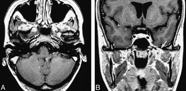
fig 5. Patient 3: 56-year-old woman with a sensory deficit in the second division of the trigeminal nerve. Blink reflex recorded to left supraorbital nerve stimulation showed the R1 to be absent and the R2 components delayed bilaterally. This lesion corresponds to the location of the left principal sensory nucleus in the pons with involvement of the descending trigeminal tract. Axial T2-weighted MR image (3500/90) confirms the location of the lesion in the left lower dorsal half of the pons, including the region of the left principal sensory nucleus (arrow).
- fig 6.
Patient 13: 25-year-old woman with pain in the first trigeminal division. Sensory loss was found in all three trigeminal divisions, and oculomotor, abducens, and facial nerve palsies were present. Blink reflex recorded to left supraorbital nerve stimulation showed a delayed R1 and normal R2 components. The left jaw-jerk reflex response was absent. The left masseter inhibitory reflex showed an afferent delay (ie, a bilateral delay of the first and second silent periods). All reflex responses were normal after stimulation of the right side. The abnormal reflex findings corresponded to a proximal trigeminal nerve lesion, probably at the root level.
A and B, Axial (A) and coronal (B) contrast-enhanced T1-weighted MR images show enhancement and thickening of the trigeminal ganglion and proximal third division of the trigeminal nerve in the foramen ovale (arrows). Follow-up MR imaging studies (not shown) and clinical findings showed resolution of the abnormalities, indicating an inflammatory lesion.


Tables
 Table 1:
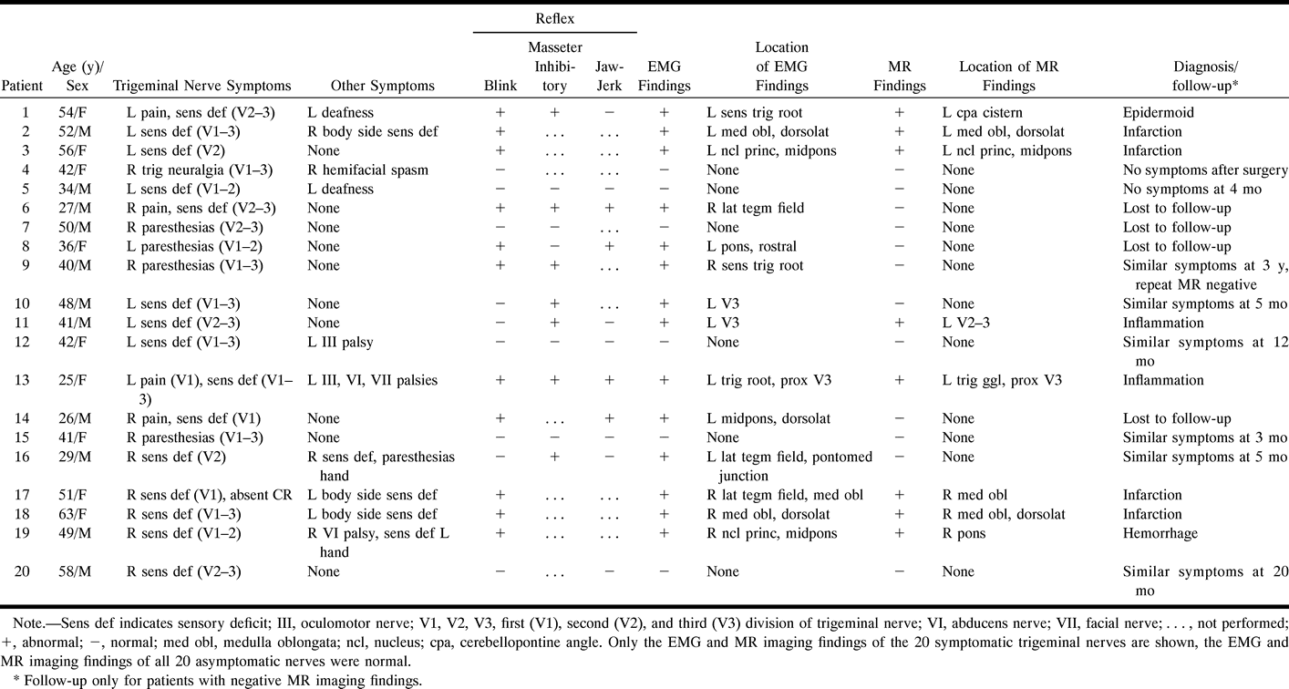
Clinical, EMG reflex, and MR imaging findings in 20 patients with symptoms related to the trigeminal nerve
- Table 2:
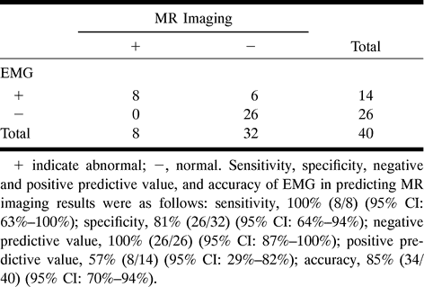
EMG versus MR imaging results in 40 trigeminal nerves (20 symptomatic)


