Advertisement

Main menu

  • Home
  • Content
    • Current Issue
    • Accepted Manuscripts
    • Article Preview
    • Past Issue Archive
    • Video Articles
    • AJNR Case Collection
    • Case of the Week Archive
    • Case of the Month Archive
    • Classic Case Archive
  • Special Collections
    • AJNR Awards
    • Low-Field MRI
    • Alzheimer Disease
    • ASNR Foundation Special Collection
    • Photon-Counting CT
    • View All
  • Multimedia
    • AJNR Podcasts
    • AJNR SCANtastic
    • Trainee Corner
    • MRI Safety Corner
    • Imaging Protocols
  • For Authors
    • Submit a Manuscript
    • Submit a Video Article
    • Submit an eLetter to the Editor/Response
    • Manuscript Submission Guidelines
    • Statistical Tips
    • Fast Publishing of Accepted Manuscripts
    • Graphical Abstract Preparation
    • Imaging Protocol Submission
    • Author Policies
  • About Us
    • About AJNR
    • Editorial Board
    • Editorial Board Alumni
  • More
    • Become a Reviewer/Academy of Reviewers
    • Subscribers
    • Permissions
    • Alerts
    • Feedback
    • Advertisers
    • ASNR Home

User menu

  • Alerts
  • Log in

Search

  • Advanced search
American Journal of Neuroradiology

American Journal of Neuroradiology

ASHNR American Society of Functional Neuroradiology ASHNR American Society of Pediatric Neuroradiology ASSR
  • Alerts
  • Log in

Advanced Search

  • Home
  • Content
    • Current Issue
    • Accepted Manuscripts
    • Article Preview
    • Past Issue Archive
    • Video Articles
    • AJNR Case Collection
    • Case of the Week Archive
    • Case of the Month Archive
    • Classic Case Archive
  • Special Collections
    • AJNR Awards
    • Low-Field MRI
    • Alzheimer Disease
    • ASNR Foundation Special Collection
    • Photon-Counting CT
    • View All
  • Multimedia
    • AJNR Podcasts
    • AJNR SCANtastic
    • Trainee Corner
    • MRI Safety Corner
    • Imaging Protocols
  • For Authors
    • Submit a Manuscript
    • Submit a Video Article
    • Submit an eLetter to the Editor/Response
    • Manuscript Submission Guidelines
    • Statistical Tips
    • Fast Publishing of Accepted Manuscripts
    • Graphical Abstract Preparation
    • Imaging Protocol Submission
    • Author Policies
  • About Us
    • About AJNR
    • Editorial Board
    • Editorial Board Alumni
  • More
    • Become a Reviewer/Academy of Reviewers
    • Subscribers
    • Permissions
    • Alerts
    • Feedback
    • Advertisers
    • ASNR Home
  • Follow AJNR on Twitter
  • Visit AJNR on Facebook
  • Follow AJNR on Instagram
  • Join AJNR on LinkedIn
  • RSS Feeds

AJNR Awards, New Junior Editors, and more. Read the latest AJNR updates

Research ArticleBRAIN

Decreased BOLD Functional MR Activation of the Motor and Sensory Cortices Adjacent to a Glioblastoma Multiforme: Implications for Image-Guided Neurosurgery

Andrei I. Holodny, Michael Schulder, Wen Ching Liu, Joseph A. Maldjian and Andrew J. Kalnin
American Journal of Neuroradiology April 1999, 20 (4) 609-612;
Andrei I. Holodny
  • Find this author on Google Scholar
  • Find this author on PubMed
  • Search for this author on this site
Michael Schulder
  • Find this author on Google Scholar
  • Find this author on PubMed
  • Search for this author on this site
Wen Ching Liu
  • Find this author on Google Scholar
  • Find this author on PubMed
  • Search for this author on this site
Joseph A. Maldjian
  • Find this author on Google Scholar
  • Find this author on PubMed
  • Search for this author on this site
Andrew J. Kalnin
  • Find this author on Google Scholar
  • Find this author on PubMed
  • Search for this author on this site
  • Article
  • Figures & Data
  • Info & Metrics
  • Responses
  • References
  • PDF
Loading

Article Figures & Data

Figures

  • fig 1.
    • Download figure
    • Open in new tab
    • Download powerpoint
    fig 1.

    Axial T1-weighted images with coregistered functional MR images obtained during a bilateral motor paradigm show a larger volume of activation on the normal side than on the side with the tumor (arrows). This effect is seen for different correlation coefficients (r). The red areas indicate significant activation for r = .48, P < .01. The yellow areas indicate significant activation for r = .60, P < .01. Notwithstanding the difference in the volume of activation, one is still able to identify the motor cortex on the side with the tumor. The motor cortex on the right is displaced anteriorly and superiorly by the tumor mass. The accessory motor area is seen at the midline in the superiormost image (bottom right)
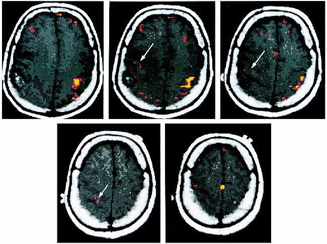
  • fig 2.
    • Download figure
    • Open in new tab
    • Download powerpoint
    fig 2.

    Axial T1-weighted images with coregistered functional MR images obtained during right-hand (top row, left sensory cortex) and left-hand (bottom row, right sensory cortex) sensory paradigms. The right-hand sensory paradigm shows robust activation in the left postcentral gyrus at different correlation coefficients (r). The red areas indicate activation for r = .48, P < .01. The yellow areas indicate activation for r = .60, P < .01. The left-hand sensory paradigm fails to show activation in the right postcentral gyrus (located just anterior to the tumor mass), even with P < .10

PreviousNext
Back to top

In this issue

American Journal of Neuroradiology
Vol. 20, Issue 4
1 Apr 1999
  • Table of Contents
  • Index by author
Advertisement
Print
Download PDF
Email Article

Thank you for your interest in spreading the word on American Journal of Neuroradiology.

NOTE: We only request your email address so that the person you are recommending the page to knows that you wanted them to see it, and that it is not junk mail. We do not capture any email address.

Enter multiple addresses on separate lines or separate them with commas.
Decreased BOLD Functional MR Activation of the Motor and Sensory Cortices Adjacent to a Glioblastoma Multiforme: Implications for Image-Guided Neurosurgery
(Your Name) has sent you a message from American Journal of Neuroradiology
(Your Name) thought you would like to see the American Journal of Neuroradiology web site.
CAPTCHA
This question is for testing whether or not you are a human visitor and to prevent automated spam submissions.
Cite this article
Andrei I. Holodny, Michael Schulder, Wen Ching Liu, Joseph A. Maldjian, Andrew J. Kalnin
Decreased BOLD Functional MR Activation of the Motor and Sensory Cortices Adjacent to a Glioblastoma Multiforme: Implications for Image-Guided Neurosurgery
American Journal of Neuroradiology Apr 1999, 20 (4) 609-612;

Citation Manager Formats

  • BibTeX
  • Bookends
  • EasyBib
  • EndNote (tagged)
  • EndNote 8 (xml)
  • Medlars
  • Mendeley
  • Papers
  • RefWorks Tagged
  • Ref Manager
  • RIS
  • Zotero
0 Responses
Respond to this article
Share
Bookmark this article
Decreased BOLD Functional MR Activation of the Motor and Sensory Cortices Adjacent to a Glioblastoma Multiforme: Implications for Image-Guided Neurosurgery
Andrei I. Holodny, Michael Schulder, Wen Ching Liu, Joseph A. Maldjian, Andrew J. Kalnin
American Journal of Neuroradiology Apr 1999, 20 (4) 609-612;
del.icio.us logo Twitter logo Facebook logo Mendeley logo
  • Tweet Widget
  • Facebook Like
  • Google Plus One

Jump to section

  • Article
    • Abstract
    • Case Report
    • Discussion
    • Conclusion
    • Footnotes
    • References
  • Figures & Data
  • Info & Metrics
  • Responses
  • References
  • PDF

Related Articles

  • No related articles found.
  • PubMed
  • Google Scholar

Cited By...

  • Glioma-Induced Disruption of Resting-State Functional Connectivity and Amplitude of Low-Frequency Fluctuations in the Salience Network
  • A measure of vascular reactivity to overcome neurovascular uncoupling in functional imaging of brain tumors: initial results
  • Local Glioma Cells Are Associated with Vascular Dysregulation
  • Anatomic Location of Tumor Predicts the Accuracy of Motor Function Localization in Diffuse Lower-Grade Gliomas Involving the Hand Knob Area
  • Clinical applications of imaging biomarkers. Part 2. The neurosurgeon's perspective
  • Isolated Translocation of Wernicke's Area to the Right Hemisphere in a 62-Year-Man with a Temporo-Parietal Glioma
  • Imaging of intracranial tumours
  • Perimetric visual field and functional MRI correlation: implications for image-guided surgery in occipital brain tumours
  • MEG versus BOLD MR Imaging: Functional Imaging, the Next Generation?
  • The Effect of Brain Tumors on BOLD Functional MR Imaging Activation in the Adjacent Motor Cortex: Implications for Image-guided Neurosurgery
  • Crossref
  • Google Scholar

This article has not yet been cited by articles in journals that are participating in Crossref Cited-by Linking.

More in this TOC Section

  • Usefulness of Quantitative Susceptibility Mapping for the Diagnosis of Parkinson Disease
  • White Matter Alterations in the Brains of Patients with Active, Remitted, and Cured Cushing Syndrome: A DTI Study
  • Qualitative and Quantitative Analysis of MR Imaging Findings in Patients with Middle Cerebral Artery Stroke Implanted with Mesenchymal Stem Cells
Show more BRAIN

Similar Articles

Advertisement

Indexed Content

  • Current Issue
  • Accepted Manuscripts
  • Article Preview
  • Past Issues
  • Editorials
  • Editor's Choice
  • Fellows' Journal Club
  • Letters to the Editor
  • Video Articles

Cases

  • Case Collection
  • Archive - Case of the Week
  • Archive - Case of the Month
  • Archive - Classic Case

More from AJNR

  • Trainee Corner
  • Imaging Protocols
  • MRI Safety Corner

Multimedia

  • AJNR Podcasts
  • AJNR Scantastics

Resources

  • Turnaround Time
  • Submit a Manuscript
  • Submit a Video Article
  • Submit an eLetter to the Editor/Response
  • Manuscript Submission Guidelines
  • Statistical Tips
  • Fast Publishing of Accepted Manuscripts
  • Graphical Abstract Preparation
  • Imaging Protocol Submission
  • Evidence-Based Medicine Level Guide
  • Publishing Checklists
  • Author Policies
  • Become a Reviewer/Academy of Reviewers
  • News and Updates

About Us

  • About AJNR
  • Editorial Board
  • Editorial Board Alumni
  • Alerts
  • Permissions
  • Not an AJNR Subscriber? Join Now
  • Advertise with Us
  • Librarian Resources
  • Feedback
  • Terms and Conditions
  • AJNR Editorial Board Alumni

American Society of Neuroradiology

  • Not an ASNR Member? Join Now

© 2025 by the American Society of Neuroradiology All rights, including for text and data mining, AI training, and similar technologies, are reserved.
Print ISSN: 0195-6108 Online ISSN: 1936-959X

Powered by HighWire