
Article Figures & Data
Figures
- fig 1.
Life cycle of Coccidioides immitis.
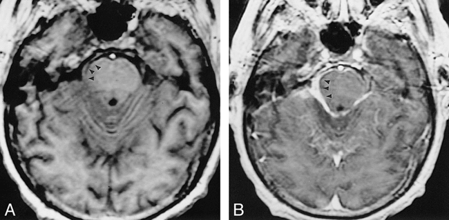
- fig 2.
Case 7: 28-year-old woman.
A, Coronal contrast-enhanced T1-weighted image shows diffuse abnormal meningeal enhancement (arrow) along the course of the middle and anterior cerebral arteries. A shunt catheter (arrowheads) has been placed for acute hydrocephalus.
B, Axial T2-weighted image shows edema in the deep nuclei bilaterally (arrows). This is believed to represent areas of infarction/ischemia consequent to coccidioidal vasculitis.
fig 3. Case 1: 36-year-old man. Findings on initial study were normal (not shown). Follow-up axial contrast-enhanced T1-weighted image 2 months later shows discrete nodules of abnormal enhancement (arrow) in the perimesencephalic cistern.
- fig 4.
Case 2: 22-year-old woman with disease progression.
A, Initial coronal contrast-enhanced T1-weighted MR image with focal enhancement in the left sylvian fissure (arrow).
B, Follow-up study 2 weeks later shows a new small focus of abnormal meningeal enhancement (arrow) on the medial aspect of the uncus.
C and D, Another week later, there is marked progression of disease to diffusely involve the basilar cisterns (arrows) as well as the course of the middle cerebral arteries (arrowheads).
- fig 5.
Case 4: 70-year-old man.
A, Unenhanced T1-weighted image shows abnormal soft-tissue mass in the ambient cistern (arrows).
B, Dense enhancement (arrowheads) is seen after administration of gadolinium chelate.
- fig 6.
Case 10: Low signal on T2-weighted images.
A, Axial T2-weighted image shows decreased signal (arrow) in the prepontine cistern.
B, Contrast-enhanced T1-weighted sequence shows marked enhancement (arrow) of the same area.


Tables

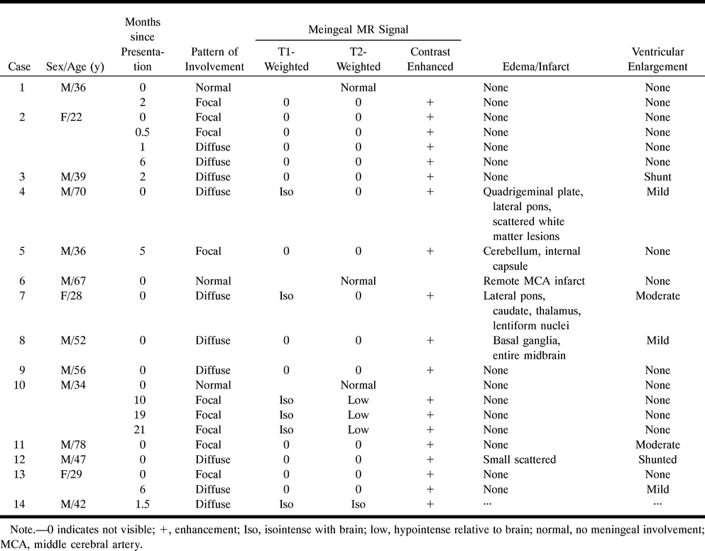
- TABLE 2:
Summary of imaging findings in 14 patients at presentation (one patient had an initial CT scan)
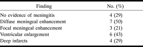
- TABLE 3:
Summary of findings in 13 patients on the first abnormal MR examination (no follow-up examination was available for one patient with an initially normal study)


