Advertisement

Main menu

  • Home
  • Content
    • Current Issue
    • Accepted Manuscripts
    • Article Preview
    • Past Issue Archive
    • Video Articles
    • AJNR Case Collection
    • Case of the Week Archive
    • Case of the Month Archive
    • Classic Case Archive
  • Special Collections
    • AJNR Awards
    • Low-Field MRI
    • Alzheimer Disease
    • ASNR Foundation Special Collection
    • Photon-Counting CT
    • View All
  • Multimedia
    • AJNR Podcasts
    • AJNR SCANtastic
    • Trainee Corner
    • MRI Safety Corner
    • Imaging Protocols
  • For Authors
    • Submit a Manuscript
    • Submit a Video Article
    • Submit an eLetter to the Editor/Response
    • Manuscript Submission Guidelines
    • Statistical Tips
    • Fast Publishing of Accepted Manuscripts
    • Graphical Abstract Preparation
    • Imaging Protocol Submission
    • Author Policies
  • About Us
    • About AJNR
    • Editorial Board
    • Editorial Board Alumni
  • More
    • Become a Reviewer/Academy of Reviewers
    • Subscribers
    • Permissions
    • Alerts
    • Feedback
    • Advertisers
    • ASNR Home

User menu

  • Alerts
  • Log in

Search

  • Advanced search
American Journal of Neuroradiology

American Journal of Neuroradiology

ASHNR American Society of Functional Neuroradiology ASHNR American Society of Pediatric Neuroradiology ASSR
  • Alerts
  • Log in

Advanced Search

  • Home
  • Content
    • Current Issue
    • Accepted Manuscripts
    • Article Preview
    • Past Issue Archive
    • Video Articles
    • AJNR Case Collection
    • Case of the Week Archive
    • Case of the Month Archive
    • Classic Case Archive
  • Special Collections
    • AJNR Awards
    • Low-Field MRI
    • Alzheimer Disease
    • ASNR Foundation Special Collection
    • Photon-Counting CT
    • View All
  • Multimedia
    • AJNR Podcasts
    • AJNR SCANtastic
    • Trainee Corner
    • MRI Safety Corner
    • Imaging Protocols
  • For Authors
    • Submit a Manuscript
    • Submit a Video Article
    • Submit an eLetter to the Editor/Response
    • Manuscript Submission Guidelines
    • Statistical Tips
    • Fast Publishing of Accepted Manuscripts
    • Graphical Abstract Preparation
    • Imaging Protocol Submission
    • Author Policies
  • About Us
    • About AJNR
    • Editorial Board
    • Editorial Board Alumni
  • More
    • Become a Reviewer/Academy of Reviewers
    • Subscribers
    • Permissions
    • Alerts
    • Feedback
    • Advertisers
    • ASNR Home
  • Follow AJNR on Twitter
  • Visit AJNR on Facebook
  • Follow AJNR on Instagram
  • Join AJNR on LinkedIn
  • RSS Feeds

AJNR Awards, New Junior Editors, and more. Read the latest AJNR updates

Research ArticleReview Articles

Newer Sequences for Spinal MR Imaging: Smorgasbord or Succotash of Acronyms?

Jeffrey S. Ross
American Journal of Neuroradiology March 1999, 20 (3) 361-373;
Jeffrey S. Ross
  • Find this author on Google Scholar
  • Find this author on PubMed
  • Search for this author on this site
  • Article
  • Figures & Data
  • Info & Metrics
  • Responses
  • References
  • PDF
Loading

Article Figures & Data

Figures

  • Tables
  • fig 1.
    • Download figure
    • Open in new tab
    • Download powerpoint
    fig 1.

    EPI diffusion-sequence structure. The 90° and 180° RF pulses are followed by a bipolar, trapezoidal frequency encode gradient (Gx) for rapid collection of multiple echos. DWI is applied by symmetrical gradients along a frequency-encoded direction (black rectangles). Subsequent sequence acquisitions would apply diffusion weighting along phase (Gy) and slice-select (Gz) directions

  • fig 2.
    • Download figure
    • Open in new tab
    • Download powerpoint
    fig 2.

    Half Fourier schematic. Slightly more than half of the data can be collected and used to “fill in” the remainder of k-space because the data is assumed to be symmetrical. This partial data acquisition shortens imaging time.

    fig 3. Interpolation schematic. Matrix is expanded by addition of “place holder” data, allowing reconstruction, for example, of a 512 matrix from a 256 data set. Appearance of image will be filtered due to more heavy weighting from central k-space data.
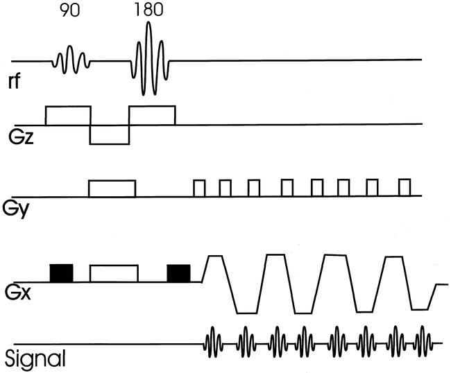
  • fig 4.
    • Download figure
    • Open in new tab
    • Download powerpoint
    fig 4.

    3D GE with slice interpolation (36/15/1). Three contiguous slices reconstructed at 1.5 mm and acquired at 3 mm with slice interpolation. The advantage is that neural foramina are encompassed by multiple images with very-thin-slice reconstruction.

  • fig 5.
    • Download figure
    • Open in new tab
    • Download powerpoint
    fig 5.

    Fast FLAIR sequence structure. Typical FSE sequence structure of multiple 180° pulses is modified by addition of a 180° inversion pulse, followed by a delay time until the alpha pulse (inversion time or TI). CSF is suppressed by appropriate selection of inversion time, which for FLAIR is approximately 2000 ms. Effective TE is determined by low-amplitude phase-encoding steps (central k-space). S = slice-select direction, R = “read” or frequency-encode direction, P = phase-encode direction

  • fig 6.
    • Download figure
    • Open in new tab
    • Download powerpoint
    fig 6.

    False-negative fast FLAIR for demyelinating disease.

    A, Sagittal T1-weighted image (500/12/2) demonstrates a markedly enlarged cord with slighted decreased signal centrally.

    B, Sagittal T2-weighted FSE (4620/112/ 3) shows diffuse increased signal throughout cervical cord.

    C, Sagittal FSE FLAIR (6000/105/ 2) shows low signal at C7-T1 level, but no abnormal increased signal as on the FSE.
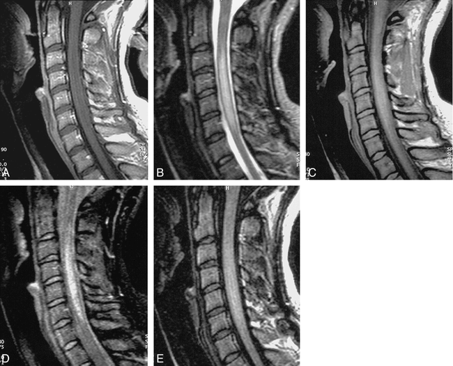
  • fig 7.
    • Download figure
    • Open in new tab
    • Download powerpoint
    fig 7.

    Fast STIR sequence structure. This is analogous to FLAIR sequence, except that TI time is shorter to null fat signal, and low-amplitude phase-encode steps are acquired earlier. S = slice-select direction, R = “read” or frequency- encode direction, P = phase-encode direction

  • fig 8.
    • Download figure
    • Open in new tab
    • Download powerpoint
    fig 8.

    T2W vs. FLAIR vs. STIR in demyelinating disease.

    A, Fusiform enlargement of cord without enhancement is shown on sagittal T1-weighted sequence (500/12/2).

    B, Abnormal high signal within cord is shown on sagittal FSE T2-weighted sequence (4620/112/ 3).

    C and D, FSE spin-density weighted (2000/10/2), and FSE STIR (1200/14/4), respectively.

    E, Abnormal cord signal is not revealed by fast FLAIR sequence (6000/105/ 2). Lesion is most conspicuous on FSE T2-weighted and fast STIR sequences.
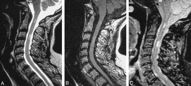
  • fig 9.
    • Download figure
    • Open in new tab
    • Download powerpoint
    fig 9.

    Chronic demyelinating disease.

    A, Sagittal T2-weighted FSE (4620/112/ 3) shows faint focal increased signal in cervical cord at C1 and C3 levels.

    B, Sagittal FSE FLAIR (6000/105/2) also shows very indistinct abnormal signal at those two levels.

    C, High lesion-to-cord contrast is achieved with fast STIR sequence (1200/14/4).

  • fig 10.
    • Download figure
    • Open in new tab
    • Download powerpoint
    fig 10.

    Echo-planar diffusion imaging of the normal cervical cord. Three orthogonal directions of diffusion gradients are applied: anteroposterior (A); transverse (B); and through-plane (C). Notice the least signal from the cord with through-plane diffusion encoding (parallel to white matter tracts) reflecting direction of relatively fastest water diffusion

  • fig 11.
    • Download figure
    • Open in new tab
    • Download powerpoint
    fig 11.

    MT contrast. Axial gradient echo slice without (A) and with (B) application of off-resonance MT pulse. Application of MT dramatically improves cord/CSF contrast.
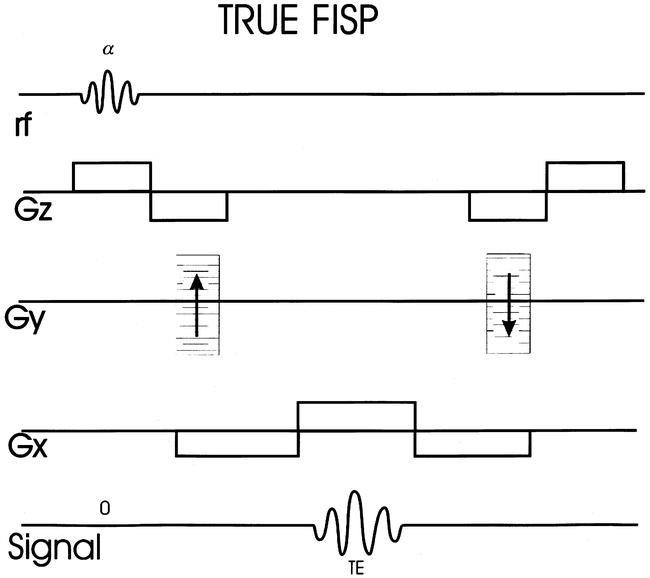
  • fig 12.
    • Download figure
    • Open in new tab
    • Download powerpoint
    fig 12.

    True FISP sequence structure with balanced gradients. Net effect of gradients allows spins that are stationary as well as those moving with constant velocity to reach a steady state. Gz = slice select gradient, Gy = phase encode gradient, and Gx = frequency encode gradient.
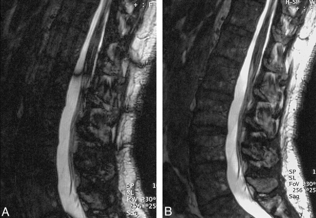
  • fig 13.
    • Download figure
    • Open in new tab
    • Download powerpoint
    fig 13.

    True FISP (17/8/2, 70° flip angle).

    A, Sagittal 2-mm slice from one of the two sequences acquired with different RF phase (combined to produce final image) demonstrates areas of banding or signal loss related to nonuniform resonant offset.

    B, Combined final sequence shows more uniform high-signal CSF with relative suppression of soft-tissue signal.
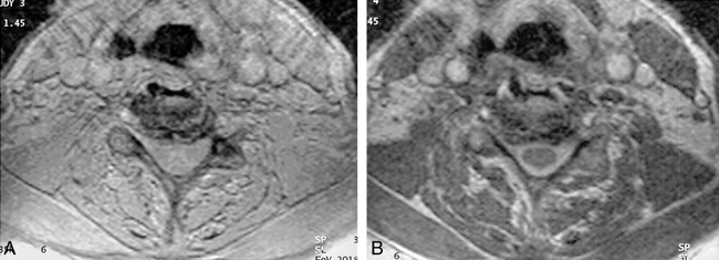
  • fig 14.
    • Download figure
    • Open in new tab
    • Download powerpoint
    fig 14.

    3D CISS (12/6/3, 70° flip angle). Axial 2-mm section through cervical spine shows sharp interface between cord/intradural dorsal and ventral roots (arrows) and the CSF. There is slight truncation artifact surrounding the cord, manifest as curvilinear low signal.

    fig 15. PSIF diffusion-sequence structure (aka, FISP backwards).Diffusion weighting is applied as a single gradient along slice-select direction. Acquired signal is an RF echo. Echo occurs prior to alpha pulse because it is generated by the refoccussing of magnetization that has resided in transverse plane over at least one previous complete TR cycle. Gz = slice-select gradient, Gy = phase- encode gradient, Gx = frequency-encode gradient.

  • fig 16.
    • Download figure
    • Open in new tab
    • Download powerpoint
    fig 16.

    Diffusion true positive in patient with myeloma (PSIF 22/2/10, 75° flip angle).

    A, Sagittal T1-weighted image shows diffuse abnormal marrow signal with mild compression fracture.

    B, Sagittal PSIF sequence with diffusion gradient shows high signal from compression fracture comfirming malignant origin.
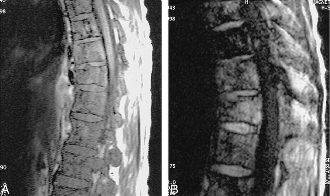
  • fig 17.
    • Download figure
    • Open in new tab
    • Download powerpoint
    fig 17.

    Diffusion false positive in trauma (PSIF 22/2/10, 75° flip angle) found in a 17-year-old who sustained a flexion injury at C3–4 after going over handlebars of waterski.

    A and B, Sagittal T1-weighted (A) and T2-weighted (B) images show anterior wedge deformities of C3 and C4 bodies.

    C, Diffusion sequence shows slight increased signal from bodies, falsely suggesting a cellular infiltrate.

Tables

  • Figures
  • TABLE 1:
    • View popup
    • Download powerpoint
    TABLE 1:

    FLAIR and MS in the cord

  • TABLE 2:
    • View popup
    • Download powerpoint
    TABLE 2:

    STIR and cord disease

  • TABLE 3:
    • View popup
    • Download powerpoint
    TABLE 3:

    Bright CSF GE imaging and the cervical spine

PreviousNext
Back to top

In this issue

American Journal of Neuroradiology
Vol. 20, Issue 3
1 Mar 1999
  • Table of Contents
  • Index by author
Advertisement
Print
Download PDF
Email Article

Thank you for your interest in spreading the word on American Journal of Neuroradiology.

NOTE: We only request your email address so that the person you are recommending the page to knows that you wanted them to see it, and that it is not junk mail. We do not capture any email address.

Enter multiple addresses on separate lines or separate them with commas.
Newer Sequences for Spinal MR Imaging: Smorgasbord or Succotash of Acronyms?
(Your Name) has sent you a message from American Journal of Neuroradiology
(Your Name) thought you would like to see the American Journal of Neuroradiology web site.
CAPTCHA
This question is for testing whether or not you are a human visitor and to prevent automated spam submissions.
Cite this article
Jeffrey S. Ross
Newer Sequences for Spinal MR Imaging: Smorgasbord or Succotash of Acronyms?
American Journal of Neuroradiology Mar 1999, 20 (3) 361-373;

Citation Manager Formats

  • BibTeX
  • Bookends
  • EasyBib
  • EndNote (tagged)
  • EndNote 8 (xml)
  • Medlars
  • Mendeley
  • Papers
  • RefWorks Tagged
  • Ref Manager
  • RIS
  • Zotero
0 Responses
Respond to this article
Share
Bookmark this article
Newer Sequences for Spinal MR Imaging: Smorgasbord or Succotash of Acronyms?
Jeffrey S. Ross
American Journal of Neuroradiology Mar 1999, 20 (3) 361-373;
del.icio.us logo Twitter logo Facebook logo Mendeley logo
  • Tweet Widget
  • Facebook Like
  • Google Plus One

Jump to section

  • Article
    • Techniques
    • Spinal Cord Abnormalities
    • Extramedullary/Bony Abnormalities
    • Conclusion
    • Acknowledgments
    • Footnotes
    • References
  • Figures & Data
  • Info & Metrics
  • Responses
  • References
  • PDF

Related Articles

  • No related articles found.
  • PubMed
  • Google Scholar

Cited By...

  • Are T1 weighted images helpful in MRI of cervical radiculopathy?
  • Diagnostic and therapeutic radiology of the spine: an overview
  • The American Journal of Neuroradiology 1980-1999 Where We Have Been: Where We Are Going
  • Crossref
  • Google Scholar

This article has not yet been cited by articles in journals that are participating in Crossref Cited-by Linking.

More in this TOC Section

  • An Atlas of Neonatal Neurovascular Imaging Anatomy as Depicted with Microvascular Imaging: The Intracranial Arteries
  • An Atlas of Neonatal Neurovascular Imaging Anatomy as Depicted with Microvascular Imaging: The Intracranial Veins
  • Clinical Translation of Hyperpolarized 13C Metabolic Probes for Glioma Imaging
Show more Review Articles

Similar Articles

Advertisement

Indexed Content

  • Current Issue
  • Accepted Manuscripts
  • Article Preview
  • Past Issues
  • Editorials
  • Editor's Choice
  • Fellows' Journal Club
  • Letters to the Editor
  • Video Articles

Cases

  • Case Collection
  • Archive - Case of the Week
  • Archive - Case of the Month
  • Archive - Classic Case

More from AJNR

  • Trainee Corner
  • Imaging Protocols
  • MRI Safety Corner
  • Book Reviews

Multimedia

  • AJNR Podcasts
  • AJNR Scantastics

Resources

  • Turnaround Time
  • Submit a Manuscript
  • Submit a Video Article
  • Submit an eLetter to the Editor/Response
  • Manuscript Submission Guidelines
  • Statistical Tips
  • Fast Publishing of Accepted Manuscripts
  • Graphical Abstract Preparation
  • Imaging Protocol Submission
  • Evidence-Based Medicine Level Guide
  • Publishing Checklists
  • Author Policies
  • Become a Reviewer/Academy of Reviewers
  • News and Updates

About Us

  • About AJNR
  • Editorial Board
  • Editorial Board Alumni
  • Alerts
  • Permissions
  • Not an AJNR Subscriber? Join Now
  • Advertise with Us
  • Librarian Resources
  • Feedback
  • Terms and Conditions
  • AJNR Editorial Board Alumni

American Society of Neuroradiology

  • Not an ASNR Member? Join Now

© 2025 by the American Society of Neuroradiology All rights, including for text and data mining, AI training, and similar technologies, are reserved.
Print ISSN: 0195-6108 Online ISSN: 1936-959X

Powered by HighWire