
Abstract
BACKGROUND AND PURPOSE: An outbreak of enterovirus infection occurred in Taiwan from late spring to early fall of 1998. Most of the pediatric infections presented as hand-foot-mouth disease (HFMD) and herpangina. A small portion of patients had symptoms of polio-like encephalitis and paralysis. The purpose of this study was to review the MR imaging findings in CNS involvement of enterovirus infection.
METHODS: Twenty patients who had HFMD and clinical encephalitis were examined with MR imaging. T1-weighted and T2-weighted MR images were obtained. From the rectum, throat, CSF, and peripheral blood, the presence of enterovirus 71 (EV 71) was determined by virus culture, immunofluorescent microscopy, immunologic dot blotting, and reverse-transcription polymerase chain reaction.
RESULTS: MR imaging studies of 20 patients showed hyperintensity in the brain stem and spinal cord in 15 patients, as seen on T2-weighted images. The major CNS lesions were in the medulla oblongata, pons, midbrain, and the dentate nuclei of the cerebellum. In some cases, the lesions involved the spinal cord (three cases) as well as the thalamus (two cases) and putamina (one case). Five patients had normal MR imaging results. After the appropriate management for tachycardia and tachypnea, 18 patients recovered within 1 to 2 weeks. In the follow-up MR imaging examination of five patients, the lesions completely disappeared within 2 weeks to 2 months. In two patients who were still respirator-dependent, MR imaging showed the tissue destruction in the posterior portions of the medulla, pons, and the ventral horns of cervical spinal cord. In one patient, most of midbrain was damaged. The presence of EV 71 was detected in specimens from 18 patients.
CONCLUSION: Because EV 71 was identified in 18 patients, and no other virus was detected, EV 71 was determined to be the major causative agent of this encephalomyelitis. Brain stem and cervical spinal cord involvement are characteristic findings of enteroviral encephalomyelitis.
An outbreak of enterovirus infection occurred in Taiwan from April to October 1998. A large number of children contracted hand-foot-and-mouth disease (HFMD) or herpangina. Most of the children had fever, vesicles on the hands, feet, elbows and knees, and vesicles with ulceration on the mucosa of the lips (HFMD). Some had erythematous ulcers on the palate and pharynx (herpangina). A number of children were hospitalized with suspicious meningitis or encephalitis. Seventy-two children had a short febrile illness, decompensated suddenly, went into respiratory distress, and suddenly died within 12 to 48 hours. Virus isolation proved that enterovirus 71 (EV 71) was the causative agent of these serious CNS complications.
The location of the EV 71 encephalitis was suspected to be in the brain stem because of the clinical manifestations of lethargy, cranial nerve palsies (cranial nerves VI, VII, IX, X, XI, and XII), conjugated disturbance of ocular movement, dyspnea, and tachycardia. The brain stem involvement by EV 71 was proved in autopsy reports (1, 2); however, to our knowledge there are no MR imaging reports of EV 71 encephalitis in the literature. The early detection of the locations of CNS involvement by EV 71 was helpful for the management of cardiopulmonary complication and decreased mortality.
Methods
There were 5632 children brought to the pediatric outpatient department or emergency unit of our institution for management of HFMD and herpangina. Of these, 126 were admitted because of restlessness, drowsiness, and dyspnea. Forty-one children were admitted into the pediatric intensive care unit (PICU) because of more serious CNS symptoms that included drowsiness, tachycardia, dyspnea, shock, semicoma, and coma. Two patients died in the emergency unit, and three died in the PICU.
According to the clinical manifestations, we classified five grades of symptoms:
Grade I (5506 Patients)
Fever, anorexia, with skin manifestations of erythematous vesicles on the feet, hands, elbows, and trunk, and oral ulcers on the mucosa of the lips (HFMD) or herpangina. The clinical course persisted for 2 to 7 days, averaging 3 days. Most patients recovered completely without sequela, but some patients worsened to grade II.
Grade II (83 Patients)
Fever, restlessness, photophobia, vomiting, headache, and abdominal pain. Most patients recovered, whereas the condition in some progressed to grade III. Patients younger than 2 years old may proceed directly into grade IV.
Grade III (20 Patients)
Fever, vomiting, lethargy, apathy, drowsiness, tachycardia, cranial nerve palsies (nerves VI, VII, IX, X, XI, XII), auditory hallucination, myoclonic jerks, hemiparesis or monoparesis, conjugated disturbance of ocular movement, dyspnea, and ataxia in older children. The above clinical manifestations were suspected brain stem symptoms owing to encephalitis. Some patients needed endotracheal tubes with cardiorespiratory monitoring. The symptoms may persist for 3 to 7 days, with recovery in 1 to 2 weeks. Those older than 2 years might recover completely, while those younger than 2 years had a tendency to proceed into grade IV.
Grade IV (18 Patients)
Hypothermia, pulmonary edema, respiratory failure, neurogenic shock, and semicoma.
Grade V (Five Patients)
Pulmonary hemorrhage, respiratory distress syndrome, cardiorespiratory failure, coma, and death.
Patients for MR Imaging Study
Patients with symptoms of grade III, IV, and V were suspected of having enteroviral encephalitis or meningitis. Although 126 patients were admitted, most were in grade II, so no MR imaging was arranged. Some were in grade IV but were under endotracheal tubes since their arrival to our emergency unit; therefore, MR imaging could not be performed. The five patients who expired were categorized as grade V when they were sent to the emergency room or PICU. Because these patients were on respirators, cardiac and pulmonary functions were monitored. The patients died after a very short time, so there was no opportunity for MR imaging. Fifteen patients categorized as grade III and five patients categorized as grade IV underwent MR imaging of the brain.
The ages of the 20 patients who underwent MR imaging ranged from 2 months to 7 years, and the mean age was 25 months. The MR imaging sequences were spin-echo T1-weighted (500–600/20/2 [TR/TE/excitations]) in the axial plane and T2-weighted (TR/TE/E 3200–3600/120/4–6) in the axial, sagittal, and coronal plane. No contrast medium was given.
Virus Identification
EV 71 was identified using the immunologic dot-blotting method. A total of 200 μL of filter-prepared sample collected from the throat swab, rectal swab, or CSF was siphoned through a well of the Bio-Dot apparatus (Bio-Rad, Hercules, CA) that was lined with a nitrocellulose membrane (MSI, Westboro, MA). The membrane was then incubated with antibodies specific to EV 71 (Chemicon, Temecula, CA) and probed with biotinylated rabbit-antimouse antibodies, together with alkaline phosphatase-conjugated streptavidin (Dako, Kyoto, Japan). A positive reaction was identified by developing with a chromogen containing nitroblue tetrazolium/5-bromo-4-chloro-3-inodolyl phosphate (NBT/BCIP; Boehringer Mannheim, Germany). Individual samples were tested in duplicate. Rectal and throat swabs from patients with bacteria-induced diarrhea and patients with human herpes simplex type I or cytomegalovirus infection were used as negative controls. A portion of the specimen was subjected to virus culture and immunofluorescent microscopy together with reverse-transcription polymerase chain reaction for confirming the EV 71 infection.
Results
Findings from MR imaging of the brains of the 15 patients with grade III clinical symptoms were normal in five patients, whereas 10 patients had positive findings in the brain stem (Table). All lesions presented as significantly hyperintense lesions in the posterior portions of the medulla oblongata (Fig 1A) and pons (Fig 1B), as seen in T2-weighted images. Six patients had lesions in most of the midbrain (Fig 1C), except in the corticospinal tracts of bilateral cerebral peduncles and tectum. Seven patients had lesions in the dentate nuclei of the cerebellum (Fig 1A). None had lesions in the spinal cord. The chest films were negative, except for one obtained from a patient who had consolidation in the right upper lobe, presumed to be pneumonia. These patients were admitted to the PICU. After appropriate management for tachycardia and tachypnea, they recovered well in 1 to 2 weeks, except for one patient who had left-leg monoplegia that persisted for 1 month. This patient also recovered completely. Five patients underwent MR imaging follow-up from 2 weeks to 2 months after recovery and the lesions had resolved (Fig 1D–F).
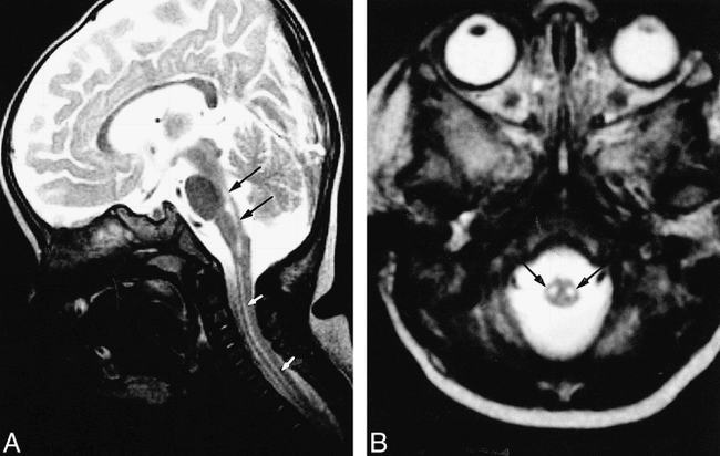
MR imaging lesion sites in EV 71 encephalomyelitis

Male patient, 4 years old. Acute EV 71 encephalitis. Patient presented with HFMD on June 10, 1998. Two days later, patient developed somnolence, tachycardia, and tachypnea. All MR images are T2-weighted images (3567/120/6 [TR/TE/excitations]).
A, Hyperintense lesions in the posterior portion of the medulla oblongata (arrow) and the bilateral dentate nuclei (arrowheads) of the cerebellum.
B, Hyperintense lesions in the posterior portion of the pons (arrow).
C, Hyperintense lesions in central-most portion of the midbrain (arrows).
D–F, Patient completely recovered without any sequelae. Follow-up MR imaging on July 29, 1998. The hyperintense lesions in the medulla, pons, midbrain, and dentate nuclei had disappeared. (The mild high-signal intensity of the posterior portion of the pons is normal in infants, possibly because of its under-myelinated status.)
In three of the five patients with grade IV clinical symptoms, T2-weighted images in the acute stage disclosed similar findings to those seen in patients with grade III manifestations. These three patients gradually recovered after adequate cardiopulmonary supportive treatment. Two patients did not undergo MR imaging in the acute stage; they were in a semicomatose state upon arrival to our emergency unit and were intubated and oxygen-dependent. Of the two, one patient gradually awoke and had clear consciousness 3 months later, but was still respirator-dependent. MR imaging was performed 3 months after onset of EV 71 infection. MR images showed abnormal signal in the posterior portions of the medulla oblongata and pons (Fig 2A) and symmetrical lesions in the bilateral ventral horns of the whole cervical spinal cord (Fig 2B). The second patient did not undergo MR imaging in the acute stage because of the seriousness of her condition upon arrival. Follow-up MR imaging was performed 3 months later when the patient was in a deep coma and vegetative state. MR images showed discrete lesions in the posterior aspect of the medulla (Fig 3A and B), pons (Fig 3C and D), and most of the midbrain (Fig 3E and F), the bilateral putamina, and the bilateral thalami (Fig 3G); symmetrical lesions also were in the bilateral ventral horns of the whole cervical spinal cord (Fig 3H). These lesions were well defined and dark on T1-weighted images and bright on T2-weighted images, reflecting old brain-tissue destruction.

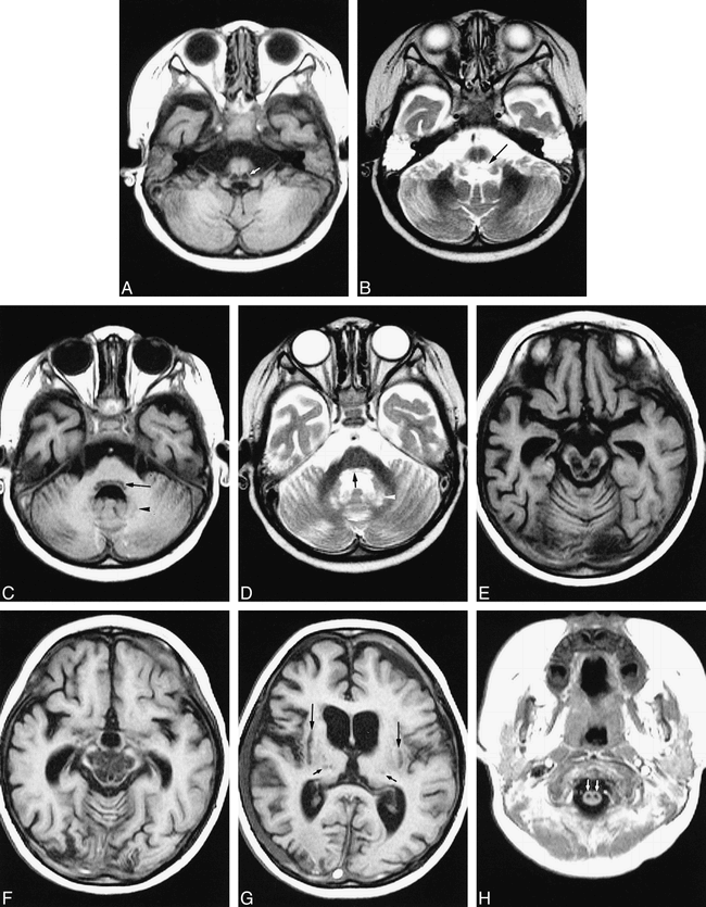
Female patient, 10 months old. Chronic stage of EV 71 encephalomyelitis. Patient presented with HFMD on June 20, 1998. Two days later, patient developed somnolence, tachycardia, tachypnea, and coma. Patient recovered very slowly, awaking in September. Patient remained ventilator- and oxygen-dependent. MR imaging was performed on September 22, 1998. All images are T2-weighted images (3560/120/6 [TR/TE/excitations]).
A, In sagittal sections, the lesions appear as a linear high signal in the posterior portions of the pons and medulla oblongata (arrows) and in the whole cervical spinal cord (white arrows).
B, Two symmetrical, well-defined hyperintense lesions in the cervical spinal cord (arrows), corresponding to the locations of ventral horns of the spinal cord.

C (T1-weighted image) and D (T2-weighted image), Discrete lesions also were seen in the posterior aspect of the pons (arrows) and dentate nuclei (arrowheads). The nuclei of V, VI, VII, IX nerves were destroyed.
E and F (T1-weighted images), Most of the midbrain was destroyed, including the red nuclei, substantia nuclei, the medial lemniscus, and the nuclei of III and IV nerves.
G (T1-weighted image), Lesions are noted in the bilateral thalami (short arrows) and the putamina (long arrows).
H (T1-weighted image), Two symmetrical lesions within the cervical spinal cord (arrows), corresponding to the locations of the ventral horns.
Female patient, 22 months old. Chronic stage of EV 71 encephalomyelitis. Patient presented with HFMD on July 17, 1998. Two days later, she developed a sudden consciousness change, tachycardia, tachypnea, then coma. MR imaging was performed on October 16, 1998, while the patient was in a vegetative state and respirator-dependent. T2-weighted images (3567/120/4 [TR/TE/exciations]) and T1-weighted images (553/20/2) were performed.
A and B, Very low-signal intensity lesions on T1-weighted image (A) and bright on T2-weighted image (B) in the posterior portion of the medulla oblongata (arrows). The dorsal nucleus of the vagus nerve, the nuclei of the solitary tract, and the medial longitudinal fasciculus were destroyed. There were fluid accumulations in the bilateral mastoids, reflecting mastoiditis.
The Table herein summarizes the MR imaging findings of the 20 patients of EV 71 encephalomyelitis with clinical symptoms of grade III and grade IV.
The five patients who expired (grade V) presented with diffuse interstitial consolidation in both lungs and reflected severe acute pulmonary edema and pulmonary hemorrhage. Because of rapid clinical demise and death, these five patients did not undergo MR imaging.
Among the 20 patients examined, 18 were confirmed with EV 71. No other virus was identified in the other two patients.
Discussion
The enteroviruses include Coxsackie viruses A and B, poliovirus, echoviruses, and enteroviruses 68 to 71. They may cause HFMD (Coxsackie virus A16, EV 71) (3), herpangina (EV 71), hemorrhagic conjunctivitis (enterovirus 70, Coxsackie virus A24) (4), poliomyelitis (poliovirus), polio-like paralysis, or radiculomyelitis (EV 70, EV 71, Coxsackie virus A7, A24) (5, 6).
Incidence of HFMD and herpangina are typical in the summer season in Taiwan. Nevertheless, an outbreak is rare, as is CNS involvement. In April and May 1998, an outbreak of HFMD began to spread rapidly around the island. This caused a panic in Taiwan because of publicity surrounding a small number of children who had a short febrile illness (2-day duration), decompensated suddenly, developed acute pulmonary edema and hemorrhage (7), and died within 12 to 24 hours.
HFMD usually is a mild, self-limiting illness that primarily affects infants and young children. The most common cause of HFMD is infection with Coxsackie virus A16 (CA16) or Coxsackie virus A10 (CA10). Usually, there are no CNS complications of HFMD caused by CA16 and CA10 infection, although aseptic meningitis occasionally may occur (8). A second common cause of HFMD is EV 71. In addition to HFMD, EV 71 also may cause aseptic or viral meningitis, encephalitis, or polio-like paralysis (9). EV 71 meningitis or encephalitis can, on occasion, be fatal (2, 10, 11).
Apparently, the typical locations of EV 71 encephalomyelitis are in the posterior aspects of the medulla oblongata and pons (Fig 1–3), most of the midbrain (Figs 1 and 3), and the bilateral dentate nuclei of the cerebellum (Figs 1 and 3). It also may involve the cervical spinal cord (Figs 2 and 3). The thalamus and putamen were involved only in a few patients (Fig 3).
During the acute stage, T2-weighted images revealed hyperintense areas that were not seen on T1-weighted images, reflecting acute inflammation of brain tissue. These lesions were reversible in most patients. All the patients with clinical symptoms of grade III and three patients with grade IV completely recovered; follow-up MR imaging disclosed total disappearance of these lesions (Fig 1D–F). Two patients with clinical symptoms of grade IV did not fully recover, and follow-up MR imaging performed 3 months later disclosed hypointense lesions on T1-weighted images and hyperintensity on T2-weighted images, reflecting tissue destruction in these areas (Figs 2 and 3). These lesions appeared to be irreversible.
EV 71 was first isolated from a small outbreak that occurred in California between 1969 and 1972 (12). Subsequently, several outbreaks of EV 71 occurred around the world (1–3, 9–11, 13–19). HFMD, aseptic meningitis, encephalitis, and polio-like myelitis were the main clinical manifestations. Serious CNS complications were uncommon (13, 14–19). In 1975, an epidemic occurred in Bulgaria, in which paralytic disease similar to poliomyelitis occurred in 21% of patients and 44 patients expired. One patient had an autopsy and a lesion was found in the spinal cord (10). In Hungary in 1978, there was an outbreak of EV 71. A total of 1550 children presented with HFMD; 724 patients had complications of encephalitis and of these, 45 patients expired (11). A small portion of patients had a polio-like syndrome (1). In Sarawak, Malaysia, large-scale HFMD epidemics started in February 1997 among young children and involved 30 deaths. Sudden death occurred in the course of HFMD, and EV 71 was the etiologic agent (2).
We searched the current medical literature and found no reports of MR imaging findings in nonpolio enteroviral encephalitis. In 1989, Hayward et al (18) reported an outbreak (five patients) of poliomyelitis-like paralysis associated with EV 71. Two patients underwent MR imaging and lesions were reported in the anterior horn cells of the spinal cord. Our MR imaging findings are similar to these (Figs 2B and 3H). Nevertheless, Hayward et al used T1-weighted image, which was not as clear as ours.
An outbreak of EV 71 occurred in Hungary 1978 and 45 children expired (11). An autopsy of the deceased patient showed inflammation in the posterior portion of the medulla and the anterior horns of the spinal cord (1). The MR imaging findings of our patients were similar to this autopsy report. The large-scale HFMD epidemics that occurred in Malaysia in 1997 caused 30 deaths. Autopsies of some patients showed similar inflammation in the medulla oblongata, pons, and midbrain (2).
Poliomyelitis caused by poliovirus has almost disappeared in developing countries, including Taiwan. Thus, reports of MR imaging of poliomyelitis are very rare. The pathologic process of poliomyelitis caused by poliovirus often affects the medulla oblongata, pons, midbrain tegmentum, and the dentate nuclei of the cerebellum (20). Wasserstrom et al (21) reported MR imaging findings in an immunodeficient girl with bulbar-type poliomyelitis. There were hyperintense lesions in the midbrain and posterior aspect of the pons and medulla oblongata. The images were very similar to those of our patients; thus, EV 71 affects the same locations of the brain as does poliovirus.
MR imaging findings of spinal cord–type poliomyelitis also are rarely reported. Malzberg et al (22) reported a case of poliomyelitis with flaccid paralysis. MR imaging disclosed hyperintense lesions in the bilateral ventral horns of the spinal cord at the C4–5 level, seen on spin-echo T2-weighted images. MR imaging of our patients also disclosed symmetrical lesions in the bilateral ventral horns of the cervical spinal cord (Fig 2B and Fig 3H) as the paralysis occurred. Thus, these locations were similar to those seen in poliomyelitis.
Because EV 71 and poliovirus are enteroviruses, their CNS involvement was located in the following: (1) posterior portion of the medulla oblongata, where the dorsal nuclei of the vagus nerve, the medial longitudinal fasciculus, the reticular formation, and the nuclei of the solitary tract were affected; (2) posterior portion of the pons, where the nuclei of cranial nerves VI, VII and IX were affected; (3) central portion of the midbrain, where the red nuclei, substantia nigra, the nuclei of cranial nerves III and IV were affected; (4) bilateral dentate nuclei of the cerebellum; (5) bilateral putamina and thalami, though these were rarely involved; and (6) bilateral ventral horns of cervical spinal cord. Why these areas were the predilection sites of CNS involvement of enteroviruses is unknown.
The patients who had clinical symptoms of grade V all proceeded to sudden death in a very short time. These patients were mildly ill in the first 2 to 3 days, then clinically manifested with HFMD, herpangina, and high fever. They then suddenly lost consciousness, went into a coma, and died soon after (within 12 to 24 hours of onset of conscious disturbance). Chest X-ray films from these patients showed diffuse consolidation in the bilateral lung fields, indicative of acute pulmonary edema. When these patients were intubated, fresh blood flowed from the endotracheal tube. In addition to pulmonary edema, the lungs hemorrhaged. Although clinically patients had tachycardia, there was no cardiomegaly. Patients died from cardiorespiratory failure. The pathogenesis of the acute pulmonary edema was mysterious; it could have been a kind of neurogenic pulmonary edema (NPE), related to the lesions in the brain stem, especially the medulla oblongata (23–25). The association of pulmonary edema with CNS disease without underlying cardiopulmonary disease is known as NPE. NPE is characterized clinically by an often-fulminant course marked by pulmonary vascular congestion, protein-rich edema fluid, and intra-alveolar hemorrhage. Several clinical and experimental observations point toward involvement of the medulla in the pathogenesis of NPE (23–25). Baker (24) reported poliomyelitis associated with pulmonary edema. In his study of NPE following poliomyelitis, Baker noted that only patients with pathologic changes in the region of the dorsal nucleus of the vagus and the medial reticular nuclei of the medulla developed NPE. His description is particularly interesting because poliovirus and EV 71 are enteroviruses. The cause of NPE in our patients with clinical symptoms of grade V may be attributed to lesions in the posterior aspect of the medulla oblongata.
We used MR imaging to evaluate the patients with CNS involvement; association of EV 71 infection was detected by quick immunologic dot blotting. When a patient fit these two criteria, the treatment priority centered on CNS involvement. In this case, a patient was sent to PICU on cardiac and respiratory monitor and on a respirator, if necessary. Manitol was administered with good result. The mortality rate decreased. A patient whose clinical symptoms neared grade V was saved and survived; however, the patient was in vegetative status and on a respirator. Follow-up MR imaging showed destruction of the characteristic sites of enteroviral encephalomyelitis (Fig 3). Most patients recovered well after the appropriate management of the CNS involvement.
Conclusions
The characteristic lesion locations of EV 71 encephalomyelitis were in the posterior portions of the medulla oblongata and pons, and also may have involved the most central part of the midbrain. Some were in the bilateral dentate nuclei of the cerebellum, the putamina, thalami, and the ventral horns of the cervical spinal cord. Under adequate cardiopulmonary support, most patients recovered well, and the lesions disappeared. Patients who suddenly died and those with serious clinical symptoms showed acute pulmonary edema and hemorrhage in both lungs, possibly arising from NPE caused by involvement of the nucleus of the vagus and the medial reticular nuclei of the medulla oblongata.
Footnotes
↵1 Address reprint requests to Wu-Chung Shen, MD, Department of Radiology, China Medical College Hospital, No. 2, Yuh-Der Road, 404, Taichung, Taiwan.
2 Presented at the 37th annual meeting of the American Society of Neuroradiology, San Diego, CA.
This study was supported by a grant from the National Scientific Council, Taiwan, ROC NSC-89-2320-8-039-025-M08.
References
- Received February 18, 1999.
- Copyright © American Society of Neuroradiology
In this issue


Jump to section
Related Articles
Cited By...
- A Single Mutation in the VP1 of Enterovirus 71 Is Responsible for Increased Virulence and Neurotropism in Adult Interferon-Deficient Mice
- MRI Findings in Children with Acute Flaccid Paralysis and Cranial Nerve Dysfunction Occurring during the 2014 Enterovirus D68 Outbreak
- Walking unsteadily: a case of acute cerebellar ataxia
- A Non-Mouse-Adapted Enterovirus 71 (EV71) Strain Exhibits Neurotropism, Causing Neurological Manifestations in a Novel Mouse Model of EV71 Infection
- Enterovirus 71: Emerging Central Nervous System Pathogen?!
- Survival after pulmonary edema due to enterovirus 71 encephalitis
- Acute Flaccid Paralysis in Infants and Young Children with Enterovirus 71 Infection: MR Imaging Findings and Clinical Correlates