
Article Figures & Data
Figures
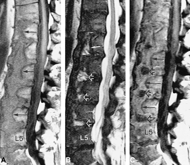
- fig 1.
Persistent notochordal canal.
A, Sagittal T1-weighted (522/15/4; TR/TE/excitations) MR image shows a vertically oriented canal (arrows) spanning T12–L5 vertebrae. The intravertebral canal is similar in signal to the intervertebral canal. Note the well-defined low signal outlining the periphery of the canal, compatible with sclerosis.
B, Sagittal T2-weighted (2700/220/4) MR image shows hyperintense, rounded central components at the disk spaces (open arrows), corresponding to the nuclei pulposi. Hyperintense signal extends for a short segment superiorly and inferiorly into the intravertebral portions of the canal (solid arrow).
C, Sagittal contrast-enhanced T1-weighted (522/15/4) MR image shows enhancement of the canal (solid arrows). The rounded central components at the disk spaces (open arrows), corresponding to the nuclei pulposi, do not enhance.
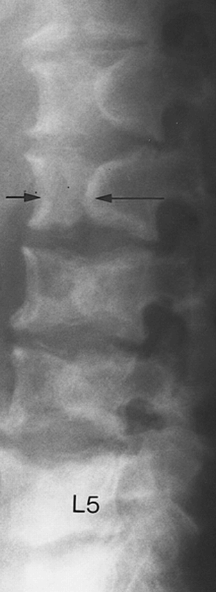
- fig 2.
Lateral radiograph of the lumbar spine shows the vertically oriented sclerotic rimmed clefts (arrows) within the anterior aspects of the T12–L5 vertebra, corresponding to the notochordal canal
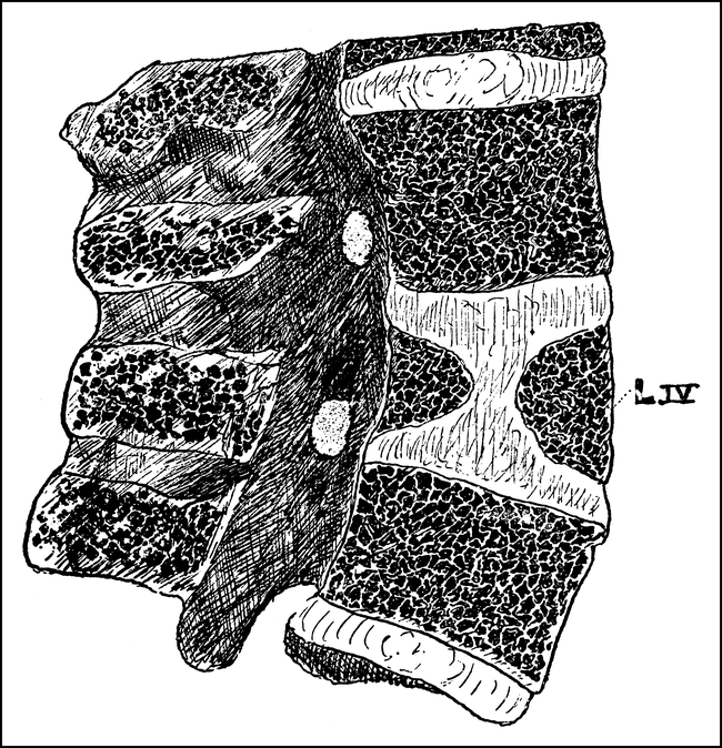
- fig 3.
Drawing of persistent notochordal canal from original description by Musgrove (3)
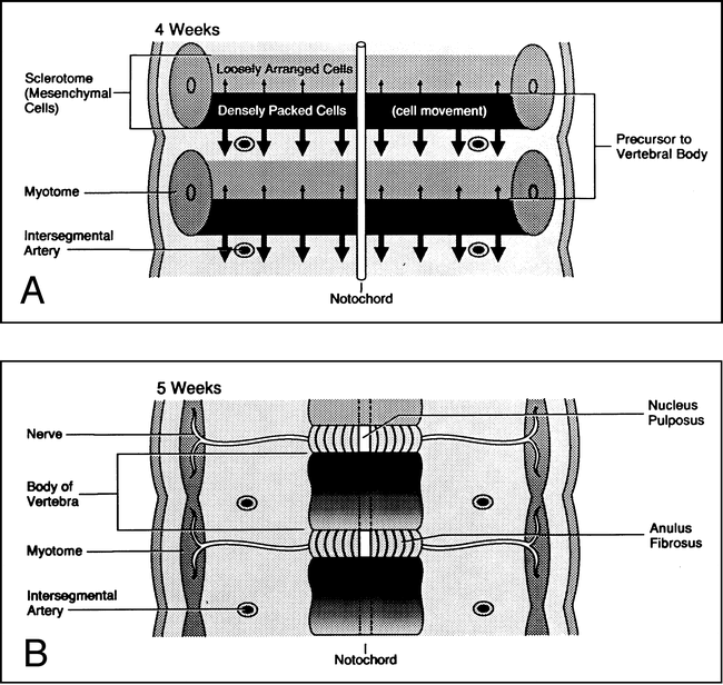
- fig 4.
Diagrams showing normal development of the vertebral column (adapted from Moore [4]).
A, At 4 weeks' gestation, the vertebral body develops around the notochord. Each mesenchymal segment contributes to the formation of the vertebrae above and below and to the formation of the intervertebral disk.
B, At 5 weeks' gestation, development of the vertebral centrum is complete and the notochord regresses.
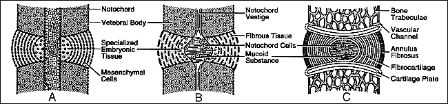
- fig 5.
Schematic showing normal development of the intervertebral disk (adapted from Peacock [5]).
A, 15-mm embryo: mesenchymal cells that will form the intervertebral disks are arranged in parallel rows. The cells toward the center are more rounded and are irregularly arranged around the notochord. They have more intercellular substance between them and contain small capsules of cartilage, referred to as specialized embryonic cartilage.
B, 29-mm embryo: at the disk periphery, fibers of the annulus fibrosus are now present. These fibers run in parallel rows and cross the disk space; their terminations are lost in the adjacent vertebrae. More mature fibrocartilage is now present at the periphery of the specialized embryonic cartilage. Polygonal-shaped notochordal cells centrally are loosely arranged around an amorphous mucoid matrix.
C, Full term: bony trabeculae and intertrabecular marrow have developed within the vertebral body. No vestige of the intravertebral notochord is present. The notochordal region within the center of the intervertebral disk contains a faintly basophilic homogeneous mucoid substance. Small groups of residual notochordal cells can be found. At the circumference of the notochordal area, small groups of fibrocartilage are surrounded by the fibers of the annulus fibrosus.


