
Abstract
Summary: The CT, MR, and histologic findings of three patients with surgically proved lumbar extradural cavernous and arteriovenous hemangiomas are reported. All three patients suffered from radicular and low back pain that disappeared completely or nearly so after total surgical excision. In each case, neuroimaging studies showed a well-defined ventrally located extradural mass with no bone involvement. On MR images, all lesions were homogeneous and isointense on noncontrast T1-weighted images and hyperintense on T2-weighted images relative to the intervertebral disk. Homogeneous enhancement was seen in one of the two cases in which contrast-enhanced T1-weighted images were obtained. Purely extradural hemangiomas should be included in the differential diagnosis of lumbar extradural soft-tissue lesions. Features that may help to distinguish this entity from the more common extruded disk herniation or neurogenic tumors are its homogeneous high signal intensity on T2-weighted images, ovoid shape, and lack of anatomic relationship with the adjacent intervertebral disk or exiting nerve root.
Benign vascular lesions located primarily in the extradural space within the spine are uncommon. They constitute less than 6% of all spinal neoplasms and classically are represented by cavernous hemangioma, angiolipoma, and arteriovenous hemangioma. This spectrum of hamartomas comprises vascular malformations that may be found in any part of the neuraxis. The most frequent type is cavernous hemangioma, which is generally seen as single or multiple intracranial lesions or as lesions arising within the vertebral bodies from where they may extend secondarily into the extradural space (1). Purely extradural lesions with no bone involvement, which represent 12% of all intraspinal hemangiomas (2–4), are uncommon. They may cause lumbar radiculopathy mimicking clinically and radiologically the presence of disk herniation (5). Only a few reports contain descriptions of the CT and MR findings of this vascular lesion (6–8). We present the CT and MR features and the histopathologic appearance of two cavernous hemangiomas and one arteriovenous hemangioma, all located exclusively in the lumbar extradural space.
Case Reports
Case 1
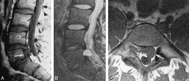
A 51-year-old woman reported progressive low back pain and right-sided sciatica over 6 months. Neurologic examination and EMG were normal. A CT study of the lumbar spine revealed an irregular but well-defined ventral extradural soft-tissue mass with attenuation similar to that of the adjacent intervertebral disk, extending laterally toward the L3–L4 left foramen. The posterior wall of the L3 vertebral body was sharply eroded (Fig 1A). MR imaging showed a ventral soft-tissue mass compressing the adjacent thecal sac, with no direct connection with the adjacent intervertebral disk. The mass was isointense on T1-weighted images and hyperintense on gradient-echo T2-weighted images relative to the intervertebral disk (Fig 1B and D). Contrast-enhanced T1-weighted images showed homogeneous enhancement of the mass, which was clearly separate from the exiting nerve root (Fig 1C). A specific preoperative diagnosis of the extradural soft-tissue mass could not be established, but extruded disk herniation and neurinoma were ruled out on the basis of imaging findings. A left hemilaminectomy at L3 revealed a red-purple rounded extradural soft-tissue mass with a yellowish border. Active bleeding requiring coagulation occurred during excision. Three years after surgery, the patient still had occasional mild low back pain. Histologic analysis found different-sized endothelium-lined vessels within abundant fibrous stroma and intermixed isolated mature adipose tissue, which was more abundant in the marginal areas, leading to the diagnosis of cavernous hemangioma (Fig 1E).

Case 1: 51-year-old woman with low back pain and right-sided sciatica for 6 months.
A, CT scan of the lumbar spine shows a left-sided ventrolateral extradural mass isodense with respect to the intervertebral disk extending through the left foramen (wide arrow, right) and slightly eroding the posterior wall of the L3 vertebral body (thin arrow, left).
B, Transverse T1-weighted (660/15/3) image at L3–L4 level confirms the presence of an extradural soft-tissue mass (arrow), which is isointense relative to the intervertebral disk and extends laterally through the left intervertebral foramen.
C, Contrast-enhanced T1-weighted (570/15/3) image at the same level shows intense and homogeneous enhancement of the extradural lesion (arrow). The left L3 nerve root is slightly displaced posteriorly by the infiltrating mass (arrowhead).
D, Gradient-echo (660/12/1; 25°) image shows the lateroforaminal mass to be isointense with respect to the contents of the adjacent dural sac.
E, Photomicrograph shows the typical findings of cavernous hemangioma. Note many dilated blood-filled vessels lined with flattened endothelium (asterisk), without muscular layer, immersed in stroma that contains adipose tissue (arrow). No fresh or organized interstitial hemorrhage was observed (hematoxylin-eosin, original magnification ×40).
Case 2
A 16-year-old boy had left leg pain that had gradually worsened over 1 year. Neurologic findings were normal. Plain films of the lumbar spine showed unilateral L5 spondylolysis with L5–S1 grade I spondylolisthesis. MR imaging revealed a slight posterior bulging of the L5–S1 intervertebral disk associated with a rounded left ventrolateral extradural soft-tissue mass that was nearly isointense relative to the intervertebral disk on T1-weighted images (Fig 2A and C) but hyperintense on T2-weighted images (Fig 2B). No contrast-enhanced images were obtained owing to the preliminary diagnosis of free-fragment disk herniation. A left hemilaminectomy at L5 revealed the presence of a well-defined rounded extradural mass, which was completely removed. Histopathologic examination of the mass showed variably sized endothelium-lined vessels within a fibrous stroma, consistent with a typical cavernous hemangioma. Five years after surgery, the patient reported subjective resolution of all clinical symptoms.

Case 2: 16-year-old boy with left leg pain for 1 year.
A and B, Sagittal MR images show a well-defined ovoid extradural mass (arrow) with intermediate signal intensity on T1-weighted (500/15/3) image (A) and high signal intensity on T2-weighted (5000/90/2) image (B).
C, Transverse T1-weighted (660/15/3) image through L5 level confirms the location of the left ventrolateral extradural mass (arrow). At surgery, a cavernous hemangioma was found and resected.
Case 3
A 19-year-old woman was admitted because of a 3-month history of left-sided radiculopathy and pollakiuria. Neurologic examination revealed lower left extremity hypalgesia and dysesthesia, but without sphincter disturbances or motor deficits. A lumbar CT examination showed a slight posterior bulging of the L3–L4 intervertebral disk that effaced the left ventrolateral epidural fat adjacent to the L4 vertebral body. MR imaging showed a well-defined ovoid extradural mass filling the L4 left lateral recess and compressing the thecal sac. The mass appeared isointense relative to the intervertebral disk on T1-weighted images, with increased signal intensity on T2-weighted images (Fig 3A–C). Contrast-enhanced T1-weighted images showed no significant enhancement (Fig 3D). A preoperative diagnosis of extruded disk herniation was made, but surgery revealed an extradural soft-tissue mass that was completely resected without major bleeding. Histologic study of the resected polypoid red-purple epidural mass showed ectatic vascular elements with interstitial hemosiderin deposits and hemorrhagic foci, consistent with an arteriovenous hemangioma (Fig 3E). Symptoms disappeared gradually after surgery, and the patient was nearly asymptomatic 1 year later.

Case 3: 19-year-old woman with 3-month history of left-sided radiculopathy and pollakiuria.
A, Left parasagittal T2-weighted (5000/90/2) image shows the ovoid shape of the mass (arrow), which is hyperintense with respect to CSF in the thecal sac. Note the bulging disk at L3–L4 level.
B, Transverse T2-weighted (5000/90/2) image shows a well-demarcated uniformly hyperintense lesion (arrow) without remodeling of the posterior vertebral body.
C, Transverse T1-weighted (802/12/4) image at the superior aspect of L4 shows an extradural mass occupying the left lateral recess (arrow) whose signal intensity is slightly greater than that of the contents of the adjacent dural sac.
D, Contrast-enhanced T1-weighted (802/12/4) image shows no significant changes in the signal intensity of the extradural lesion.
E, Pathologic findings of arteriovenous hemangioma included a large vascular element (asterisk) with thickened wall in association with interstitial hemosiderin deposits (arrow) and hemorrhagic foci (hematoxylin-eosin, original magnification ×100).
Discussion
Hemangiomas are congenital vascular malformations of unknown origin. Current classification schemes define them as vascular malformations, occurring throughout the body and CNS, that are clearly differentiated from true vascular neoplasms, such as hemangioblastomas (5). It has been suggested that lipomas and vascular malformations represent the ends of a wide and continuous overlapping spectrum of hamartomas in which cavernous hemangiomas, angiolipomas, and arteriovenous hemangiomas constitute intermediate and undifferentiated entities (8). None of these lesions undergo spontaneous regression. Although they have no mitotic activity, they grow slowly, probably because of recurrent hemorrhaging and thrombotic phenomena with organization and recanalization (2). A purely extradural location of cavernous or arteriovenous hemangiomas is rare, representing less than 4% of all extradural spinal tumors (3). The rarity of these lesions as a cause of lumbar radiculopathy (4, 6, 9, 10) may be attributed to the uncommon nature of the lesion, but difficulties in their preoperative diagnosis with either CT or MR imaging may also contribute.
Histologically, immunohistochemically, and electron microscopically (11), cavernous hemangiomas are nearly identical at every location throughout the body. Histologically, they are composed of irregular sinusoidal vascular channels with thin collagenous walls lined by a single layer of flattened endothelium. Elastic fibers and smooth muscle fibers within the walls are scant if not absent. The fatty component of cavernous hemangioma can vary depending on its location (3, 12). For instance, lesions found at the thoracic level contain a higher proportion of fat, although usually this fat is restricted to the periphery of the lesion or sometimes intermingled with vessels within fibrous stroma. The only histologic characteristic that would allow differentiation between cavernous hemangioma and angiolipoma is a fat tissue: cavernous vessel ratio of less than 1:10 (8). Extradural cavernous hemangiomas display a paucity of interstitial hemosiderin deposits in comparison with their intraparenchymal counterparts. This feature is presumably caused by easier removal of blood products outside the blood-brain barrier (7). Arteriovenous hemangiomas correspond to clusters of abnormal arteries and veins, with the vessel walls containing elastin and smooth muscle. Hemosiderin deposits are always seen as well as various degenerative changes, such as thrombosis or fibrosis. Owing to the small size of the vessels involved, arteriovenous shunting may be absent (8). Along with angiolipomas, cavernous and arteriovenous hemangiomas probably constitute a spectrum of hamartomas, with various pathologic components and different hemodynamic features.
In purely extradural hemangiomas, plain films may be normal or show sharp bone erosion of the vertebral body or enlargement of the intervertebral foramen. Bone erosion was seen in only one of our cases. CT and MR imaging are the best diagnostic imaging procedures, the latter being more valuable in assessing the extent of the anatomic relationship between the lesion and the intervertebral disk and thecal sac. Few reports of the CT or MR features of extradural cavernous or arteriovenous hemangiomas of the lumbar spine without direct bone involvement are found in the literature. In contrast to intracerebral cavernous hemangiomas, the patterns of density on CT scans and signal intensity on MR images are more homogeneous within extradural hemangiomas. On CT studies, these neoplasms appear as intermediate or slightly hyperdense extradural masses, frequently found beyond or beneath the intervertebral disk. At MR imaging, they display high signal intensity on T2-weighted images, which may be explained by the high content of stagnant blood. Slow blood flow may contribute substantially to the signal, conferring a low or intermediate signal intensity on T1-weighted images (6–10). The morphologic characteristics of extradural hemangiomas described in the literature (6, 8, 10) and as seen in our cases include the round or ovoid shape of the lesion, their tendency to extend through the intervertebral foramen, a location in the ventral extradural space of the lumbar spine, and the absence of a direct anatomic relationship with the intervertebral disk or exiting nerve root. The preoperative radiologic diagnosis of extradural hemangioma is nonetheless difficult, probably because of the rarity of this lesion and the few cases studied with CT or MR imaging described in the literature, which may lead to an erroneous preoperative diagnosis of the more common extruded disk herniations. This misdiagnosis occurred in two of our patients, because of the concomitant presence of degenerative changes and posterior bulging of the intervertebral disk adjacent to the extradural mass. Contrast-enhanced CT or MR imaging is generally not performed as part of the routine work-up in a patient with lumbar radiculopathy, but it may be useful for clarifying the vascular nature of this lesion and for differentiating it from extruded disk herniation, which presents an enhancement that envelopes the extradural lesion, increasing its conspicuity and illuminating frequently associated annular tears and enhancement of disk margins. We found, however, no significant contrast enhancement in the arteriovenous hemangioma on MR images, probably because the intralesional blood flow was too slow. Angiographic findings are usually negative, as occurs in intraparenchymal lesions, whereas in vertebral bone lesions they are frequently positive (3, 9).
The CT and MR characteristics of these two types of hemangiomas are similar to those observed in spinal angiolipoma, but in this lesion the presence of calcifications, hemorrhagic foci, and a marked fat component are features that, when present, help in the differential diagnosis (8). The MR differential diagnosis for extradural hemangioma also includes free-fragment herniated disk, epidural tumor (most commonly the nerve sheath tumor group), epidural infiltrating disease, epidural abscess, and congenital or acquired epidural cyst. Especially in the case of dumbbell-shaped spinal cavernous hemangiomas, the differential diagnosis should include schwannoma or neurofibroma, but close inspection of the MR images in these conditions can usually establish the absence of an anatomic relationship between the mass and the exiting nerve root. Lymphoma and metastases could be considered, but in both conditions a multiplicity of lesions and body bone marrow involvement will usually be found. An epidural abscess will frequently produce marrow changes and disk space abnormalities that indicate infection rather than degenerative disk disease.
Conclusion
Although a precise preoperative diagnosis of extradural hemangiomas cannot be established in many cases, the presence of a well-defined ovoid mass that shows a homogeneous high signal intensity on T2-weighted images relative to the intervertebral disk, that has a tendency to extend through the intervertebral foramen, and that is well separated from the adjacent intervertebral disk and exiting nerve root should arouse suspicion of this unusual extradural lesion. A preoperative diagnosis is important, as intraoperative hemorrhage may occur. Extradural hemangiomas show no tendency to regress, and, as suggested in previous studies (7–10), complete resection of the lesion seems to be the best treatment, producing relief of patients' symptoms and good prognosis in most instances, as exemplified in our three cases.
Footnotes
↵1 Address reprint requests to Dr. Antoni Rovira, SDI-UDIAT, Parc-Taulí s/n, 08208 Sabadell, Spain.
References
- Received September 12, 1997.
- Accepted after revision April 14, 1998.
- Copyright © American Society of Neuroradiology


