
Article Figures & Data
Figures
- fig 1.
Axial RARE moderately T2-weighted images obtained from scanner A (A) (3800/22/1) and scanner B (B) (3800/25/1). Several hyperintense lesions are visible in both the cerebral hemispheres
- fig 2.
Axial FLAIR images obtained from scanner A (A) (9999/105/1; TI, 2200) and scanner B (B) (6500/150/2; TI, 2000) at the same anatomic level as in figure 1


Tables
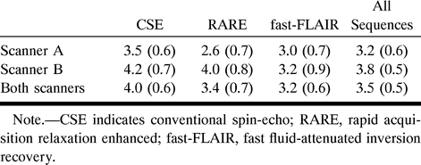
 TABLE 1:
Mean (standard error) lesion volumes (mL) obtained for different scanners and sequences
- TABLE 2:
Mean (standard error) coefficients of variation (%) obtained for different scanners and sequences
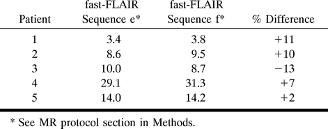
- TABLE 3:
Lesion volumes (mL) obtained from five patients with MS undergoing imaging with the two fast-FLAIR sequences on scanner B


