
This article requires a subscription to view the full text. If you have a subscription you may use the login form below to view the article. Access to this article can also be purchased.
Graphical Abstract

Abstract
BACKGROUND AND PURPOSE: The imaging of inflammatory myelopathies has advanced significantly across time, with MRI techniques playing a pivotal role in enhancing lesion detection. However, the impact of deep learning (DL)-based reconstruction on 3D double inversion recovery (DIR) imaging for inflammatory myelopathies remains unassessed. This study aimed to compare the acquisition time, image quality, diagnostic confidence, and lesion detection rates among sagittal T2WI, standard DIR, and DL-reconstructed DIR in patients with inflammatory myelopathies.
MATERIALS AND METHODS: In this observational study, patients diagnosed with inflammatory myelopathies were recruited between June 2023 and March 2024. Each patient underwent sagittal conventional TSE sequences and standard 3D DIR (T2WI and standard 3D DIR were used as references for comparison), followed by an undersampled accelerated double inversion recovery deep learning (DIRDL) examination. Three neuroradiologists evaluated the images using a 4-point Likert scale (from 1 to 4) for overall image quality, perceived SNR, sharpness, artifacts, and diagnostic confidence. The acquisition times and lesion detection rates were also compared among the acquisition protocols.
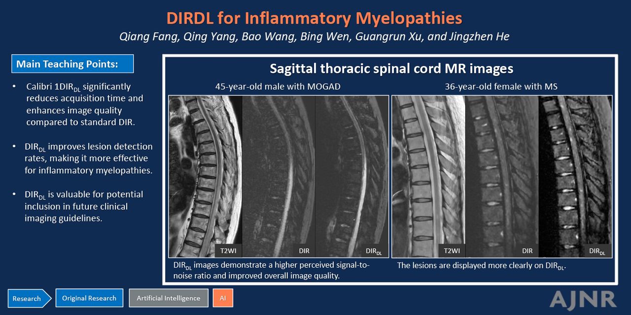
RESULTS: A total of 149 participants were evaluated (mean age, 40.6 [SD, 16.8] years; 71 women). The median acquisition time for DIRDL was significantly lower than for standard DIR (298 seconds [interquartile range, 288–301 seconds] versus 151 seconds [interquartile range, 148–155 seconds]; P < .001), showing a 49% time reduction. DIRDL images scored higher in overall quality, perceived SNR, and artifact noise reduction (all P < .001). There were no significant differences in sharpness (P = .07) or diagnostic confidence (P = .06) between the standard DIR and DIRDL protocols. Additionally, DIRDL detected 37% more lesions compared with T2WI (300 versus 219; P < .001).
CONCLUSIONS: DIRDL significantly reduces acquisition time and improves image quality compared with standard DIR, without compromising diagnostic confidence. Additionally, DIRDL enhances lesion detection in patients with inflammatory myelopathies, making it a valuable tool in clinical practice. These findings underscore the potential for incorporating DIRDL into future imaging guidelines.
ABBREVIATIONS:
- AQP4+NMOSD
- AQP4-IgG positive neuromyelitis optica spectrum disorders
- DIR
- double inversion recovery
- DL
- deep learning
- ICC
- intraclass correlation coefficient
- IQR
- interquartile range
- MOG
- myelin oligodendrocyte glycoprotein
- MOGAD
- MOG antibody-associated diseases
- NEX
- number of excitations
Footnotes
Qiang Fang and Qing Yang contributed equally to this work and should be considered as co-first authors.
This work was supported by the Shandong Provincial Natural Science Foundation (ZR2021MH237) and the National Natural Science Foundation of China (82202114).
Disclosure forms provided by the authors are available with the full text and PDF of this article at www.ajnr.org.
- © 2025 by American Journal of Neuroradiology
Log in using your username and password
Log in through your institution


Jump to section
Related Articles
Cited By...
- No citing articles found.