
Article Figures & Data
Figures
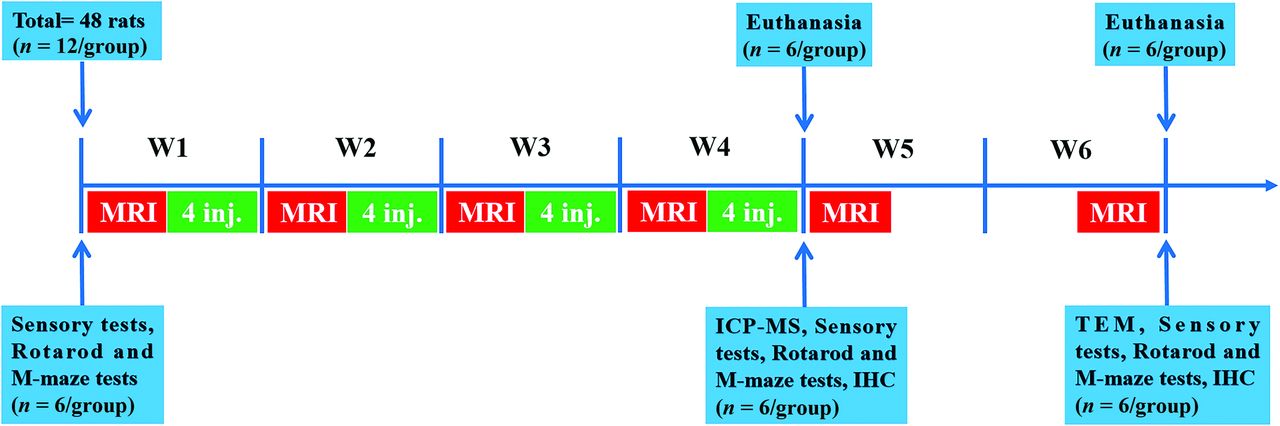
- FIG 1.
Flow chart of experimental design. The experimental timeline of GBCA injections, MR scanning (days 0, 7, 14, 21, 28, and 42), inductively coupled plasma mass-spectroscopy (day 28), TEM (day 42), heat and mechanical hyperalgesia tests (days 0, 28, and 42), the accelerated rotarod test and the Morris water maze test (days 0, 28, and 42), immunohistochemical test (days 28 and 42), and euthanasia time point. In total, 48 rats were used in the experiment. The number of rats used in each experiment is indicated in the diagram. M-maze indicates the Morris water maze test; inj., injection.
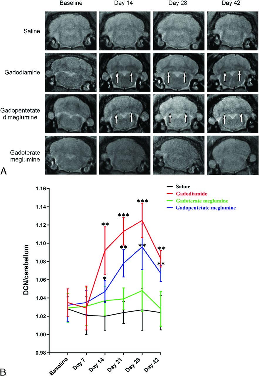
- FIG 2.
Representative T1-weighted MR images of the DCN in mice on days 14, 28, and 42 after the injections of saline, gadodiamide, gadopentetate dimeglumine, and gadoterate meglumine (A). High signal intensity in the DCN on unenhanced T1-weighted imaging is indicated by white arrows (B). Bar graph depicts the quantitative analysis of the T1-weighted DCN to cerebellum signal ratio on MR images. Error bar represents the SD. Single asterisk, P < .05; double asterisks, P < .01; and triple asterisks, P < .001 compared with the saline group.
- FIG 3.
Bar graph depicting the Gd deposition in the cerebellum, spinal cord, and sciatic nerve after the administration of saline, gadodiamide, gadopentetate dimeglumine, and gadoterate meglumine. Error bar represents the SD. Double asterisks indicate P < .01; and triple asterisks, P < .001.
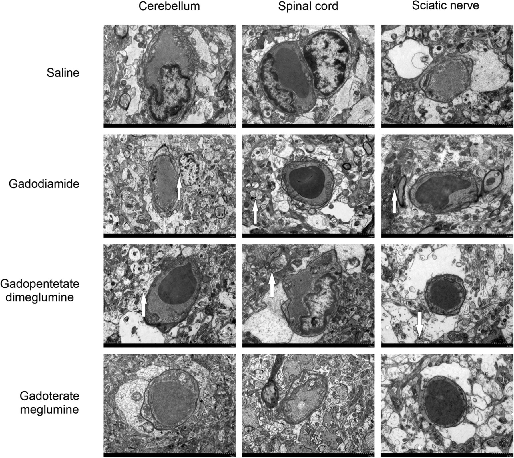
- FIG 4.
Localization of Gd deposits in the cerebellum, spinal cord, and sciatic nerve on day 28 by TEM. Electron-dense granules are indicated by white arrows.
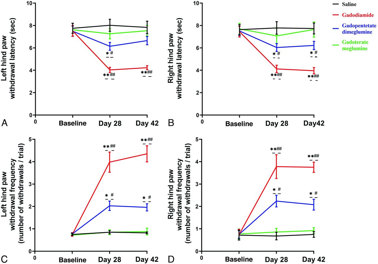
- FIG 5.
Bar graph depicting the effect of GBCA exposure on heat and mechanical hyperalgesia. The time course of heat hyperalgesia in the left (A) and right (B) hind paws of rats treated with GBCAs or saline on days 28 and 42 of the GBCA or saline exposure period. Time course of mechanical hyperalgesia in the left (C) and right (D) hind paws of rats treated with GBCAs or saline. Single asterisk, P < .05; double asterisks, P < .01, compared with the saline group; # indicates P < .05; ##, P < .01, compared with the baseline.
- FIG 6.
Effect of GBCA exposure on the Morris water maze test (A) and the accelerated rotarod test (B). The Morris water maze test in rats treated with 0.9% saline solution, gadodiamide, gadopentetate dimeglumine, and gadoterate meglumine before the injection of GBCAs (training trail 1), 28 and 42 days after the injection of GBCA exposure period (A). The tests were conducted on days 28 (training trail 2 and 3) and 42 (training trail 4 and 5) after the injection of GBCAs twice daily. The accelerated rotarod test in rats treated with 0.9% saline solution, gadodiamide, gadopentetate dimeglumine, and gadoterate meglumine before the injection of GBCAs and 28 and 42 days after the injection of GBCA exposure period (B). Error bar represents the SD. Single asterisk, P < .05; double asterisks, P < .01 compared with the saline group.


