Advertisement
  • Main menu
  • User menu
  • Search
  • English ▼
    • English
    • Afrikaans
    • Albanian
    • Amharic
    • Arabic
    • Armenian
    • Azerbaijani
    • Basque
    • Belarusian
    • Bengali
    • Bosnian
    • Bulgarian
    • Catalan
    • Cebuano
    • Chichewa
    • Chinese (Simplified)
    • Chinese (Traditional)
    • Corsican
    • Croatian
    • Czech
    • Danish
    • Dutch
    • Esperanto
    • Estonian
    • Filipino
    • Finnish
    • French
    • Frisian
    • Galician
    • Georgian
    • German
    • Greek
    • Gujarati
    • Haitian Creole
    • Hausa
    • Hawaiian
    • Hebrew
    • Hindi
    • Hmong
    • Hungarian
    • Icelandic
    • Igbo
    • Indonesian
    • Irish
    • Italian
    • Japanese
    • Javanese
    • Kannada
    • Kazakh
    • Khmer
    • Korean
    • Kurdish (Kurmanji)
    • Kyrgyz
    • Lao
    • Latin
    • Latvian
    • Lithuanian
    • Luxembourgish
    • Macedonian
    • Malagasy
    • Malay
    • Malayalam
    • Maltese
    • Maori
    • Marathi
    • Mongolian
    • Myanmar (Burmese)
    • Nepali
    • Norwegian
    • Pashto
    • Persian
    • Polish
    • Portuguese
    • Punjabi
    • Romanian
    • Russian
    • Samoan
    • Scottish Gaelic
    • Serbian
    • Sesotho
    • Shona
    • Sindhi
    • Sinhala
    • Slovak
    • Slovenian
    • Somali
    • Spanish
    • Sudanese
    • Swahili
    • Swedish
    • Tajik
    • Tamil
    • Telugu
    • Thai
    • Turkish
    • Ukrainian
    • Urdu
    • Uzbek
    • Vietnamese
    • Welsh
    • Xhosa
    • Yiddish
    • Yoruba
    • Zulu

Main menu

  • Home
  • Content
    • Current Issue
    • Accepted Manuscripts
    • Article Preview
    • Past Issue Archive
    • Video Articles
    • AJNR Case Collection
    • Case of the Week Archive
    • Case of the Month Archive
    • Classic Case Archive
  • Special Collections
    • AJNR Awards
    • Low-Field MRI
    • Alzheimer Disease
    • ASNR Foundation Special Collection
    • Photon-Counting CT
    • View All
  • Multimedia
    • AJNR Podcasts
    • AJNR SCANtastic
    • Trainee Corner
    • MRI Safety Corner
    • Imaging Protocols
  • For Authors
    • Submit a Manuscript
    • Submit a Video Article
    • Submit an eLetter to the Editor/Response
    • Manuscript Submission Guidelines
    • Statistical Tips
    • Fast Publishing of Accepted Manuscripts
    • Graphical Abstract Preparation
    • Imaging Protocol Submission
    • Author Policies
  • About Us
    • About AJNR
    • Editorial Board
    • Editorial Board Alumni
  • More
    • Become a Reviewer/Academy of Reviewers
    • Subscribers
    • Permissions
    • Alerts
    • Feedback
    • Advertisers
    • ASNR Home

User menu

  • Alerts
  • Log in

Search

  • Advanced search
American Journal of Neuroradiology

American Journal of Neuroradiology

ASHNR American Society of Functional Neuroradiology ASHNR American Society of Pediatric Neuroradiology ASSR
  • Alerts
  • Log in

Advanced Search

English ▼
  • English
  • Afrikaans
  • Albanian
  • Amharic
  • Arabic
  • Armenian
  • Azerbaijani
  • Basque
  • Belarusian
  • Bengali
  • Bosnian
  • Bulgarian
  • Catalan
  • Cebuano
  • Chichewa
  • Chinese (Simplified)
  • Chinese (Traditional)
  • Corsican
  • Croatian
  • Czech
  • Danish
  • Dutch
  • Esperanto
  • Estonian
  • Filipino
  • Finnish
  • French
  • Frisian
  • Galician
  • Georgian
  • German
  • Greek
  • Gujarati
  • Haitian Creole
  • Hausa
  • Hawaiian
  • Hebrew
  • Hindi
  • Hmong
  • Hungarian
  • Icelandic
  • Igbo
  • Indonesian
  • Irish
  • Italian
  • Japanese
  • Javanese
  • Kannada
  • Kazakh
  • Khmer
  • Korean
  • Kurdish (Kurmanji)
  • Kyrgyz
  • Lao
  • Latin
  • Latvian
  • Lithuanian
  • Luxembourgish
  • Macedonian
  • Malagasy
  • Malay
  • Malayalam
  • Maltese
  • Maori
  • Marathi
  • Mongolian
  • Myanmar (Burmese)
  • Nepali
  • Norwegian
  • Pashto
  • Persian
  • Polish
  • Portuguese
  • Punjabi
  • Romanian
  • Russian
  • Samoan
  • Scottish Gaelic
  • Serbian
  • Sesotho
  • Shona
  • Sindhi
  • Sinhala
  • Slovak
  • Slovenian
  • Somali
  • Spanish
  • Sudanese
  • Swahili
  • Swedish
  • Tajik
  • Tamil
  • Telugu
  • Thai
  • Turkish
  • Ukrainian
  • Urdu
  • Uzbek
  • Vietnamese
  • Welsh
  • Xhosa
  • Yiddish
  • Yoruba
  • Zulu
  • Home
  • Content
    • Current Issue
    • Accepted Manuscripts
    • Article Preview
    • Past Issue Archive
    • Video Articles
    • AJNR Case Collection
    • Case of the Week Archive
    • Case of the Month Archive
    • Classic Case Archive
  • Special Collections
    • AJNR Awards
    • Low-Field MRI
    • Alzheimer Disease
    • ASNR Foundation Special Collection
    • Photon-Counting CT
    • View All
  • Multimedia
    • AJNR Podcasts
    • AJNR SCANtastic
    • Trainee Corner
    • MRI Safety Corner
    • Imaging Protocols
  • For Authors
    • Submit a Manuscript
    • Submit a Video Article
    • Submit an eLetter to the Editor/Response
    • Manuscript Submission Guidelines
    • Statistical Tips
    • Fast Publishing of Accepted Manuscripts
    • Graphical Abstract Preparation
    • Imaging Protocol Submission
    • Author Policies
  • About Us
    • About AJNR
    • Editorial Board
    • Editorial Board Alumni
  • More
    • Become a Reviewer/Academy of Reviewers
    • Subscribers
    • Permissions
    • Alerts
    • Feedback
    • Advertisers
    • ASNR Home
  • Follow AJNR on Twitter
  • Visit AJNR on Facebook
  • Follow AJNR on Instagram
  • Join AJNR on LinkedIn
  • RSS Feeds

AJNR Awards, New Junior Editors, and more. Read the latest AJNR updates

Research ArticleHEAD AND NECK IMAGING

Visualization of the Extracranial Branches of the Trigeminal Nerve Using Improved Motion-Sensitized Driven Equilibrium–Prepared 3D Inversion Recovery TSE Sequence

Dejun She, Hao Huang, Dongmei Jiang, Junhuan Hong, Peiying You, Lu Li, Xiance Zhao and Dairong Cao
American Journal of Neuroradiology July 2024, DOI: https://doi.org/10.3174/ajnr.A8273
Dejun She
aFrom the Department of Radiology (D.S., H.H., D.J., J.H., P.Y., D.C.), First Affiliated Hospital of Fujian Medical University, Fuzhou, China
bDepartment of Radiology (D.S., H.H., D.J., J.H., P.Y., D.C), National Regional Medical Center, Binhai Campus of the First Affiliated Hospital, Fujian Medical University, Fuzhou, P.R. China
cKey Laboratory of Radiation Biology of Fujian Higher Education Institutions (D.S., D.C.), the First Affiliated Hospital, Fujian Medical University, Fuzhou, Fujian, China
  • Find this author on Google Scholar
  • Find this author on PubMed
  • Search for this author on this site
  • ORCID record for Dejun She
Hao Huang
aFrom the Department of Radiology (D.S., H.H., D.J., J.H., P.Y., D.C.), First Affiliated Hospital of Fujian Medical University, Fuzhou, China
bDepartment of Radiology (D.S., H.H., D.J., J.H., P.Y., D.C), National Regional Medical Center, Binhai Campus of the First Affiliated Hospital, Fujian Medical University, Fuzhou, P.R. China
  • Find this author on Google Scholar
  • Find this author on PubMed
  • Search for this author on this site
  • ORCID record for Hao Huang
Dongmei Jiang
aFrom the Department of Radiology (D.S., H.H., D.J., J.H., P.Y., D.C.), First Affiliated Hospital of Fujian Medical University, Fuzhou, China
bDepartment of Radiology (D.S., H.H., D.J., J.H., P.Y., D.C), National Regional Medical Center, Binhai Campus of the First Affiliated Hospital, Fujian Medical University, Fuzhou, P.R. China
  • Find this author on Google Scholar
  • Find this author on PubMed
  • Search for this author on this site
  • ORCID record for Dongmei Jiang
Junhuan Hong
aFrom the Department of Radiology (D.S., H.H., D.J., J.H., P.Y., D.C.), First Affiliated Hospital of Fujian Medical University, Fuzhou, China
bDepartment of Radiology (D.S., H.H., D.J., J.H., P.Y., D.C), National Regional Medical Center, Binhai Campus of the First Affiliated Hospital, Fujian Medical University, Fuzhou, P.R. China
  • Find this author on Google Scholar
  • Find this author on PubMed
  • Search for this author on this site
  • ORCID record for Junhuan Hong
Peiying You
aFrom the Department of Radiology (D.S., H.H., D.J., J.H., P.Y., D.C.), First Affiliated Hospital of Fujian Medical University, Fuzhou, China
bDepartment of Radiology (D.S., H.H., D.J., J.H., P.Y., D.C), National Regional Medical Center, Binhai Campus of the First Affiliated Hospital, Fujian Medical University, Fuzhou, P.R. China
  • Find this author on Google Scholar
  • Find this author on PubMed
  • Search for this author on this site
  • ORCID record for Peiying You
Lu Li
dThe School of Medical Imaging (L.L.), Fujian Medical University, Fuzhou, Fujian, China
  • Find this author on Google Scholar
  • Find this author on PubMed
  • Search for this author on this site
  • ORCID record for Lu Li
Xiance Zhao
ePhilips Healthcare (X.Z.), Shanghai, P.R. China
  • Find this author on Google Scholar
  • Find this author on PubMed
  • Search for this author on this site
  • ORCID record for Xiance Zhao
Dairong Cao
aFrom the Department of Radiology (D.S., H.H., D.J., J.H., P.Y., D.C.), First Affiliated Hospital of Fujian Medical University, Fuzhou, China
bDepartment of Radiology (D.S., H.H., D.J., J.H., P.Y., D.C), National Regional Medical Center, Binhai Campus of the First Affiliated Hospital, Fujian Medical University, Fuzhou, P.R. China
cKey Laboratory of Radiation Biology of Fujian Higher Education Institutions (D.S., D.C.), the First Affiliated Hospital, Fujian Medical University, Fuzhou, Fujian, China
fDepartment of Radiology (D.C.), Fujian Key Laboratory of Precision Medicine for Cancer, First Affiliated Hospital, Fujian Medical University, Fuzhou, Fujian, China
  • Find this author on Google Scholar
  • Find this author on PubMed
  • Search for this author on this site
  • ORCID record for Dairong Cao
  • Article
  • Figures & Data
  • Supplemental
  • Info & Metrics
  • Responses
  • References
  • PDF
Loading

Article Figures & Data

Figures

  • Tables
  • FIG 1.
    • Download figure
    • Open in new tab
    • Download powerpoint
    FIG 1.

    A 32-year-old patient with no lesion in the head and neck. Curved planar reformation MIP (2-mm layer thickness) of the mandibular nerve (blue triangle), inferior alveolar nerve (red thin arrow), and lingual nerve (yellow arrowhead) at the oblique coronary position. Both iMSDE 3D IR-TSE and CE 3D IR-TSE significantly improved visualization in assessing the inferior alveolar nerve and lingual nerve. CE indicates contrast-enhanced.

  • FIG 2.
    • Download figure
    • Open in new tab
    • Download powerpoint
    FIG 2.

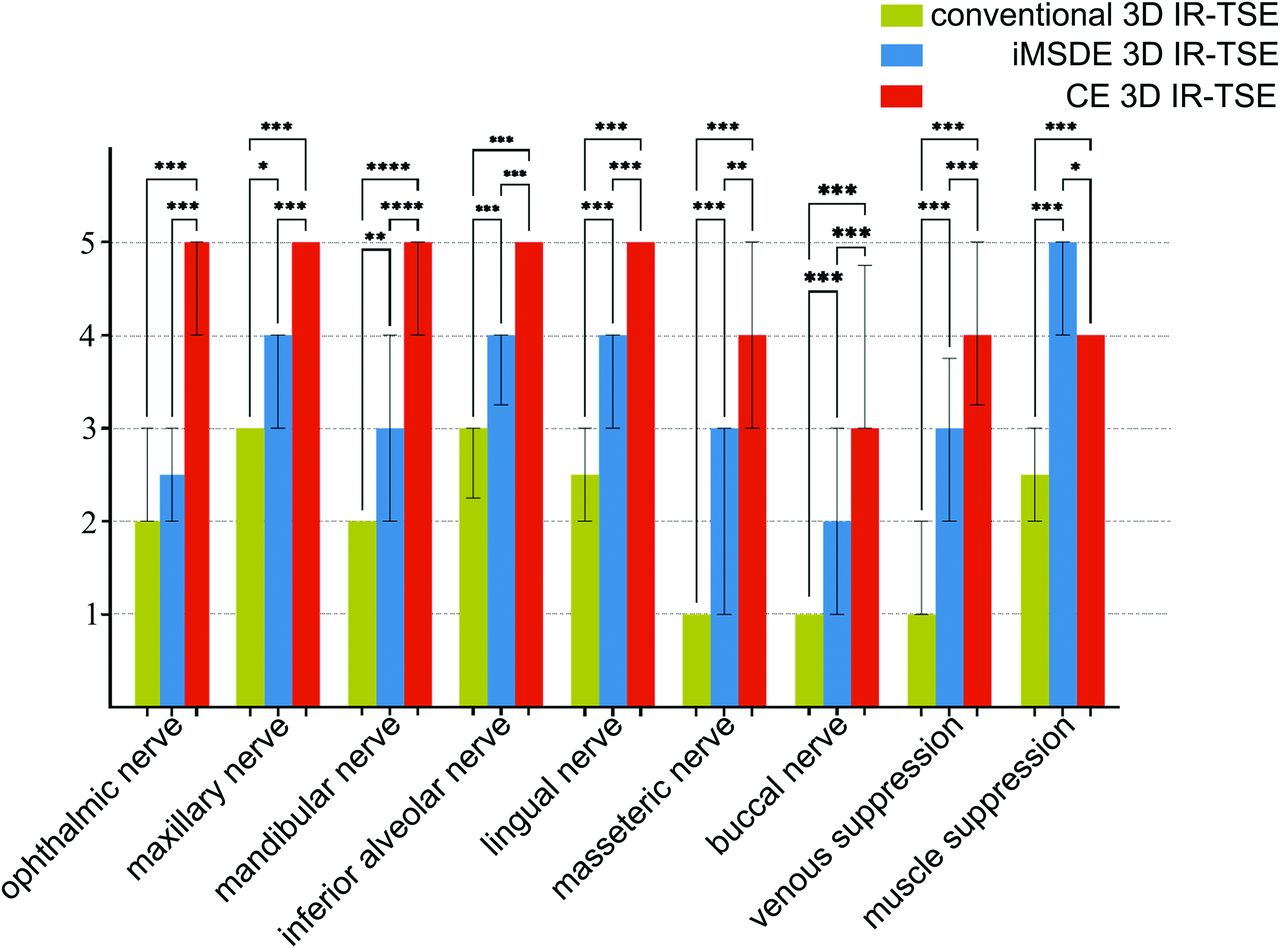
    Comparisons of the visibility of 7 major peripheral branches of the trigeminal nerve and muscle/venous suppression among 3 sequences in 20 healthy subjects. The asterisk indicates P < .05; double asterisks, P < .01; triple asterisks, P < .001; CE, contrast-enhanced.

  • FIG 3.
    • Download figure
    • Open in new tab
    • Download powerpoint
    FIG 3.

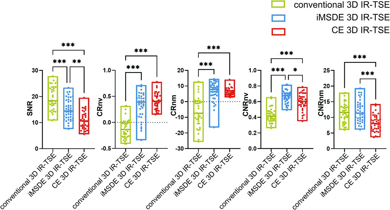
    Comparisons of SNR, CRnv, CNRnv, CRnm, and CNRnm among 3 sequences in 20 subjects. The asterisk indicates P < .05; double asterisks, P < .01; triple asterisks, P < .001; CE, contrast-enhanced.

  • FIG 4.
    • Download figure
    • Open in new tab
    • Download powerpoint
    FIG 4.

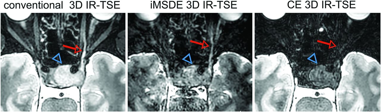
    A 29-year-old patient with neurilemmoma. The sagittal image after MPR and MIP (3-mm layer thickness) shows that the neurilemmoma (blue triangle) compressed and moved the inferior alveolar nerve (red thin arrow) forward and the lingual nerve (yellow arrowhead) was not involved. Venous signals make nerves challenging to visualize on conventional 3D IR-TSE. CE indicates contrast-enhanced.

  • FIG 5.
    • Download figure
    • Open in new tab
    • Download powerpoint
    FIG 5.

    A 46-year-old patient with a large pituitary adenoma. The transverse image after MPR and MIP (3-mm layer thickness) demonstrates a large pituitary adenoma (blue triangle) invasion of the ophthalmic nerve (red thin arrow). The involved nerve and tumor are shown as low signal, which is difficult to identify on contrast-enhanced 3D IR-TSE. CE indicates contrast-enhanced.

Tables

  • Figures
  • Parameters of MR imaging

    ParameterConventional 3D IR-TSEiMSDE 3D IR-TSECE 3D IR-TSE
    TR/TE (ms)2800/1812800/1812800/181
    Flip angle (degrees)Variable flip angleVariable flip angleVariable flip angle
    Field of view (mm)200 × 200200 × 200200 × 200
    Voxel size (mm)0.9 × 0.9 × 0.90.9 × 0.9 × 0.90.9 × 0.9 × 0.9
    No. of slices210210210
    Averages222
    Fat-suppressionSTIRSTIRSTIR
    iMSDE TEprep (ms)60/
    iMSDE VENC (cm/s)0.35
    Acquisition time (min/sec)8 min 27 sec8 min 27 sec8 min 27 sec
    • Note:—TEprep indicates magnetization preparation TE; VENC, velocity-encoded; CE, contrast-enhanced.

PreviousNext
Back to top
Advertisement
Print
Download PDF
Email Article
Cite this article
0 Responses
Respond to this article
Share
Bookmark this article
Visualization of the Extracranial Branches of the Trigeminal Nerve Using Improved Motion-Sensitized Driven Equilibrium–Prepared 3D Inversion Recovery TSE Sequence
Dejun She, Hao Huang, Dongmei Jiang, Junhuan Hong, Peiying You, Lu Li, Xiance Zhao, Dairong Cao
American Journal of Neuroradiology Jul 2024, DOI: 10.3174/ajnr.A8273
del.icio.us logo Twitter logo Facebook logo Mendeley logo
  • Tweet Widget
Purchase

Jump to section

  • Article
    • Abstract
    • ABBREVIATIONS:
    • MATERIALS AND METHODS
    • RESULTS
    • DISCUSSION
    • CONCLUSIONS
    • Footnotes
    • References
  • Figures & Data
  • Supplemental
  • Info & Metrics
  • Responses
  • References
  • PDF

Related Articles

  • PubMed
  • Google Scholar

Cited By...

  • No citing articles found.
  • Crossref
  • Google Scholar

This article has not yet been cited by articles in journals that are participating in Crossref Cited-by Linking.

More in this TOC Section

  • Hydrops Herniation into the Semicircular Canals
  • DIVr of NPC Predicted Outcome
  • MRI of the Parasellar Ligaments
Show more HEAD AND NECK IMAGING

Similar Articles

Advertisement

Indexed Content

  • Current Issue
  • Accepted Manuscripts
  • Article Preview
  • Past Issues
  • Editorials
  • Editor's Choice
  • Fellows' Journal Club
  • Letters to the Editor
  • Video Articles

Cases

  • Case Collection
  • Archive - Case of the Week
  • Archive - Case of the Month
  • Archive - Classic Case

More from AJNR

  • Trainee Corner
  • Imaging Protocols
  • MRI Safety Corner
  • Book Reviews

Multimedia

  • AJNR Podcasts
  • AJNR Scantastics

Resources

  • Turnaround Time
  • Submit a Manuscript
  • Submit a Video Article
  • Submit an eLetter to the Editor/Response
  • Manuscript Submission Guidelines
  • Statistical Tips
  • Fast Publishing of Accepted Manuscripts
  • Graphical Abstract Preparation
  • Imaging Protocol Submission
  • Evidence-Based Medicine Level Guide
  • Publishing Checklists
  • Author Policies
  • Become a Reviewer/Academy of Reviewers
  • News and Updates

About Us

  • About AJNR
  • Editorial Board
  • Editorial Board Alumni
  • Alerts
  • Permissions
  • Not an AJNR Subscriber? Join Now
  • Advertise with Us
  • Librarian Resources
  • Feedback
  • Terms and Conditions
  • AJNR Editorial Board Alumni

American Society of Neuroradiology

  • Not an ASNR Member? Join Now

© 2025 by the American Society of Neuroradiology All rights, including for text and data mining, AI training, and similar technologies, are reserved.
Print ISSN: 0195-6108 Online ISSN: 1936-959X

Powered by HighWire
Email this Article

Thank you for your interest in spreading the word on American Journal of Neuroradiology.

NOTE: We only request your email address so that the person you are recommending the page to knows that you wanted them to see it, and that it is not junk mail. We do not capture any email address.

Enter multiple addresses on separate lines or separate them with commas.
Visualization of the Extracranial Branches of the Trigeminal Nerve Using Improved Motion-Sensitized Driven Equilibrium–Prepared 3D Inversion Recovery TSE Sequence
(Your Name) has sent you a message from American Journal of Neuroradiology
(Your Name) thought you would like to see the American Journal of Neuroradiology web site.
CAPTCHA
This question is for testing whether or not you are a human visitor and to prevent automated spam submissions.
Cite this article
Dejun She, Hao Huang, Dongmei Jiang, Junhuan Hong, Peiying You, Lu Li, Xiance Zhao, Dairong Cao
Visualization of the Extracranial Branches of the Trigeminal Nerve Using Improved Motion-Sensitized Driven Equilibrium–Prepared 3D Inversion Recovery TSE Sequence
American Journal of Neuroradiology Jul 2024, DOI: 10.3174/ajnr.A8273

Citation Manager Formats

  • BibTeX
  • Bookends
  • EasyBib
  • EndNote (tagged)
  • EndNote 8 (xml)
  • Medlars
  • Mendeley
  • Papers
  • RefWorks Tagged
  • Ref Manager
  • RIS
  • Zotero

We use cookies on this site to enhance your user experience. By clicking any link on this page you are giving your consent for us to set cookies.