Advertisement

Main menu

  • Home
  • Content
    • Current Issue
    • Accepted Manuscripts
    • Article Preview
    • Past Issue Archive
    • Video Articles
    • AJNR Case Collection
    • Case of the Week Archive
    • Case of the Month Archive
    • Classic Case Archive
  • Special Collections
    • Advancing NeuroMRI with High-Relaxivity Contrast Agents
    • AJNR Awards
    • Low-Field MRI
    • Alzheimer Disease
    • ASNR Foundation Special Collection
    • Photon-Counting CT
    • View All
  • Multimedia
    • AJNR Podcasts
    • AJNR SCANtastic
    • Trainee Corner
    • MRI Safety Corner
    • Imaging Protocols
  • For Authors
    • Submit a Manuscript
    • Submit a Video Article
    • Submit an eLetter to the Editor/Response
    • Manuscript Submission Guidelines
    • Statistical Tips
    • Fast Publishing of Accepted Manuscripts
    • Graphical Abstract Preparation
    • Imaging Protocol Submission
    • Author Policies
  • About Us
    • About AJNR
    • Editorial Board
    • Editorial Board Alumni
  • More
    • Become a Reviewer/Academy of Reviewers
    • Subscribers
    • Permissions
    • Alerts
    • Feedback
    • Advertisers
    • ASNR Home

User menu

  • Alerts
  • Log in

Search

  • Advanced search
American Journal of Neuroradiology

American Journal of Neuroradiology

ASHNR American Society of Functional Neuroradiology ASHNR American Society of Pediatric Neuroradiology ASSR
  • Alerts
  • Log in

Advanced Search

  • Home
  • Content
    • Current Issue
    • Accepted Manuscripts
    • Article Preview
    • Past Issue Archive
    • Video Articles
    • AJNR Case Collection
    • Case of the Week Archive
    • Case of the Month Archive
    • Classic Case Archive
  • Special Collections
    • Advancing NeuroMRI with High-Relaxivity Contrast Agents
    • AJNR Awards
    • Low-Field MRI
    • Alzheimer Disease
    • ASNR Foundation Special Collection
    • Photon-Counting CT
    • View All
  • Multimedia
    • AJNR Podcasts
    • AJNR SCANtastic
    • Trainee Corner
    • MRI Safety Corner
    • Imaging Protocols
  • For Authors
    • Submit a Manuscript
    • Submit a Video Article
    • Submit an eLetter to the Editor/Response
    • Manuscript Submission Guidelines
    • Statistical Tips
    • Fast Publishing of Accepted Manuscripts
    • Graphical Abstract Preparation
    • Imaging Protocol Submission
    • Author Policies
  • About Us
    • About AJNR
    • Editorial Board
    • Editorial Board Alumni
  • More
    • Become a Reviewer/Academy of Reviewers
    • Subscribers
    • Permissions
    • Alerts
    • Feedback
    • Advertisers
    • ASNR Home
  • Follow AJNR on Twitter
  • Visit AJNR on Facebook
  • Follow AJNR on Instagram
  • Join AJNR on LinkedIn
  • RSS Feeds

AJNR Awards, New Junior Editors, and more. Read the latest AJNR updates


Improved Turnaround Times | Median time to first decision: 12 days

Review ArticleReview Articles

Photon-Counting CT in the Head and Neck: Current Applications and Future Prospects

John C. Benson, Norbert G. Campeau, Felix E. Diehn, John I. Lane, Shuai Leng and Gul Moonis on behalf of the ASHNR Research Committee
American Journal of Neuroradiology July 2024, DOI: https://doi.org/10.3174/ajnr.A8265
John C. Benson
aFrom the Department of Neuroradiology (J.C.B., N.G.C., F.E.D., J.I.L.), Mayo Clinic, Rochester, MN USA
  • Find this author on Google Scholar
  • Find this author on PubMed
  • Search for this author on this site
  • ORCID record for John C. Benson
Norbert G. Campeau
aFrom the Department of Neuroradiology (J.C.B., N.G.C., F.E.D., J.I.L.), Mayo Clinic, Rochester, MN USA
  • Find this author on Google Scholar
  • Find this author on PubMed
  • Search for this author on this site
  • ORCID record for Norbert G. Campeau
Felix E. Diehn
aFrom the Department of Neuroradiology (J.C.B., N.G.C., F.E.D., J.I.L.), Mayo Clinic, Rochester, MN USA
  • Find this author on Google Scholar
  • Find this author on PubMed
  • Search for this author on this site
  • ORCID record for Felix E. Diehn
John I. Lane
aFrom the Department of Neuroradiology (J.C.B., N.G.C., F.E.D., J.I.L.), Mayo Clinic, Rochester, MN USA
  • Find this author on Google Scholar
  • Find this author on PubMed
  • Search for this author on this site
  • ORCID record for John I. Lane
Shuai Leng
bDepartment of Radiology (S.L.), Mayo Clinic, Rochester, MN USA
  • Find this author on Google Scholar
  • Find this author on PubMed
  • Search for this author on this site
  • ORCID record for Shuai Leng
Gul Moonis
cDepartment of Radiology (G.M.), Columbia University Irving Medical Center, New York, New York.
  • Find this author on Google Scholar
  • Find this author on PubMed
  • Search for this author on this site
  • ORCID record for Gul Moonis
  • Article
  • Figures & Data
  • Info & Metrics
  • Responses
  • References
  • PDF
Loading

Article Figures & Data

Figures

  • FIG 1.
    • Download figure
    • Open in new tab
    • Download powerpoint
    FIG 1.

    Utility of iodine map in detecting invasion of tracheal cartilage in metastatic melanoma. PCD-CT scan of the neck with contrast (A, W/L = −350/−40) and VNC image (B) show a metastatic mass (short arrow in A) with no visible cartilage invasion. On corresponding iodine map image (C), there is involvement of the left first tracheal ring (arrow). Subsequent follow-up image on an EID scanner (D) shows tumor progression at the site of the cartilage invasion with a fistula (arrow). VNC indicates virtual non-contrast. Figure courtesy of Dr. Nitesh Shekrajka.

  • FIG 2.
    • Download figure
    • Open in new tab
    • Download powerpoint
    FIG 2.

    Visualization of the chorda tympani nerve on PCD-CT. Reformatted sagittal images of the left temporal bone show the mastoid segment of the left facial nerve canal (asterisks). From this arises the chorda tympani: first in its posterior canaliculus segment (solid arrow), and then its tympanic segment in the middle ear (dashed arrows), where it transverses between the malleus and incus. W/L = 1000/4000.

  • FIG 3.
    • Download figure
    • Open in new tab
    • Download powerpoint
    FIG 3.

    PCD-CT in the evaluation of SSCD. Reformatted coronal EID-CT (A) and PCD-CT (B) images are shown. EID-CT image is highly suggestive of SSCD. PCD-CT image, however, shows a tiny ridge of bone overlying the SSCD (curved arrows on both). W/L = 1000/4000.

  • FIG 4.
    • Download figure
    • Open in new tab
    • Download powerpoint
    FIG 4.

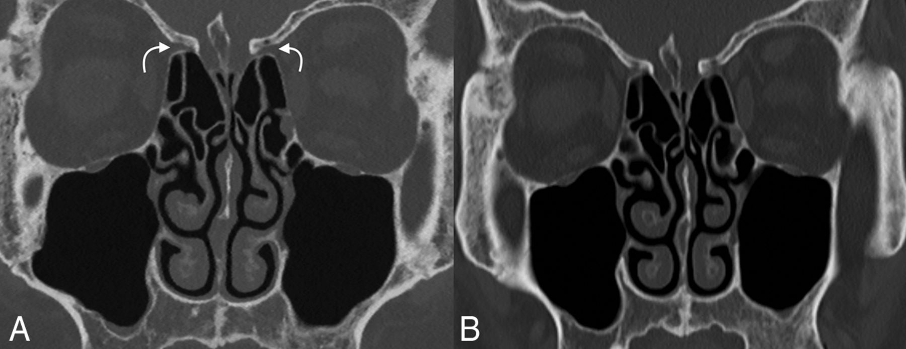
    Comparison of PCD-CT to EID-CT in paranasal sinus imaging. Although both are useful for the relatively larger anatomic structures of the paranasal sinuses, PCD-CT image (A) has substantially greater spatial resolution at less radiation doses than EID-CT image (B). For example, the anterior ethmoidal artery notch is seen much clearer on PCD-CT image (curved arrows). W/L = 575/3630.

  • FIG 5.
    • Download figure
    • Open in new tab
    • Download powerpoint
    FIG 5.

    Example of an adenoid cystic carcinoma with invasion of the skull base in a 69-year-old man. A large soft tissue mass was seen centered in the right parotid gland (asterisk). Coronal bone kernel images demonstrated invasion into the right temporal bone with involvement of the external auditory canal (arrows), abutting the tympanic membrane (dashed arrows), and abutting the handle of the malleus. A = soft tissue kernel, W/L = 51/337. B and C = bone kernel, W/L = 1000/4000.

  • FIG 6.
    • Download figure
    • Open in new tab
    • Download powerpoint
    FIG 6.

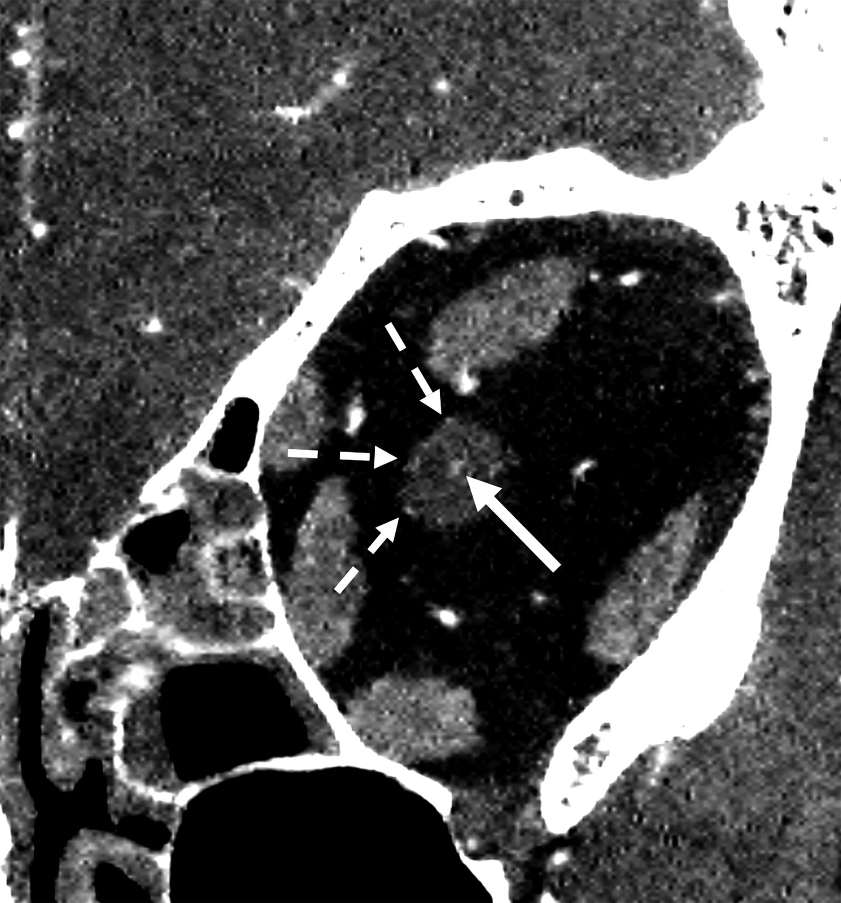
    Evaluation of a central retinal artery on PCD-CT. Coronal CTA image of the midleft orbit from a PCD-CT scanner demonstrates both the central retinal artery in the distal optic nerve (solid arrow) and branches of the posterior ciliary artery (dashed arrows). W/L = 219/48.

  • FIG 7.
    • Download figure
    • Open in new tab
    • Download powerpoint
    FIG 7.

    Arteries in multiple skull base foramina on PCD-CT. A reformatted coronal CTA image through the sphenoid sinuses exemplifies the ability of PCD-CT to evaluate the left ophthalmic artery (solid white arrow), foramen rotundum artery (dashed white arrow), and pterygoid canal artery (solid black arrows). W/L = 262/88.

  • FIG 8.
    • Download figure
    • Open in new tab
    • Download powerpoint
    FIG 8.

    Example of improved vessel lumen conspicuity in the setting of a calcified atherosclerotic plaque. Blooming artifact–related densely calcified plaque at the origin of the right ICA (arrow) somewhat obscures the adjacent lumen (dashed arrow) on EID-CT, scanned with 0.6 slice thickness (A). PCD-CT image (at 0.2 mm slice thickness) provides substantially better evaluation of the lumen (B). W/L = 810/216 on EID-CT, 868/2220 on PCD-CT.

PreviousNext
Back to top
Advertisement
Print
Download PDF
Email Article

Thank you for your interest in spreading the word on American Journal of Neuroradiology.

NOTE: We only request your email address so that the person you are recommending the page to knows that you wanted them to see it, and that it is not junk mail. We do not capture any email address.

Enter multiple addresses on separate lines or separate them with commas.
Photon-Counting CT in the Head and Neck: Current Applications and Future Prospects
(Your Name) has sent you a message from American Journal of Neuroradiology
(Your Name) thought you would like to see the American Journal of Neuroradiology web site.
CAPTCHA
This question is for testing whether or not you are a human visitor and to prevent automated spam submissions.
Cite this article
John C. Benson, Norbert G. Campeau, Felix E. Diehn, John I. Lane, Shuai Leng, Gul Moonis
Photon-Counting CT in the Head and Neck: Current Applications and Future Prospects
American Journal of Neuroradiology Jul 2024, DOI: 10.3174/ajnr.A8265

Citation Manager Formats

  • BibTeX
  • Bookends
  • EasyBib
  • EndNote (tagged)
  • EndNote 8 (xml)
  • Medlars
  • Mendeley
  • Papers
  • RefWorks Tagged
  • Ref Manager
  • RIS
  • Zotero
0 Responses
Respond to this article
Share
Bookmark this article
Photon-Counting CT in the Head and Neck: Current Applications and Future Prospects
John C. Benson, Norbert G. Campeau, Felix E. Diehn, John I. Lane, Shuai Leng, Gul Moonis
American Journal of Neuroradiology Jul 2024, DOI: 10.3174/ajnr.A8265
del.icio.us logo Twitter logo Facebook logo Mendeley logo
  • Tweet Widget
  • Facebook Like
  • Google Plus One
Purchase

Jump to section

  • Article
    • SUMMARY:
    • ABBREVIATIONS:
    • TEMPORAL BONE
    • Facial Bones, Skull Base, Paranasal Sinuses
    • HEAD AND NECK ARTERIAL VASCULATURE
    • CONCLUSIONS
    • Footnotes
    • References
  • Figures & Data
  • Info & Metrics
  • Responses
  • References
  • PDF

Related Articles

  • No related articles found.
  • PubMed
  • Google Scholar

Cited By...

  • No citing articles found.
  • Crossref
  • Google Scholar

This article has not yet been cited by articles in journals that are participating in Crossref Cited-by Linking.

More in this TOC Section

  • An Atlas of Neonatal Neurovascular Imaging Anatomy as Depicted with Microvascular Imaging: The Intracranial Arteries
  • An Atlas of Neonatal Neurovascular Imaging Anatomy as Depicted with Microvascular Imaging: The Intracranial Veins
  • Clinical Translation of Hyperpolarized 13C Metabolic Probes for Glioma Imaging
Show more Review Articles

Similar Articles

Advertisement

Indexed Content

  • Current Issue
  • Accepted Manuscripts
  • Article Preview
  • Past Issues
  • Editorials
  • Editor's Choice
  • Fellows' Journal Club
  • Letters to the Editor
  • Video Articles

Cases

  • Case Collection
  • Archive - Case of the Week
  • Archive - Case of the Month
  • Archive - Classic Case

More from AJNR

  • Trainee Corner
  • Imaging Protocols
  • MRI Safety Corner
  • Book Reviews

Multimedia

  • AJNR Podcasts
  • AJNR Scantastics

Resources

  • Turnaround Time
  • Submit a Manuscript
  • Submit a Video Article
  • Submit an eLetter to the Editor/Response
  • Manuscript Submission Guidelines
  • Statistical Tips
  • Fast Publishing of Accepted Manuscripts
  • Graphical Abstract Preparation
  • Imaging Protocol Submission
  • Evidence-Based Medicine Level Guide
  • Publishing Checklists
  • Author Policies
  • Become a Reviewer/Academy of Reviewers
  • News and Updates

About Us

  • About AJNR
  • Editorial Board
  • Editorial Board Alumni
  • Alerts
  • Permissions
  • Not an AJNR Subscriber? Join Now
  • Advertise with Us
  • Librarian Resources
  • Feedback
  • Terms and Conditions
  • AJNR Editorial Board Alumni

American Society of Neuroradiology

  • Not an ASNR Member? Join Now

© 2025 by the American Society of Neuroradiology All rights, including for text and data mining, AI training, and similar technologies, are reserved.
Print ISSN: 0195-6108 Online ISSN: 1936-959X

Powered by HighWire