
Article Figures & Data
Figures
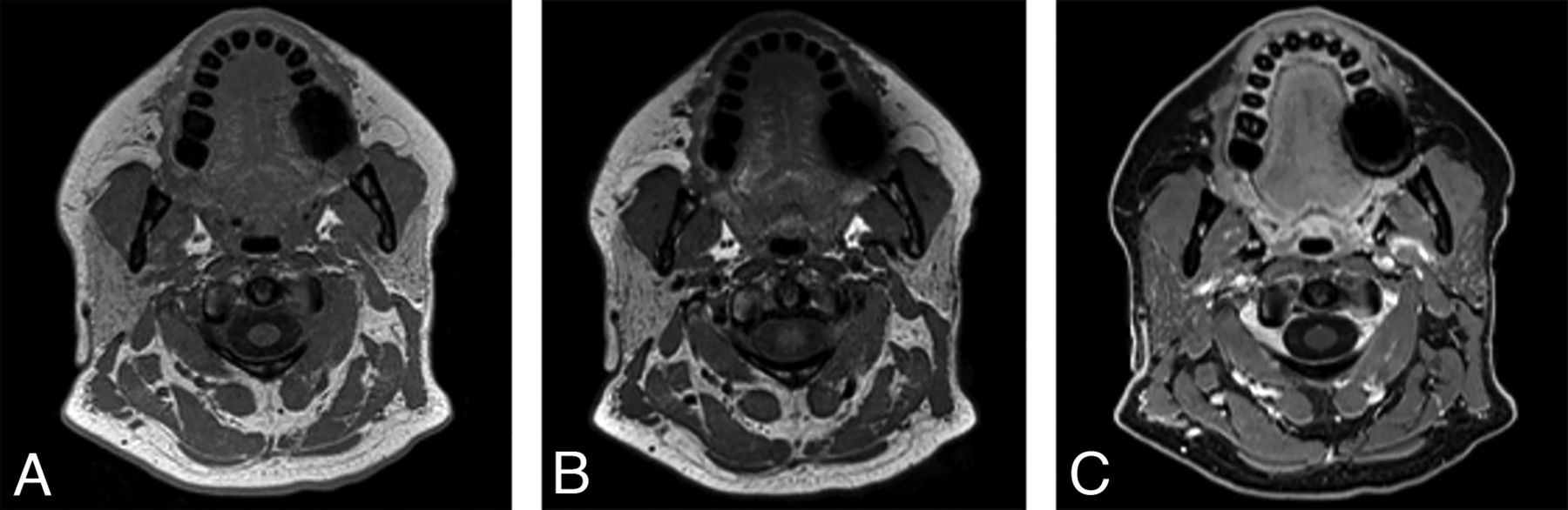
- FIG 1.
Sample 3D T1 VIBE (A), 3D T1 SPACE (B), and postcontrast 3D T1 VIBE Dixon fat-suppression (C) images acquired in the same session on a 3T Magnetom Vida system (Siemens) at the level of the oral cavity, retromolar trigone, and parotid glands in a 44-year-old woman with neck pain show excellent discrimination of soft-tissue structure boundaries with subjectively better soft-tissue contrasts on the SPACE than on VIBE and robust fat suppression on the VIBE Dixon.
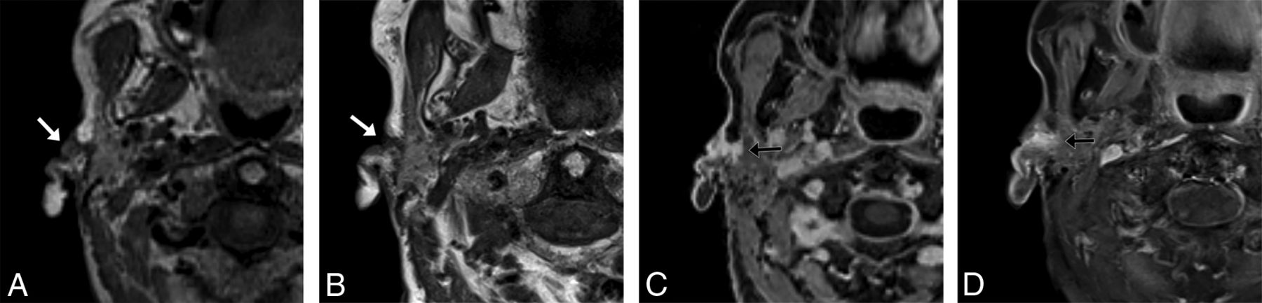
- FIG 2.
A 92-year-old woman with squamous cell carcinoma. A, T1 SPACE sequence performed in the full-neck MR imaging protocol with 2.7-mm3 (1.4 mm isotropic) voxels during early optimization iterations reformatted into the axial plane clearly shows a cutaneous preauricular lesion (arrow) that is not visible due to volume averaging on the 2D T1-weighted image (B) with 1-mm3 (4 × 0.5× 0.5 mm) voxels. The T1 VIBE Dixon (C) reformatted image in the axial plane shows the associated infiltrating tumor into the parotid gland (arrow) to advantage compared with the 2D T1-weighted image (D) with spectral fat suppression. The case convinced us that the SPACE sequence was a reasonable alternative to the 2D standard-of-care and led us to improve the spatial resolution of the SPACE sequence. These images were obtained contemporaneously on a 3T Magnetom Vida system.
- FIG 3.
A 27-year-old man with right supraclavicular and chest wall desmoid tumor images on a 1.5T Magnetom Aera system (Siemens). The tumor (arrows) is shown on a general neck protocol, 1.0-mm isotropic sagittal acquisition T1 SPACE axial reformat (A) and T1 VIBE Dixon (B) postcontrast axial reformatted images. Image quality and fat suppression remain good at the level of the thoracic inlet. In addition, T1 SPACE axial reformat (C) and T1 VIBE Dixon (D) postcontrast axial reformat images through the face are provided to show image quality in that region.
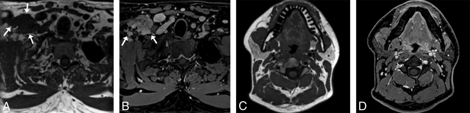
- FIG 4.
A 44-year-old woman with newly diagnosed oral cavity squamous cell carcinoma (arrows) shown on oropharynx protocol T1 VIBE Dixon 0.8 × 0.9 × 0.9 mm resolution axial acquisition (A) and coronal reformat images (B) and a corresponding precontrast T1 SPACE axial acquisition (C) and coronal reformat images (D) obtained on a 3T Magnetom Vida system.
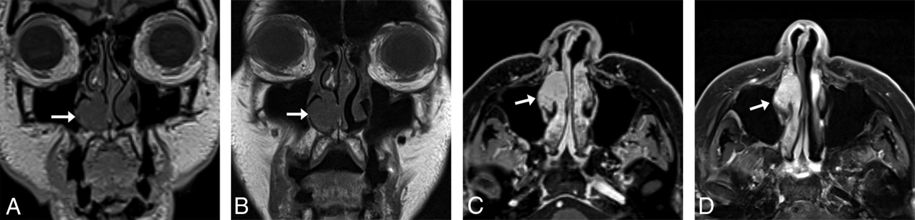
- FIG 5.
An 83-year-old woman with melanoma and a stable untreated nasal cavity mass of unknown pathology with sinus protocol MR imaging. T1 SPACE coronal acquisition (A) performed with 1-mm3 (1.0 mm isotropic) voxels shows a mass (arrow) along the anterior aspect of the right inferior turbinate, seen similarly on the 2D T1-weighted image (B) with 0.8-mm3 (3 × 0.5 × 0.52 mm) voxels. The T1 VIBE Dixon axial reformat (C) shows the mass (arrow) to advantage compared with the 2D T1-weighted image (D) with spectral fat suppression; the former shows the extent of the mass relative to the more heterogeneously enhancing turbinate. The 3D images were acquired on a 3T Magnetom Vida system 6 months following the acquisition of the 2D images on a 3T Magnetom Skyra system (Siemens).
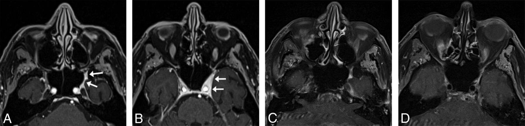
- FIG 6.
An 85-year-old man with spindle cell sarcoma with sinus protocol MR imaging. T1 VIBE Dixon axial acquisition performed with 0.9-mm3 (0.74 mm isotropic) voxels clearly shows tumor (arrows) along the left V2 (A) and in the left lateral cavernous sinus wall (B), neither of which is clearly seen on the 2D T1-weighted images (C and D) with spectral fat suppression with 2-mm3 (4 × 0.7 × 0.7 mm) voxels. The 3D examination shown was performed on a 3T Magnetom Prisma Fit system (Siemens) 3 weeks before the 2D examination, which was performed on a Siemens 3T Vida system, with no intervening treatment.


Tables
Gradient time savings from MR imaging protocols on 3T Magnetom Prisma and 1.5T Magnetom Aera systems for reference
Protocol 2D T1 Version 3D T1 Version Time Savings Savings 3T general 23:05 16:42 6:23 27.7% 3T nasopharynx 23:42 13:38 10:04 42.5% 3T oropharynx 26:37 16:33 10:04 37.8% 3T sinus 24:00 10:42 13:18 55.4% Mean 24:21 14:24 9:57 40.9% 1.5T general 23:15 18:12 5:03 21.7% 1.5T nasopharynx 24:11 15:26 8:45 36.2% 1.5T oropharynx 27:33 18:48 8:45 31.8% 1.5T sinus 23:14 9:28 13:46 59.3% Mean 24:33 15:29 9:05 37.0% Note:—Times listed as minutes:seconds.


Jump to section
Related Articles
Cited By...
- No citing articles found.