Advertisement
  • Main menu
  • User menu
  • Search
  • English ▼
    • English
    • Afrikaans
    • Albanian
    • Amharic
    • Arabic
    • Armenian
    • Azerbaijani
    • Basque
    • Belarusian
    • Bengali
    • Bosnian
    • Bulgarian
    • Catalan
    • Cebuano
    • Chichewa
    • Chinese (Simplified)
    • Chinese (Traditional)
    • Corsican
    • Croatian
    • Czech
    • Danish
    • Dutch
    • Esperanto
    • Estonian
    • Filipino
    • Finnish
    • French
    • Frisian
    • Galician
    • Georgian
    • German
    • Greek
    • Gujarati
    • Haitian Creole
    • Hausa
    • Hawaiian
    • Hebrew
    • Hindi
    • Hmong
    • Hungarian
    • Icelandic
    • Igbo
    • Indonesian
    • Irish
    • Italian
    • Japanese
    • Javanese
    • Kannada
    • Kazakh
    • Khmer
    • Korean
    • Kurdish (Kurmanji)
    • Kyrgyz
    • Lao
    • Latin
    • Latvian
    • Lithuanian
    • Luxembourgish
    • Macedonian
    • Malagasy
    • Malay
    • Malayalam
    • Maltese
    • Maori
    • Marathi
    • Mongolian
    • Myanmar (Burmese)
    • Nepali
    • Norwegian
    • Pashto
    • Persian
    • Polish
    • Portuguese
    • Punjabi
    • Romanian
    • Russian
    • Samoan
    • Scottish Gaelic
    • Serbian
    • Sesotho
    • Shona
    • Sindhi
    • Sinhala
    • Slovak
    • Slovenian
    • Somali
    • Spanish
    • Sudanese
    • Swahili
    • Swedish
    • Tajik
    • Tamil
    • Telugu
    • Thai
    • Turkish
    • Ukrainian
    • Urdu
    • Uzbek
    • Vietnamese
    • Welsh
    • Xhosa
    • Yiddish
    • Yoruba
    • Zulu

Main menu

  • Home
  • Content
    • Current Issue
    • Accepted Manuscripts
    • Article Preview
    • Past Issue Archive
    • Video Articles
    • AJNR Case Collection
    • Case of the Week Archive
    • Case of the Month Archive
    • Classic Case Archive
  • Special Collections
    • AJNR Awards
    • Low-Field MRI
    • Alzheimer Disease
    • ASNR Foundation Special Collection
    • Photon-Counting CT
    • View All
  • Multimedia
    • AJNR Podcasts
    • AJNR SCANtastic
    • Trainee Corner
    • MRI Safety Corner
    • Imaging Protocols
  • For Authors
    • Submit a Manuscript
    • Submit a Video Article
    • Submit an eLetter to the Editor/Response
    • Manuscript Submission Guidelines
    • Statistical Tips
    • Fast Publishing of Accepted Manuscripts
    • Graphical Abstract Preparation
    • Imaging Protocol Submission
    • Author Policies
  • About Us
    • About AJNR
    • Editorial Board
    • Editorial Board Alumni
  • More
    • Become a Reviewer/Academy of Reviewers
    • Subscribers
    • Permissions
    • Alerts
    • Feedback
    • Advertisers
    • ASNR Home

User menu

  • Alerts
  • Log in

Search

  • Advanced search
American Journal of Neuroradiology

American Journal of Neuroradiology

ASHNR American Society of Functional Neuroradiology ASHNR American Society of Pediatric Neuroradiology ASSR
  • Alerts
  • Log in

Advanced Search

English ▼
  • English
  • Afrikaans
  • Albanian
  • Amharic
  • Arabic
  • Armenian
  • Azerbaijani
  • Basque
  • Belarusian
  • Bengali
  • Bosnian
  • Bulgarian
  • Catalan
  • Cebuano
  • Chichewa
  • Chinese (Simplified)
  • Chinese (Traditional)
  • Corsican
  • Croatian
  • Czech
  • Danish
  • Dutch
  • Esperanto
  • Estonian
  • Filipino
  • Finnish
  • French
  • Frisian
  • Galician
  • Georgian
  • German
  • Greek
  • Gujarati
  • Haitian Creole
  • Hausa
  • Hawaiian
  • Hebrew
  • Hindi
  • Hmong
  • Hungarian
  • Icelandic
  • Igbo
  • Indonesian
  • Irish
  • Italian
  • Japanese
  • Javanese
  • Kannada
  • Kazakh
  • Khmer
  • Korean
  • Kurdish (Kurmanji)
  • Kyrgyz
  • Lao
  • Latin
  • Latvian
  • Lithuanian
  • Luxembourgish
  • Macedonian
  • Malagasy
  • Malay
  • Malayalam
  • Maltese
  • Maori
  • Marathi
  • Mongolian
  • Myanmar (Burmese)
  • Nepali
  • Norwegian
  • Pashto
  • Persian
  • Polish
  • Portuguese
  • Punjabi
  • Romanian
  • Russian
  • Samoan
  • Scottish Gaelic
  • Serbian
  • Sesotho
  • Shona
  • Sindhi
  • Sinhala
  • Slovak
  • Slovenian
  • Somali
  • Spanish
  • Sudanese
  • Swahili
  • Swedish
  • Tajik
  • Tamil
  • Telugu
  • Thai
  • Turkish
  • Ukrainian
  • Urdu
  • Uzbek
  • Vietnamese
  • Welsh
  • Xhosa
  • Yiddish
  • Yoruba
  • Zulu
  • Home
  • Content
    • Current Issue
    • Accepted Manuscripts
    • Article Preview
    • Past Issue Archive
    • Video Articles
    • AJNR Case Collection
    • Case of the Week Archive
    • Case of the Month Archive
    • Classic Case Archive
  • Special Collections
    • AJNR Awards
    • Low-Field MRI
    • Alzheimer Disease
    • ASNR Foundation Special Collection
    • Photon-Counting CT
    • View All
  • Multimedia
    • AJNR Podcasts
    • AJNR SCANtastic
    • Trainee Corner
    • MRI Safety Corner
    • Imaging Protocols
  • For Authors
    • Submit a Manuscript
    • Submit a Video Article
    • Submit an eLetter to the Editor/Response
    • Manuscript Submission Guidelines
    • Statistical Tips
    • Fast Publishing of Accepted Manuscripts
    • Graphical Abstract Preparation
    • Imaging Protocol Submission
    • Author Policies
  • About Us
    • About AJNR
    • Editorial Board
    • Editorial Board Alumni
  • More
    • Become a Reviewer/Academy of Reviewers
    • Subscribers
    • Permissions
    • Alerts
    • Feedback
    • Advertisers
    • ASNR Home
  • Follow AJNR on Twitter
  • Visit AJNR on Facebook
  • Follow AJNR on Instagram
  • Join AJNR on LinkedIn
  • RSS Feeds

AJNR Awards, New Junior Editors, and more. Read the latest AJNR updates

Research ArticleBRAIN TUMOR IMAGING
Open Access

Arterial Spin-Labeling and DSC Perfusion Metrics Improve Agreement in Neuroradiologists’ Clinical Interpretations of Posttreatment High-Grade Glioma Surveillance MR Imaging—An Institutional Experience

Ghiam Yamin, Eric Tranvinh, Bryan A. Lanzman, Elizabeth Tong, Syed S. Hashmi, Chirag B. Patel and Michael Iv
American Journal of Neuroradiology March 2024, DOI: https://doi.org/10.3174/ajnr.A8190
Ghiam Yamin
aFrom the Department of Radiology (G.Y., E. Tranvinh, B.A.L., E. Tong, S.S.H., M.I.), Division of Neuroimaging and Neurointervention, Stanford University Medical Center, Stanford, California
  • Find this author on Google Scholar
  • Find this author on PubMed
  • Search for this author on this site
  • ORCID record for Ghiam Yamin
Eric Tranvinh
aFrom the Department of Radiology (G.Y., E. Tranvinh, B.A.L., E. Tong, S.S.H., M.I.), Division of Neuroimaging and Neurointervention, Stanford University Medical Center, Stanford, California
  • Find this author on Google Scholar
  • Find this author on PubMed
  • Search for this author on this site
  • ORCID record for Eric Tranvinh
Bryan A. Lanzman
aFrom the Department of Radiology (G.Y., E. Tranvinh, B.A.L., E. Tong, S.S.H., M.I.), Division of Neuroimaging and Neurointervention, Stanford University Medical Center, Stanford, California
  • Find this author on Google Scholar
  • Find this author on PubMed
  • Search for this author on this site
  • ORCID record for Bryan A. Lanzman
Elizabeth Tong
aFrom the Department of Radiology (G.Y., E. Tranvinh, B.A.L., E. Tong, S.S.H., M.I.), Division of Neuroimaging and Neurointervention, Stanford University Medical Center, Stanford, California
  • Find this author on Google Scholar
  • Find this author on PubMed
  • Search for this author on this site
  • ORCID record for Elizabeth Tong
Syed S. Hashmi
aFrom the Department of Radiology (G.Y., E. Tranvinh, B.A.L., E. Tong, S.S.H., M.I.), Division of Neuroimaging and Neurointervention, Stanford University Medical Center, Stanford, California
  • Find this author on Google Scholar
  • Find this author on PubMed
  • Search for this author on this site
  • ORCID record for Syed S. Hashmi
Chirag B. Patel
bDepartment of Neuro-Oncology (C.B.P.), The University of Texas MD Anderson Cancer Center, Houston, Texas
  • Find this author on Google Scholar
  • Find this author on PubMed
  • Search for this author on this site
  • ORCID record for Chirag B. Patel
Michael Iv
aFrom the Department of Radiology (G.Y., E. Tranvinh, B.A.L., E. Tong, S.S.H., M.I.), Division of Neuroimaging and Neurointervention, Stanford University Medical Center, Stanford, California
  • Find this author on Google Scholar
  • Find this author on PubMed
  • Search for this author on this site
  • ORCID record for Michael Iv
  • Article
  • Figures & Data
  • Info & Metrics
  • Responses
  • References
  • PDF
Loading

Article Figures & Data

Figures

  • Tables
  • FIG 1.
    • Download figure
    • Open in new tab
    • Download powerpoint
    FIG 1.

    Example of rater BT-RADS scores in a 57-year-old woman with previously treated GBM and worsening findings on surveillance MR imaging. The T1 postgadolinium image demonstrates an enhancing lesion in the left mesial temporal lobe. The lesion has elevated ASL-CBF and DSC-rCBV (white arrows). The DSC-FTB image shows that the enhancing voxels are in the “high” fractional tumor burden (red voxels) category. The addition of perfusion metrics to CE-MR imaging resulted in a scoring upgrade from 3b (worsening imaging findings, indeterminate mix of treatment effects and tumor) to 3c/4 (likely tumor progression) across all raters and agreed with the consensus score of 3c/4. For 3 of 4 raters, the upgrade occurred with all perfusion metrics, and for rater 2, it occurred only with DSC-FTB.
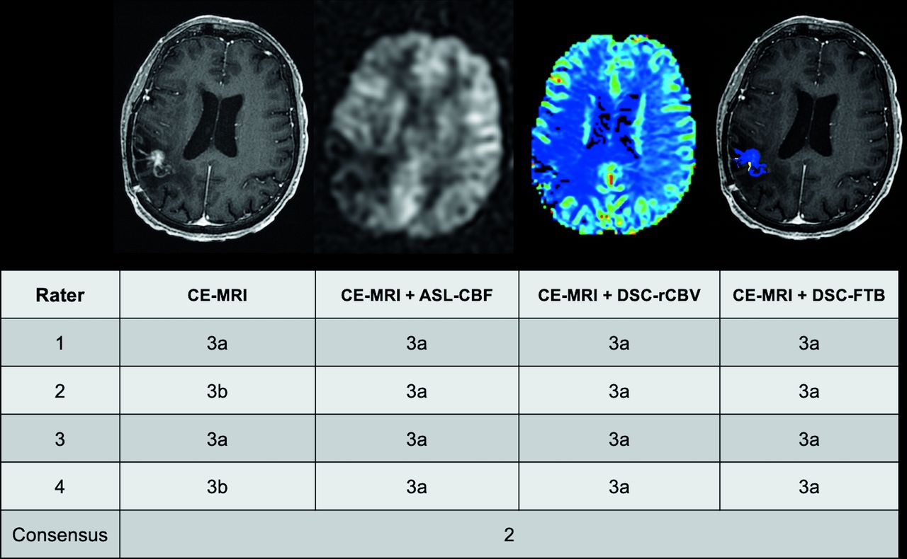
  • FIG 2.
    • Download figure
    • Open in new tab
    • Download powerpoint
    FIG 2.

    Example of rater BT-RADS scores in a 61-year-old woman with previously treated GBM and equivocally worsening findings on surveillance MR imaging. The T1 postgadolinium image demonstrates an enhancing lesion in the right temporal lobe. The lesion shows no elevated ASL-CBF or DSC-rCBV, and DSC-FTB shows that the enhancing voxels are in the “low” FTB (blue voxels) category. For two raters, the addition of perfusion metrics to CE-MR imaging resulted in a scoring downgrade from 3b (worsening imaging findings, indeterminate mix of treatment effects and tumor) to 3a (worsening imaging findings, likely treatment effects). For the other raters, perfusion metrics did not influence their assessment. The consensus score in this case was 2 (no change). The discrepancy between the consensus group and the raters was due to differences in opinion as to whether the enhancing lesion had subtly increased in size from the prior MR imaging (not shown).

  • FIG 3.
    • Download figure
    • Open in new tab
    • Download powerpoint
    FIG 3.

    Clinically meaningful changes in BT-RADS scores following the inclusion of perfusion metrics compared with conventional CE-MR imaging alone. Clinically meaningful upgrades or downgrades were defined as score changes from ≤3a⇆3b or 3b⇆3c/4 and from 3c/4⇆≤3b or 3b⇆≤3a, respectively. The numbers and arrows above the bar graph indicate the number of score upgrades (upward facing arrow) or downgrades (downward facing arrow). The greatest number of score changes was observed with the addition of DSC-FTB.
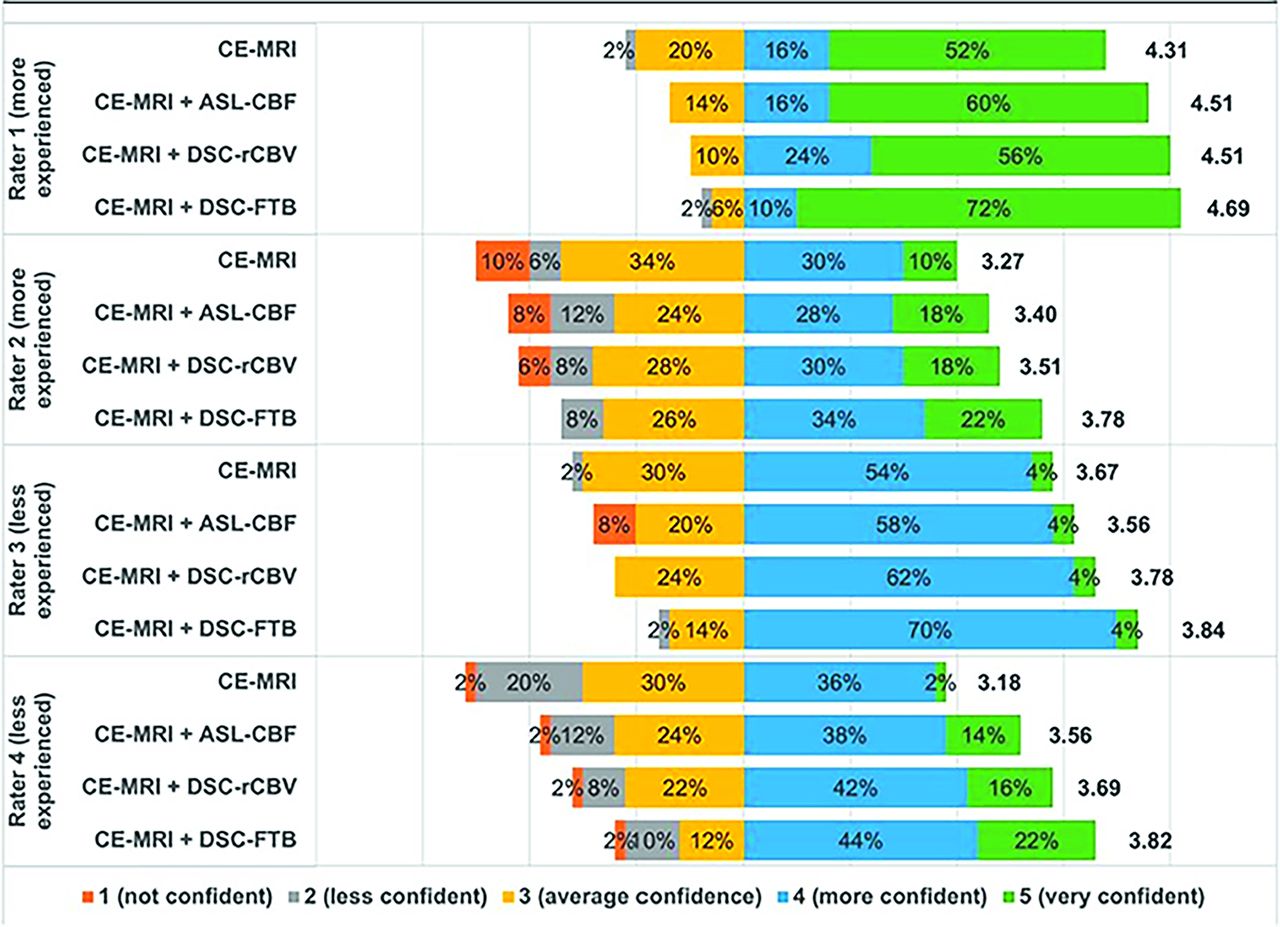
  • FIG 4.
    • Download figure
    • Open in new tab
    • Download powerpoint
    FIG 4.

    Rater confidence in MR imaging interpretation. Raters graded their confidence in interpretation and assignment of BT-RADS scores for conventional CE-MR imaging, CE-MR imaging + ASL-CBF, CE-MR imaging + DSC-rCBV, and CE-MR imaging + DSC-FTB using a 5-point Likert scale. The number to the right of each color bar represents the mean score. In general, confidence was higher with the addition of any perfusion metric but was highest with DSC-FTB in all raters.

Tables

  • Figures
    • View popup
    Table 1:

    Patient demographics

    Demographics
    Age (yr)
     Mean (SD)61 (13)
     Range31–88
    Sex
     Male25 (56%)
     Female20 (44%)
    Integrated diagnosis
     GBM, IDH wild-type, WHO 444 (98%)
     Astrocytoma, IDH wild-type, WHO 31 (2%)
    HGG molecular features
     IDH wild-type45 (100%)
     MGMT promoter-unmethylated24 (53%)
     MGMT promoter-methylated19 (42%)
     Unknown MGMT promoter methylation status2 (4%)
    • Note:—WHO indicates World Health Organization.

    • View popup
    Table 2:

    Agreement in MR imaging interpretation among 4 neuroradiologists

     Fleiss κ95% CIP Value
    Conventional CE-MR imaging0.630.56−0.69<.001
    CE-MR imaging + ASL-CBF0.670.60−0.74<.001
    CE-MR imaging + DSC-rCBV0.660.60−0.73<.001
    CE-MR imaging + DSC-FTB0.700.63−0.77<.001
    • View popup
    Table 3:

    Agreement in MR imaging interpretation between an experienced multidisciplinary consensus group and each of 4 neuroradiologistsa

     More ExperiencedLess Experienced
    Rater 1Rater 2Rater 3Rater 4
    Conventional CE-MR imaging0.53 (0.31–0.75)0.70 (0.50–0.91)0.63 (0.43–0.84)0.58 (0.36–0.80)
    CE-MR imaging + ASL-CBF0.58 (0.36–0.80)0.69 (0.48–0.89)0.61 (0.39–0.82)0.65 (0.44–0.86)
    CE-MR imaging + DSC-rCBV0.58 (0.36–0.80)0.71 (0.51–0.90)0.63 (0.42–0.84)0.68 (0.47–0.88)
    CE-MR imaging + DSC-FTB0.66 (0.46–0.87)0.80 (0.63–0.97)0.66 (0.46–0.86)0.73 (0.55–0.92)
    • ↵a All analyses showed P < .001. Values are Cohen κ with 95% confidence intervals in the parentheses.

    • View popup
    Table 4:

    Frequency of clinically meaningful changes in BT-RADS scores in 45 patients following the inclusion of perfusion metrics compared with conventional CE-MR imaging alonea

     More ExperiencedLess Experienced
    Rater 1Rater 2Rater 3Rater 4
    CE-MR imaging + ASL-CBF5 (11%)7 (16%)1 (2%)7 (16%)
    CE-MR imaging + DSC-rCBV5 (11%)4 (9%)2 (4%)8 (18%)
    CE-MR imaging + DSC-FTB8 (18%)6 (13%)3 (7%)9 (20%)
    • ↵a No significance was found among ASL-CBF, DSC-rCBV, and DSC-FTB when added to CE-MR imaging with respect to the number of clinically meaningful score changes (P = .53). Percentages are in parentheses.

PreviousNext
Back to top
Advertisement
Print
Download PDF
Email Article
Cite this article
0 Responses
Respond to this article
Share
Bookmark this article
Arterial Spin-Labeling and DSC Perfusion Metrics Improve Agreement in Neuroradiologists’ Clinical Interpretations of Posttreatment High-Grade Glioma Surveillance MR Imaging—An Institutional Experience
Ghiam Yamin, Eric Tranvinh, Bryan A. Lanzman, Elizabeth Tong, Syed S. Hashmi, Chirag B. Patel, Michael Iv
American Journal of Neuroradiology Mar 2024, DOI: 10.3174/ajnr.A8190
del.icio.us logo Twitter logo Facebook logo Mendeley logo
  • Tweet Widget
Purchase

Jump to section

  • Article
    • Abstract
    • ABBREVIATIONS:
    • MATERIALS AND METHODS
    • RESULTS
    • DISCUSSION
    • CONCLUSIONS
    • Footnotes
    • References
  • Figures & Data
  • Info & Metrics
  • Responses
  • References
  • PDF

Related Articles

  • No related articles found.
  • PubMed
  • Google Scholar

Cited By...

  • No citing articles found.
  • Crossref
  • Google Scholar

This article has not yet been cited by articles in journals that are participating in Crossref Cited-by Linking.

More in this TOC Section

  • Brain Tumor Subtyping Image-Based search via MRI
  • Neuroimaging of Erdheim-Chester Disease
  • CE MRI for Brain Metastasis Detection
Show more Brain Tumor Imaging

Similar Articles

  • Perfusion MRI-Based Fractional Tumor Burden Differentiates between Tumor and Treatment Effect in Recurrent Glioblastomas and Informs Clinical Decision-Making
See more
Advertisement

Indexed Content

  • Current Issue
  • Accepted Manuscripts
  • Article Preview
  • Past Issues
  • Editorials
  • Editor's Choice
  • Fellows' Journal Club
  • Letters to the Editor
  • Video Articles

Cases

  • Case Collection
  • Archive - Case of the Week
  • Archive - Case of the Month
  • Archive - Classic Case

More from AJNR

  • Trainee Corner
  • Imaging Protocols
  • MRI Safety Corner
  • Book Reviews

Multimedia

  • AJNR Podcasts
  • AJNR Scantastics

Resources

  • Turnaround Time
  • Submit a Manuscript
  • Submit a Video Article
  • Submit an eLetter to the Editor/Response
  • Manuscript Submission Guidelines
  • Statistical Tips
  • Fast Publishing of Accepted Manuscripts
  • Graphical Abstract Preparation
  • Imaging Protocol Submission
  • Evidence-Based Medicine Level Guide
  • Publishing Checklists
  • Author Policies
  • Become a Reviewer/Academy of Reviewers
  • News and Updates

About Us

  • About AJNR
  • Editorial Board
  • Editorial Board Alumni
  • Alerts
  • Permissions
  • Not an AJNR Subscriber? Join Now
  • Advertise with Us
  • Librarian Resources
  • Feedback
  • Terms and Conditions
  • AJNR Editorial Board Alumni

American Society of Neuroradiology

  • Not an ASNR Member? Join Now

© 2025 by the American Society of Neuroradiology All rights, including for text and data mining, AI training, and similar technologies, are reserved.
Print ISSN: 0195-6108 Online ISSN: 1936-959X

Powered by HighWire
Email this Article

Thank you for your interest in spreading the word on American Journal of Neuroradiology.

NOTE: We only request your email address so that the person you are recommending the page to knows that you wanted them to see it, and that it is not junk mail. We do not capture any email address.

Enter multiple addresses on separate lines or separate them with commas.
Arterial Spin-Labeling and DSC Perfusion Metrics Improve Agreement in Neuroradiologists’ Clinical Interpretations of Posttreatment High-Grade Glioma Surveillance MR Imaging—An Institutional Experience
(Your Name) has sent you a message from American Journal of Neuroradiology
(Your Name) thought you would like to see the American Journal of Neuroradiology web site.
CAPTCHA
This question is for testing whether or not you are a human visitor and to prevent automated spam submissions.
Cite this article
Ghiam Yamin, Eric Tranvinh, Bryan A. Lanzman, Elizabeth Tong, Syed S. Hashmi, Chirag B. Patel, Michael Iv
Arterial Spin-Labeling and DSC Perfusion Metrics Improve Agreement in Neuroradiologists’ Clinical Interpretations of Posttreatment High-Grade Glioma Surveillance MR Imaging—An Institutional Experience
American Journal of Neuroradiology Mar 2024, DOI: 10.3174/ajnr.A8190

Citation Manager Formats

  • BibTeX
  • Bookends
  • EasyBib
  • EndNote (tagged)
  • EndNote 8 (xml)
  • Medlars
  • Mendeley
  • Papers
  • RefWorks Tagged
  • Ref Manager
  • RIS
  • Zotero

We use cookies on this site to enhance your user experience. By clicking any link on this page you are giving your consent for us to set cookies.