
Article Figures & Data
Figures
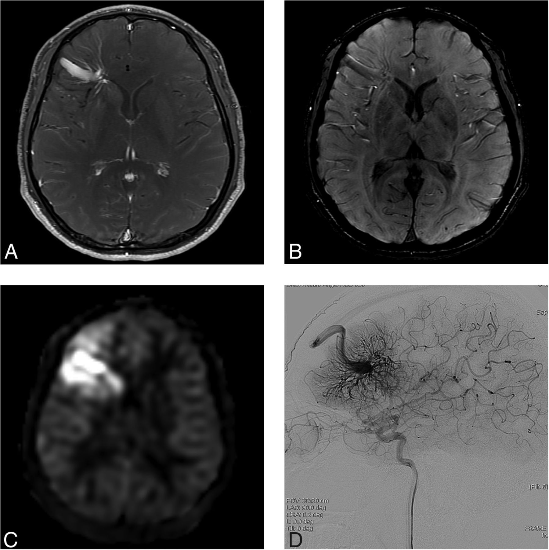
- FIG 1.
A 31-year-old man presenting with dizziness (case 4). A, Contrast-enhanced T1-weighted axial MR imaging shows a DVA-like lesion in the left frontal lobe. B, On SWI, both hyperintense and hypointense signal is present in the collecting vein, while only hypointense signal is present in the dilated medullary veins. C, The ASL quantitative CBF image demonstrates hyperintense signal intensity in the collecting vein and parenchyma corresponding to the location of the lesion, suggesting a vpAVM. D, On DSA, a caput medusa appearance of medullary veins draining to the superior sagittal sinus via a collecting cortical vein is visualized in the arterial phase.
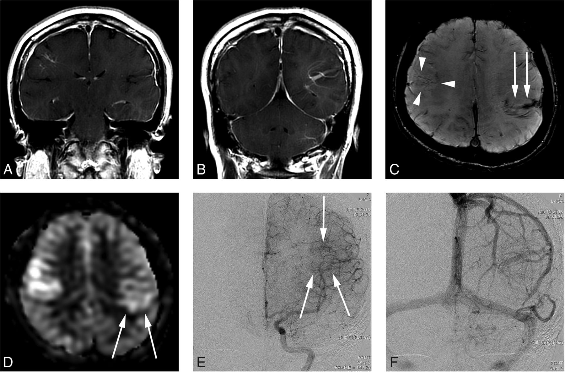
- FIG 2.
A 34-year-old woman presenting with a headache (case 6). A and B, Contrast-enhanced T1-weighted coronal MR imaging shows a DVA-like lesion in the bilateral parietal lobes. C, SWI demonstrates only hypointense signal in both the right (arrowheads) and left (arrow) lesions. D, The ASL quantitative CBF image demonstrates mildly hyperintense signal intensity in the parenchyma, corresponding to the location of the lesion. The left-sided lesion exhibits particularly subtle signal (arrows). E, In the late arterial phase of DSA, dilated medullary veins are gradually and subtly visualized (arrows). F, In the venous phase, dilated medullary veins draining to a collecting vein typical of a DVA are seen.
- FIG 3.
A 29-year-old man presenting with an AVM in the left occipital lobe (case 11). A, Contrast-enhanced T1-weighted axial MR imaging shows a DVA-like lesion in the right frontal lobe. B, SWI shows hypointense signal in the lesion. C, An ASL quantitative CBF image demonstrates no identifiable signal corresponding to the lesion. D, On DSA, the lesion is first visualized in the late venous phase (arrows), as is typically seen in a classic DVA.
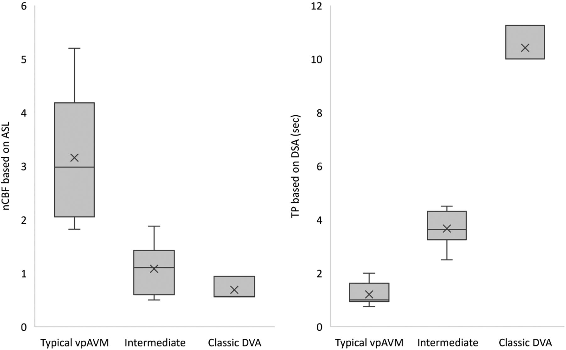
- FIG 4.
Box-and-whisker display of nCBF and TP for the 3 groups of lesions. The line across the boxes denotes the median value, whereas the ends of the boxes represent the first and third quartiles (not applied in the classic DVA group due to the small number of cases). The ends of each plot indicate the smallest and largest values. × denotes average value. Sec indicates second.


