
Article Figures & Data
Figures
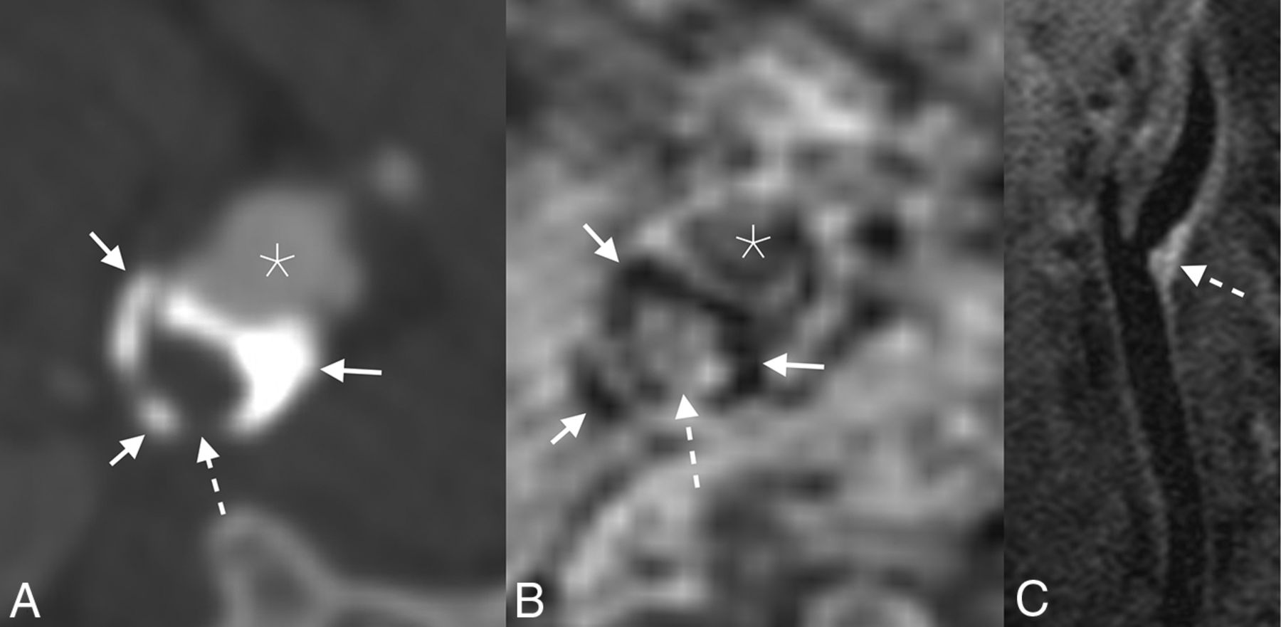
- FIG 1.
Example of an LRNC. Axial CTA image (A) demonstrates a peripherally calcified (solid arrows) plaque with a soft interior (dashed arrow). The corresponding axial-reformatted MPRAGE image (B) similarly demonstrates areas of calcifications with markedly low signal (solid arrows); the plaque interior lacks bright signal, ruling out hemorrhage (dashed arrow). T1 fat-saturated Cube image (C) shows hyperintense signal in the plaque interior (dashed arrow), compatible with an LRNC. Asterisks denote the vessel lumen.
- FIG 2.
Example of persistent IPH in a 71-year-old man who presented with acute disorientation and unsteady gait. MR imaging of the brain at the time of admission (not shown) demonstrated multiple acute left cerebral infarcts. Axial CTA image (A) shows a mixed calcified (dashed arrow) and soft (solid arrows) plaque in the left ICA. Corresponding MPRAGE image (B) demonstrates IPH throughout the soft plaque components (solid arrows); the focal calcification is also noted (dashed arrow). The patient was started on dual antiplatelet therapy (aspirin and clopidogrel). One year later (C), the appearance of the IPH (solid arrows) and calcification (dashed arrow) was unchanged. Asterisks denote the vessel lumen.
- FIG 3.
Example of pathologic plaque enhancement. Axial fat-saturated T1 Cube (A) and MPRAGE (B) images show a plaque in the left ICA, with both hemorrhagic (straight arrows) and nonhemorrhagic LRNC (curved arrows) regions. On the postgadolinium fat-saturated T1 Cube image (C), the LRNC component demonstrates marked enhancement (dashed arrow), while the hemorrhagic component does not (straight arrow). Asterisks denote the vessel lumen.
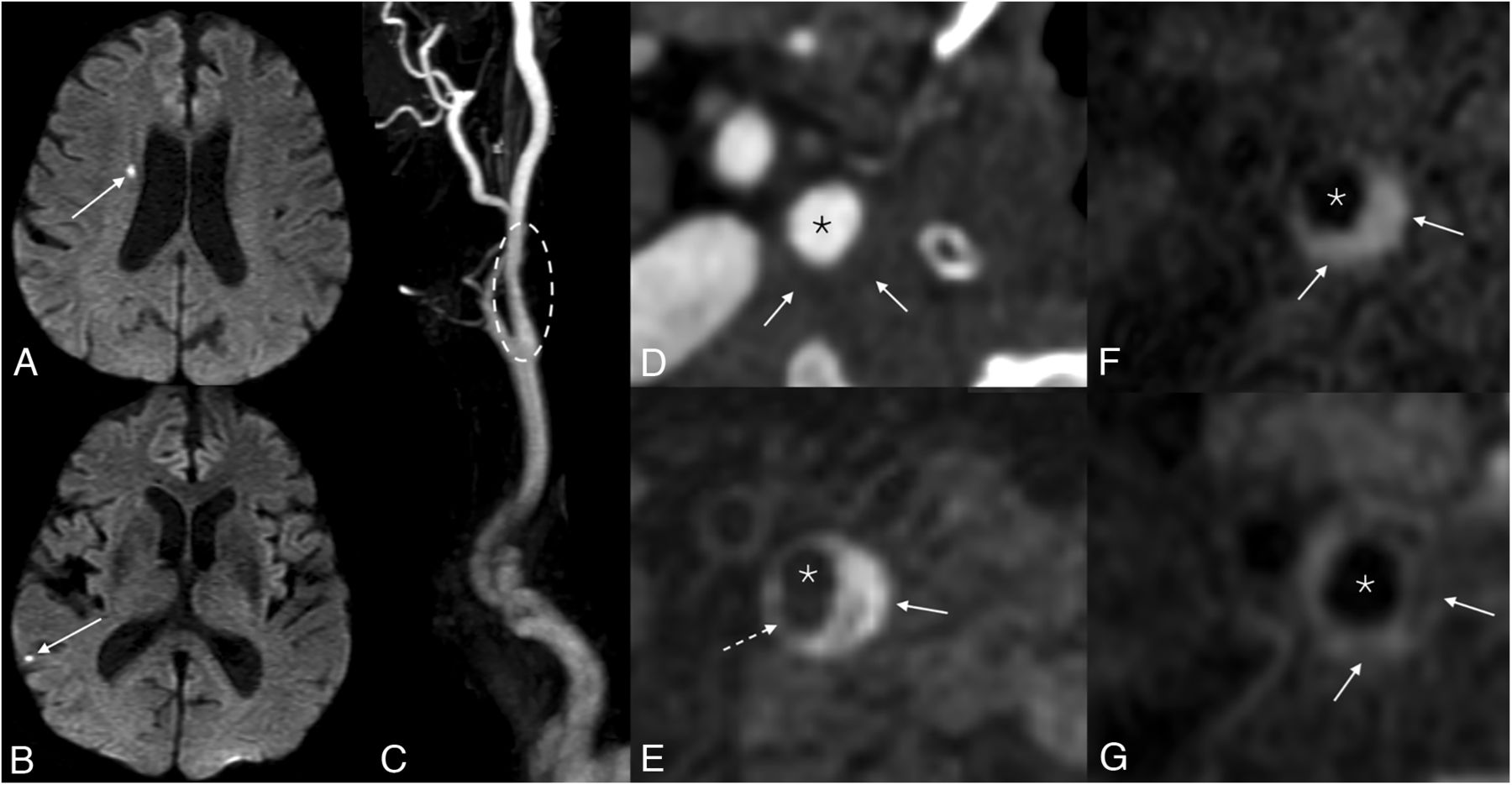
- FIG 4.
Example of a symptomatic nonstenotic plaque. Axial DWI (A and B) of the brain demonstrates multiple tiny acute infarcts in the right cerebral hemisphere (arrows). 3D reformatted gadolinium bolus image (C) demonstrates a nonstenotic plaque in the proximal right ICA (dashed oval). Axial CTA image (D) shows a soft, noncalcified plaque. On MRA, MPRAGE image (E) shows that most of this plaque is composed of hemorrhagic material (solid arrow), with a small hypointense component representing a nonhemorrhagic lipid necrotic core (dashed arrow). Pre- (F) and postcontrast (G) T1 Cube images demonstrate a LRNC (solid arrows). Asterisks denote the vessel lumen.


Jump to section
Related Articles
Cited By...
- No citing articles found.