
Article Figures & Data
Figures
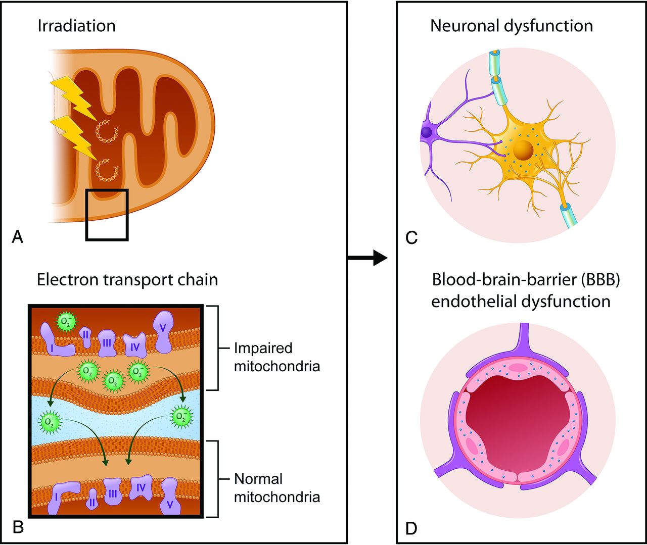
- FIG 1.
A, Ionizing irradiation causes mitochondrial DNA damage directly or secondarily via the production of reactive oxygen species (O2) and free radicals, which result in injury to the mitochondrial DNA. B, These changes alter the function of the electron transport chain, which is composed of complex proteins (I––V) and mediates creation of adenosine triphosphate, and can result in impaired mitochondria. Also, O2 can function as a signaling molecule in intermitochondrial communication and diffuse to the nearby mitochondria, resulting in further mitochondrial dysfunction. C and D, Mitochondrial dysfunction in neurons and endothelial cells can lead to a decrease of available ATP and consequent neuronal and BBB endothelial dysfunction. Inadequate availability of ATP in both neuron and BBB endothelial cells can impair the ion homeostasis within the intracellular and extracellular compartments and lead to neuronal hyperexcitability, which can trigger subsequent cortical spreading depression.
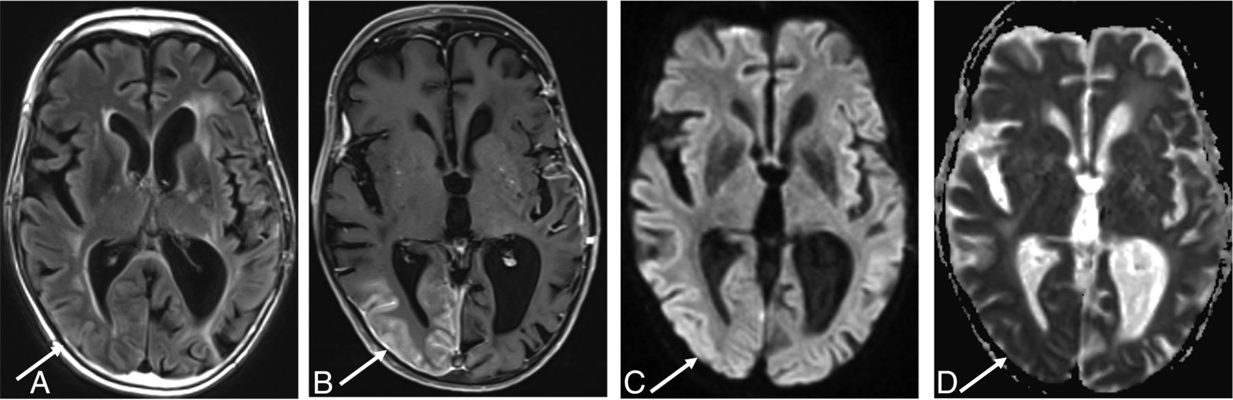
- FIG 2.
A 51-year-old man with a history of juvenile left posterior fossa tumor treated by surgery and whole-brain irradiation 45 years ago presented with acute visual changes and seizures. He was diagnosed with SMART syndrome and treated with corticosteroids and recovered from the symptoms. A FLAIR image (A) shows cortical hyperintensity, and the postcontrast T1 image (B) shows gyriform enhancement in the right temporo-occipital region (arrows), with an incidental right temporal dural-based meningioma. There is high signal on diffusion-weighted imaging (C) without low signal on ADC (D) (facilitated diffusion) (arrows).
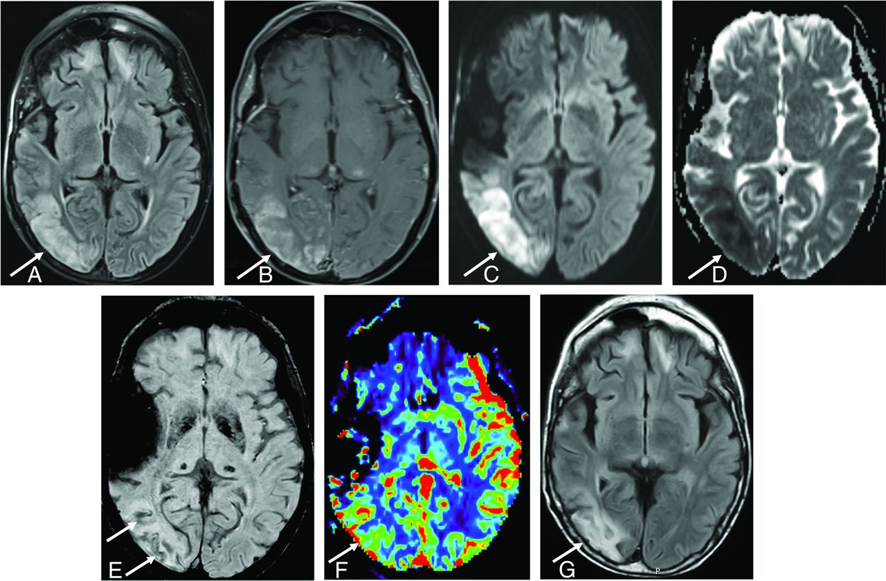
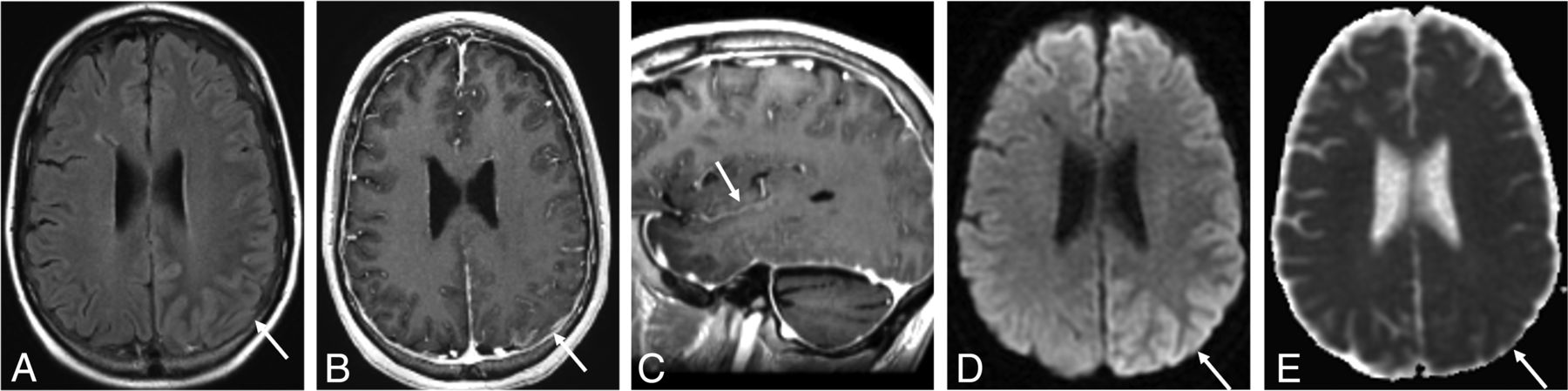
- FIG 3.
A 50-year-old man diagnosed with SMART syndrome. He had a history of pilocytic astrocytoma treated by resection and 60 Gy of radiation therapy 30 years ago and presented with left-sided hemiparesis, speech impairment, seizure, and migraine-like headache. He was diagnosed with SMART syndrome and treated by verapamil and aspirin, but residual symptoms (hemiparesis and speech impairment) remained. A, A FLAIR image shows cortical hyperintensity and involvement of subcortical white matter in the right temporo-occipital region (arrow). There is bifrontal subcortical white matter hyperintensity likely due to prior radiation injury. B, There is gyriform enhancement in the right temporo-occipital region (arrow) with restricted diffusion (C and D) (high signal on DWI and low signal on ADC) (arrow). E, SWI shows linear hypointensity along the subcortical white matter (arrow). F, Dynamic susceptibility contrast perfusion MR imaging shows an increase of CBV in the same area. G, After 3 months, FLAIR shows residual hyperintensity in the cortical and subcortical area (arrow).
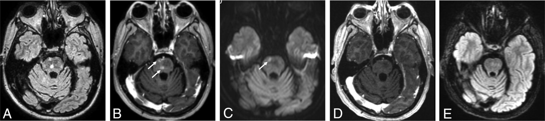
- FIG 4.
A 60-year-old man with brainstem SMART syndrome. He had a history of posterior fossa medulloblastoma treated with resection and 30 Gy of radiation therapy 16 years ago and presented with emotional lability and slurred speech. He completely recovered from the symptoms. A FLAIR image (A) shows hyperintensity in the central pons with peripheral patchy enhancement on the axial postcontrast T1-weighted image (arrows, B). C, DWI shows focal restricted diffusion in the corresponding area of the enhancing lesion (arrow). D and E, After 6 months, a postcontrast T1-weighted image shows resolution of enhancement with residual slight FLAIR hyperintensity.
- FIG 5.
A 37-year-old man diagnosed with PIPG. He had a history of pineoblastoma treated with resection and a posterior fossa meningioma treated by resection and whole-brain irradiation 12 years before, and he presented with migraine-like headache, seizure, right-sided hemiparesis, and aphasia. He was treated with verapamil, aspirin, and valproic acid. He completely recovered from the symptoms. A FLAIR image (A) shows hyperintensity (arrow) with leptomeningeal enhancement in the left temporoparietal region on the axial (B) and sagittal (C) postcontrast T1-weighted images (arrows). Diffusion-weighted imaging (D) and ADC (E) show vasogenic edema (high signal on DWI without low signal on ADC) (arrows).
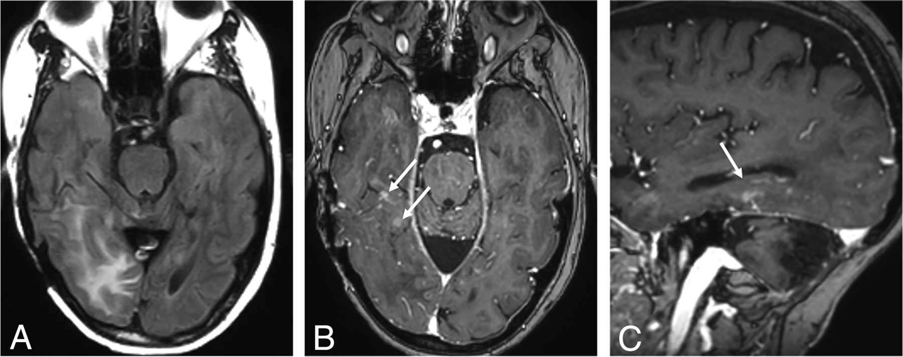
- FIG 6.
A 60-year-old man with ALERT syndrome. He had a history of atypical meningioma treated with resection and radiation therapy 12 years ago and presented with impaired consciousness, left homonymous hemianopia, and left-sided weakness. He was treated with steroids, but left-sided weakness persisted. The FLAIR (A) image shows hyperintensity with patchy enhancement in the right temporoparietal region on the axial (B) and sagittal (C) postcontrast T1-weighted images (arrows).


Tables
- Table 1:
Modified diagnostic criteria in addition to the criteria of Black et al6 for SMART syndrome
Criteria A) Remote history of external beam cranial irradiation. B) Prolonged signs and symptoms, which may be reversible or persistent, attributable to a unilateral cortical region. Clinical manifestations may include migraine-type headache with or without an aura, seizures, confusion, and stroke-like symptoms, including visuospatial deficits, hemisensory deficits, hemiparesis, and aphasia. C) Reversible or sustained unilateral gyriform enhancement with or without T2WI/FLAIR hyperintensity involving the cortex and subjacent cerebral white matter in the irradiated area. D) No definitive evidence of residual or recurrent brain tumor, and not attributable to other disorders. Category and Differential Diagnosis Neoplastic process: Tumor recurrence, leptomeningeal carcinomatosis Ischemic or vascular process: Subacute brain infarction, cortical vein thrombosis, PRES Infectious process: Cerebritis, meningoencephalitis Hyperexcitability: Hemiplegic migraine, status epilepticus Genetic disease (hyperexcitability): MELAS


Jump to section
Related Articles
Cited By...
- No citing articles found.