
Article Figures & Data
Figures
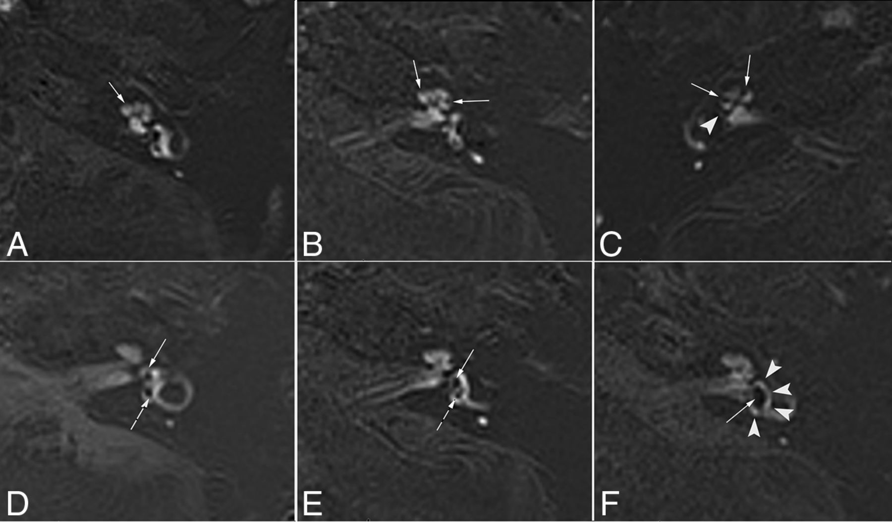
- FIG 1.
The images of different scores for the separate visualization of the cochlear and vestibular endolymph in patients with MD. A, E, and F, 3D TSE real IR images. The perilymph is slightly enhanced (arrowheads). B, C, D, and G, ZOOMit SPACE real IR images. The perilymph is markedly enhanced (arrowheads). A, Score 1 for the cochlea. It is impossible to recognize the cochlear duct (endolymph, arrows and question marks). B, Score 2 for the cochlea. The cochlear duct (endolymph, arrow) was recognized only in the basal turn of the cochlea. C, Score 3 for the cochlea. The cochlear duct (endolymph, arrows) was recognized in the basal and middle turns of the cochlea. D, Score 4 for the cochlea. The cochlear duct (endolymph, arrows) was recognized in the basal, middle, and apical turns of the cochlea. E, Score 2 for the vestibule. A small part of the boundary for the utricle (arrow) was clearly displayed (≤ one-half of vestibular endolymph), and the edge could be clearly delineated, while the rest of the boundary was blurred (saccule, arrow and question mark). F, Score 3 for the vestibule. Most of the boundary of the saccule and the whole utricle (arrows) was clearly displayed (> one-half and <1 of vestibular endolymph), and the edge could be clearly delineated. G, Score 4 for the vestibule. The boundary of the saccule and utricle (endolymph, arrows) was clearly displayed (the whole vestibular endolymph).
- FIG 2.
ZOOMit SPACE real IR images of different degrees of cochlear and vestibular EH in patients with MD. A, Normal cochlea (none). The scala media is minimally visible (solid arrow). B, Cochlear EH (I). The scala media becomes indirectly visible (solid arrows) as a nodular black cutout of the scala vestibuli, which was partially obstructed. C, Cochlear EH (II) and vestibular EH (III). The scala vestibuli is fully obliterated owing to the distended scala media (solid arrows). There is major or full obliteration of the bony vestibule; occasionally, only a few vestibular perilymphatic spaces near the cochlea remained (arrowhead). D, Normal vestibule. The saccule (solid arrow) and utricle (dotted arrow) are visibly separated and take less than one-half of the area of the vestibule. E, Vestibular EH (I). The saccule (solid arrow), normally smaller than the utricle, has become equal or larger than the utricle but is not yet confluent with the utricle (dotted arrow). F, Vestibular EH (II). There is a confluence of the saccule and utricle (solid arrow), with a continuous peripheral rim enhancement of the perilymphatic space (arrowheads).
- FIG 3.
A, ROI image of zs-3D real IR at the level of the lower cochlear basal turn. Circle 1 of 1 mm2 indicates the ROI for the perilymph in the scala tympani in the basal turn of the cochlea. B, Zs-3D real IR image at the middle level of the vestibule. Circle 1 of 1 mm2 indicates the ROI for the endolymph in the utricle. C, The zs-3D real IR image at the level of the left middle cerebellar peduncle. Circle 1 of 10 mm2 indicates the ROI for SI in the left middle cerebellar peduncle. D, The zs-3D real IR image at the level of the artifact-free air area of the ipsilateral external auditory canal. Circle 1 of 6 mm2 indicates the ROI for the SD in the artifact-free air area of the ipsilateral external ear.
- FIG 4.
Differences in the CNRs (A), SNRs (B), and SIRs (C) of the affected and asymptomatic sides are found between the 2 sequences (P < . 001).


Tables
Parameter ZOOMit SPACE Real IR TSE Real IR TR (ms) 8000 5300 TE (ms) 491 191 TI (ms) 2250 1850 Turbo factor 192 33 BW (Hz/pixel) 305 213 Voxel (mm) 0.6 × 0.6 × 1.0 0.6 × 0.6 × 0.6 Average 4 1 iPAT 2 2 Scan time 15 min 12 sec 16 min 47 sec Flip angle mode Constant 180 FOV (mm) 160 × 80 220 × 220 Matrix size 256 × 128 384 × 384 Reconstruction mode Real Real Note:—BW indicates bandwidth; iPAT, integrated parallel acquisition techniques.
- Table 2:
Scores for separate visualization of endolymphatic space in the cochlea and vestibule by 2 pulse sequences in 50 patients with MD
Visualization Sequence Score Affected side 4 3 2 1 Cochlea 3D ZOOMit SPACE real IR 37 13 0 0 3D TSE real IR 17 28 3 2 Vestibule 3D ZOOMit SPACE real IR 40 9 1 0 3D TSE real IR 27 22 1 0 Asymptomatic side Cochlea 3D ZOOMit SPACE real IR 3 45 2 0 3D TSE real IR 1 28 18 3 Vestibule 3D ZOOMit SPACE real IR 49 1 0 0 3D TSE real IR 27 23 0 0 - Table 3:
Number and percentage distribution of cochlear and vestibular EH grading by 2 pulse sequences in 50 patients with MD
3D ZOOMit SPACE Real IR 3D TSE Real IR Wilcoxon Signed-Rank Test (No.) (%) (No.) (%) Z Value P Value Affected side No cochlear EH 7 (14.0)a 15 (30.0) –2.83 .005 Cochlear EH (I) 12 (24.0)a 4 (8.0) Cochlear EH (II) 31 (62.0) 31 (62.0) No vestibular EH 3 (6.0) 3 (6.0) 0.00 1.000 Vestibular EH (I) 13 (26.0) 13 (26.0) Vestibular EH (II) 18 (36.0) 18 (36.0) Vestibular EH (III) 16 (32.0) 16 (32.0) Asymptomatic side No cochlear EH 46 (92.0) 48 (96.0) –1.41 .157 Cochlear EH (I) 3 (6.0) 1 (2.0) Cochlear EH (II) 1 (2.0) 1 (2.0) No vestibular EH 48 (96.0) 48 (96.0) 0.00 1.000 Vestibular EH (I) 0 (0.0) 0 (0.0) Vestibular EH (II) 2 (4.0) 2 (4.0) Vestibular EH (III) 0 (0.0) 0 (0.0) ↵a Compared with the 3D TSE real IR sequence, P < .017.


