Advertisement

Main menu

  • Home
  • Content
    • Current Issue
    • Accepted Manuscripts
    • Article Preview
    • Past Issue Archive
    • Video Articles
    • AJNR Case Collection
    • Case of the Week Archive
    • Case of the Month Archive
    • Classic Case Archive
  • Special Collections
    • AJNR Awards
    • Low-Field MRI
    • Alzheimer Disease
    • ASNR Foundation Special Collection
    • Photon-Counting CT
    • View All
  • Multimedia
    • AJNR Podcasts
    • AJNR SCANtastic
    • Trainee Corner
    • MRI Safety Corner
    • Imaging Protocols
  • For Authors
    • Submit a Manuscript
    • Submit a Video Article
    • Submit an eLetter to the Editor/Response
    • Manuscript Submission Guidelines
    • Statistical Tips
    • Fast Publishing of Accepted Manuscripts
    • Graphical Abstract Preparation
    • Imaging Protocol Submission
    • Author Policies
  • About Us
    • About AJNR
    • Editorial Board
    • Editorial Board Alumni
  • More
    • Become a Reviewer/Academy of Reviewers
    • Subscribers
    • Permissions
    • Alerts
    • Feedback
    • Advertisers
    • ASNR Home

User menu

  • Alerts
  • Log in

Search

  • Advanced search
American Journal of Neuroradiology

American Journal of Neuroradiology

ASHNR American Society of Functional Neuroradiology ASHNR American Society of Pediatric Neuroradiology ASSR
  • Alerts
  • Log in

Advanced Search

  • Home
  • Content
    • Current Issue
    • Accepted Manuscripts
    • Article Preview
    • Past Issue Archive
    • Video Articles
    • AJNR Case Collection
    • Case of the Week Archive
    • Case of the Month Archive
    • Classic Case Archive
  • Special Collections
    • AJNR Awards
    • Low-Field MRI
    • Alzheimer Disease
    • ASNR Foundation Special Collection
    • Photon-Counting CT
    • View All
  • Multimedia
    • AJNR Podcasts
    • AJNR SCANtastic
    • Trainee Corner
    • MRI Safety Corner
    • Imaging Protocols
  • For Authors
    • Submit a Manuscript
    • Submit a Video Article
    • Submit an eLetter to the Editor/Response
    • Manuscript Submission Guidelines
    • Statistical Tips
    • Fast Publishing of Accepted Manuscripts
    • Graphical Abstract Preparation
    • Imaging Protocol Submission
    • Author Policies
  • About Us
    • About AJNR
    • Editorial Board
    • Editorial Board Alumni
  • More
    • Become a Reviewer/Academy of Reviewers
    • Subscribers
    • Permissions
    • Alerts
    • Feedback
    • Advertisers
    • ASNR Home
  • Follow AJNR on Twitter
  • Visit AJNR on Facebook
  • Follow AJNR on Instagram
  • Join AJNR on LinkedIn
  • RSS Feeds

AJNR Awards, New Junior Editors, and more. Read the latest AJNR updates

Research ArticleNeurointervention
Open Access

Clinical Relevance of Plaque Distribution for Basilar Artery Stenosis

J. Luo, X. Bai, K. Huang, T. Wang, R. Yang, L. Li, Q. Tian, R. Xu, T. Li, Y. Wang, Y. Chen, P. Gao, J. Chen, B. Yang, Y. Ma and L. Jiao
American Journal of Neuroradiology April 2023, DOI: https://doi.org/10.3174/ajnr.A7839
J. Luo
aFrom the China International Neuroscience Institute (J.L., X.B., T.W., R.Y., L.L., R.X., T.L., Y.W., Y.C., P.G., J.C., B.Y., Y.M., L.J.), Beijing, China
bDepartment of Neurosurgery (J.L., X.B., T.W., R.Y., L.L., R.X., T.L., Y.W., Y.C., P.G., J.C., B.Y., Y.M., L.J.)
  • Find this author on Google Scholar
  • Find this author on PubMed
  • Search for this author on this site
  • ORCID record for J. Luo
X. Bai
aFrom the China International Neuroscience Institute (J.L., X.B., T.W., R.Y., L.L., R.X., T.L., Y.W., Y.C., P.G., J.C., B.Y., Y.M., L.J.), Beijing, China
bDepartment of Neurosurgery (J.L., X.B., T.W., R.Y., L.L., R.X., T.L., Y.W., Y.C., P.G., J.C., B.Y., Y.M., L.J.)
  • Find this author on Google Scholar
  • Find this author on PubMed
  • Search for this author on this site
  • ORCID record for X. Bai
K. Huang
eThe Eighth Affiliated Hospital (K.H.), SUN YAT-SEN University, Shenzhen, Guangdong Province, China
  • Find this author on Google Scholar
  • Find this author on PubMed
  • Search for this author on this site
  • ORCID record for K. Huang
T. Wang
aFrom the China International Neuroscience Institute (J.L., X.B., T.W., R.Y., L.L., R.X., T.L., Y.W., Y.C., P.G., J.C., B.Y., Y.M., L.J.), Beijing, China
bDepartment of Neurosurgery (J.L., X.B., T.W., R.Y., L.L., R.X., T.L., Y.W., Y.C., P.G., J.C., B.Y., Y.M., L.J.)
  • Find this author on Google Scholar
  • Find this author on PubMed
  • Search for this author on this site
  • ORCID record for T. Wang
R. Yang
aFrom the China International Neuroscience Institute (J.L., X.B., T.W., R.Y., L.L., R.X., T.L., Y.W., Y.C., P.G., J.C., B.Y., Y.M., L.J.), Beijing, China
bDepartment of Neurosurgery (J.L., X.B., T.W., R.Y., L.L., R.X., T.L., Y.W., Y.C., P.G., J.C., B.Y., Y.M., L.J.)
  • Find this author on Google Scholar
  • Find this author on PubMed
  • Search for this author on this site
  • ORCID record for R. Yang
L. Li
aFrom the China International Neuroscience Institute (J.L., X.B., T.W., R.Y., L.L., R.X., T.L., Y.W., Y.C., P.G., J.C., B.Y., Y.M., L.J.), Beijing, China
bDepartment of Neurosurgery (J.L., X.B., T.W., R.Y., L.L., R.X., T.L., Y.W., Y.C., P.G., J.C., B.Y., Y.M., L.J.)
  • Find this author on Google Scholar
  • Find this author on PubMed
  • Search for this author on this site
  • ORCID record for L. Li
Q. Tian
cXuanwu Hospital, Beijing Key Laboratory of Clinical Epidemiology (Q.T.), School of Public Health
  • Find this author on Google Scholar
  • Find this author on PubMed
  • Search for this author on this site
  • ORCID record for Q. Tian
R. Xu
aFrom the China International Neuroscience Institute (J.L., X.B., T.W., R.Y., L.L., R.X., T.L., Y.W., Y.C., P.G., J.C., B.Y., Y.M., L.J.), Beijing, China
bDepartment of Neurosurgery (J.L., X.B., T.W., R.Y., L.L., R.X., T.L., Y.W., Y.C., P.G., J.C., B.Y., Y.M., L.J.)
  • Find this author on Google Scholar
  • Find this author on PubMed
  • Search for this author on this site
  • ORCID record for R. Xu
T. Li
aFrom the China International Neuroscience Institute (J.L., X.B., T.W., R.Y., L.L., R.X., T.L., Y.W., Y.C., P.G., J.C., B.Y., Y.M., L.J.), Beijing, China
bDepartment of Neurosurgery (J.L., X.B., T.W., R.Y., L.L., R.X., T.L., Y.W., Y.C., P.G., J.C., B.Y., Y.M., L.J.)
  • Find this author on Google Scholar
  • Find this author on PubMed
  • Search for this author on this site
  • ORCID record for T. Li
Y. Wang
aFrom the China International Neuroscience Institute (J.L., X.B., T.W., R.Y., L.L., R.X., T.L., Y.W., Y.C., P.G., J.C., B.Y., Y.M., L.J.), Beijing, China
bDepartment of Neurosurgery (J.L., X.B., T.W., R.Y., L.L., R.X., T.L., Y.W., Y.C., P.G., J.C., B.Y., Y.M., L.J.)
  • Find this author on Google Scholar
  • Find this author on PubMed
  • Search for this author on this site
  • ORCID record for Y. Wang
Y. Chen
aFrom the China International Neuroscience Institute (J.L., X.B., T.W., R.Y., L.L., R.X., T.L., Y.W., Y.C., P.G., J.C., B.Y., Y.M., L.J.), Beijing, China
bDepartment of Neurosurgery (J.L., X.B., T.W., R.Y., L.L., R.X., T.L., Y.W., Y.C., P.G., J.C., B.Y., Y.M., L.J.)
  • Find this author on Google Scholar
  • Find this author on PubMed
  • Search for this author on this site
  • ORCID record for Y. Chen
P. Gao
aFrom the China International Neuroscience Institute (J.L., X.B., T.W., R.Y., L.L., R.X., T.L., Y.W., Y.C., P.G., J.C., B.Y., Y.M., L.J.), Beijing, China
bDepartment of Neurosurgery (J.L., X.B., T.W., R.Y., L.L., R.X., T.L., Y.W., Y.C., P.G., J.C., B.Y., Y.M., L.J.)
dDepartment of Interventional Radiology (P.G., L.J.), Xuanwu Hospital, Capital Medical University, Beijing, China
  • Find this author on Google Scholar
  • Find this author on PubMed
  • Search for this author on this site
J. Chen
aFrom the China International Neuroscience Institute (J.L., X.B., T.W., R.Y., L.L., R.X., T.L., Y.W., Y.C., P.G., J.C., B.Y., Y.M., L.J.), Beijing, China
bDepartment of Neurosurgery (J.L., X.B., T.W., R.Y., L.L., R.X., T.L., Y.W., Y.C., P.G., J.C., B.Y., Y.M., L.J.)
  • Find this author on Google Scholar
  • Find this author on PubMed
  • Search for this author on this site
B. Yang
aFrom the China International Neuroscience Institute (J.L., X.B., T.W., R.Y., L.L., R.X., T.L., Y.W., Y.C., P.G., J.C., B.Y., Y.M., L.J.), Beijing, China
bDepartment of Neurosurgery (J.L., X.B., T.W., R.Y., L.L., R.X., T.L., Y.W., Y.C., P.G., J.C., B.Y., Y.M., L.J.)
  • Find this author on Google Scholar
  • Find this author on PubMed
  • Search for this author on this site
Y. Ma
aFrom the China International Neuroscience Institute (J.L., X.B., T.W., R.Y., L.L., R.X., T.L., Y.W., Y.C., P.G., J.C., B.Y., Y.M., L.J.), Beijing, China
bDepartment of Neurosurgery (J.L., X.B., T.W., R.Y., L.L., R.X., T.L., Y.W., Y.C., P.G., J.C., B.Y., Y.M., L.J.)
  • Find this author on Google Scholar
  • Find this author on PubMed
  • Search for this author on this site
  • ORCID record for Y. Ma
L. Jiao
aFrom the China International Neuroscience Institute (J.L., X.B., T.W., R.Y., L.L., R.X., T.L., Y.W., Y.C., P.G., J.C., B.Y., Y.M., L.J.), Beijing, China
bDepartment of Neurosurgery (J.L., X.B., T.W., R.Y., L.L., R.X., T.L., Y.W., Y.C., P.G., J.C., B.Y., Y.M., L.J.)
dDepartment of Interventional Radiology (P.G., L.J.), Xuanwu Hospital, Capital Medical University, Beijing, China
  • Find this author on Google Scholar
  • Find this author on PubMed
  • Search for this author on this site
  • ORCID record for L. Jiao
  • Article
  • Figures & Data
  • Info & Metrics
  • Responses
  • References
  • PDF
Loading

Abstract

BACKGROUND AND PURPOSE: There is no clear association between plaque distribution and postoperative complications in patients with basilar artery atherosclerotic stenosis. The aim of this study was to determine whether plaque distribution and postoperative complications after endovascular treatment for basilar artery stenosis are related.

MATERIALS AND METHODS: Our study enrolled patients with severe basilar artery stenosis who were scanned with high-resolution MR imaging and followed by DSA before the intervention. According to high-resolution MR imaging, plaques can be classified as ventral, lateral, dorsal, or involved in 2 quadrants. Plaques affecting the proximal, distal, or junctional segments of the basilar artery were classified according to DSA. An experienced independent team assessed ischemic events after the intervention using MR imaging. Further analysis was conducted to determine the relationship between plaque distribution and postoperative complications.

RESULTS: A total of 140 eligible patients were included in the study, with a postoperative complication rate of 11.4%. These patients were an average age of 61.9 (SD, 7.7) years. Dorsal wall plaques accounted for 34.3% of all plaques, and plaques distal to the anterior-inferior cerebellar artery accounted for 60.7%. Postoperative complications of endovascular treatment were associated with plaques located at the lateral wall (OR = 4.00; 95% CI, 1.21–13.23; P = .023), junctional segment (OR = 8.75; 95% CI, 1.16–66.22; P = .036), and plaque burden (OR = 1.03; 95% CI, 1.01–1.06; P = .042).

CONCLUSIONS: Plaques with a large burden located at the junctional segment and lateral wall of the basilar artery may increase the likelihood of postoperative complications following endovascular therapy. A larger sample size is needed for future studies.

ABBREVIATIONS:

HR-MR imaging
high-resolution MR imaging
LA
lumen area
MLN
maximal lumen narrowing
VA
vessel area
WA
wall area

The basilar artery is the main artery for the posterior intracranial circulation, where atherosclerotic stenosis is frequently discovered in patients with ischemic events, such as stroke and TIA, which are responsible for 10.7% of strokes annually.1 Patients with symptomatic and severe basilar artery stenosis (≥70%) may benefit from endovascular treatment, including primary angiography, balloon-mounted stent placement, and self-expanding stent placement.2 Compared with the those in the anterior circulation, postoperative complications are higher in the posterior circulation with a reported risk of 21.6% in the basilar artery.3,4 Therefore, it is crucial to understand and prevent a high risk of complications occurring during endovascular treatment in patients with basilar artery stenosis.

The distribution of atherosclerotic plaques is associated with the risk of ischemic events.5⇓-7 For example, it is more likely that atherosclerotic plaques located near perforating orifices are symptomatic during stent placement and may have a “snow plowing” effect.8 Several postmortem studies have demonstrated that basilar artery perforators typically originate from the lateral and dorsal walls of the segment, distal from the anterior-inferior cerebellar artery, possibly explaining the high rate of postoperative complications.9,10 However, the relationship between the distribution of basilar artery plaque and postoperative complications has not yet been reported .

Understanding the distribution of basilar artery plaques can help in reducing the incidence of postoperative complications. Evidence from studies since the Stenting versus Aggressive Medical Management for Preventing Recurrent Stroke in Intracranial Stenosis (SAMMPRIS) trial has shown that the postoperative risk of endovascular treatment for intracranial stenosis can be reduced from 14.7% to 9.4% by excluding symptomatic patients with perforating infarction.11 The postoperative risk could be reduced excluding perforating infarction actually infers the plaque location, whether it is near perforating orifices, according to the phenotype of cerebral infarction. Presently, plaque distribution in axial and coronal positions can be precisely evaluated using high-resolution MR imaging (HR-MR imaging) and DSA.12,13 However, very few studies have explored the direct relationship between plaque distribution and postoperative complications after endovascular treatment from the axial and coronal positions for basilar artery stenosis. The current study investigated the distribution of basilar artery atherosclerotic plaques and their clinical relevance using HR-MR imaging and DSA. Our findings can help neurointerventionalists with better patient selection and, thus, lower the procedural risk.

MATERIALS AND METHODS

Subjects

This study was based on the Clinical Registration Trial of Intracranial Stenting for Patients with Symptomatic Intracranial Artery Stenosis (CRTICAS) data base (ClinicalTrials.gov identifier: NCT01994161).14 The protocol complies with ethics principles of the Declaration of Helsinki and good clinical practice and has been approved by the the review board and ethic committee of Xuanwu Hospital, Capital Medical University ([2013] 004). Written informed consent was obtained from all the patients. The results of this study have been reported in accordance with the Strengthening The Reporting Of Cohort Studies in Surgery (STROCSS) criteria.15

The study included patients with basilar artery stenosis who were enrolled between December 2013 and December 2015 in several high-volume tertiary centers in China. The inclusion criteria were as follows: 1) degree of stenosis of >70% as confirmed by DSA; 2) patients with basilar artery atherosclerotic stenosis who experienced ischemic symptoms, such as dizziness, vertigo, headache, double vision, slurred speech, and numbness or weakness; 3) treatment using endovascular therapy; 4) HR-MR imaging performed before the intervention; and 5) MR imaging performed before and within 72 hours after the intervention. The exclusion criteria were as follows: 1) stroke caused by occlusion of the basilar artery, 2) concurrent endovascular treatment of another intracranial or extracranial vessel, 3) basilar artery stenosis accompanied by moderate-to-severe vertebral artery stenosis, and 4) nonatherosclerotic causes (eg, Moyamoya disease, vasculitis, or dissection).

Imaging Protocols and Evaluation

Before the intervention, all eligible patients were examined using a 3T MR imaging scanner (Magnetom Spectra; Siemens) equipped with a standard 8-channel head coil. The scans were obtained using multiple sequences, including fast spin-echo T1-weighted imaging, TOF-MR angiography, and T1-weighted enhanced imaging. The parameters of the sequences are described in the Online Supplemental Data. HR-MR imaging was acquired in the sagittal plane covering the basilar artery vessel. Reconstruction of the axial, coronal, and sagittal views was required to analyze all the images. DSA examinations were also performed for all the eligible patients.

An image core lab (http://imagecorelabcn.com/) was used to review all the images. The clinical data of patients were undisclosed and not included in the statistical analyses. Before the formal assessment, 5% of data from the analyzed cohort was used to train raters. Whenever the agreement was excellent (reliability, >0.75) between 2 raters, the imaging data were formally evaluated. The maximal lumen narrowing (MLN) or reference site, lumen diameter, vessel area (VA), and lumen area (LA) at the MLN or reference site were manually evaluated. Reference sites were defined according to Warfarin versus Aspirin for Symptomatic Intracranial Disease (WASID) trial as normal segments proximal to the stenosis or distal vessels if the proximal segment was diseased.16

On the basis of HR-MR imaging and DSA, we observed the following vessel parameters:

  • Wall area (WA): difference between VA and LA (VA – LA).

  • Plaque burden: [(WAMLN – WAreference) / VAMLN] × 100%.

  • Remodeling index: VA MLN/VA reference.

Stenosis degree is (1-Luminal Diameter at MLN/Luminal Diameter at the Reference Site) × 100% from DSA.

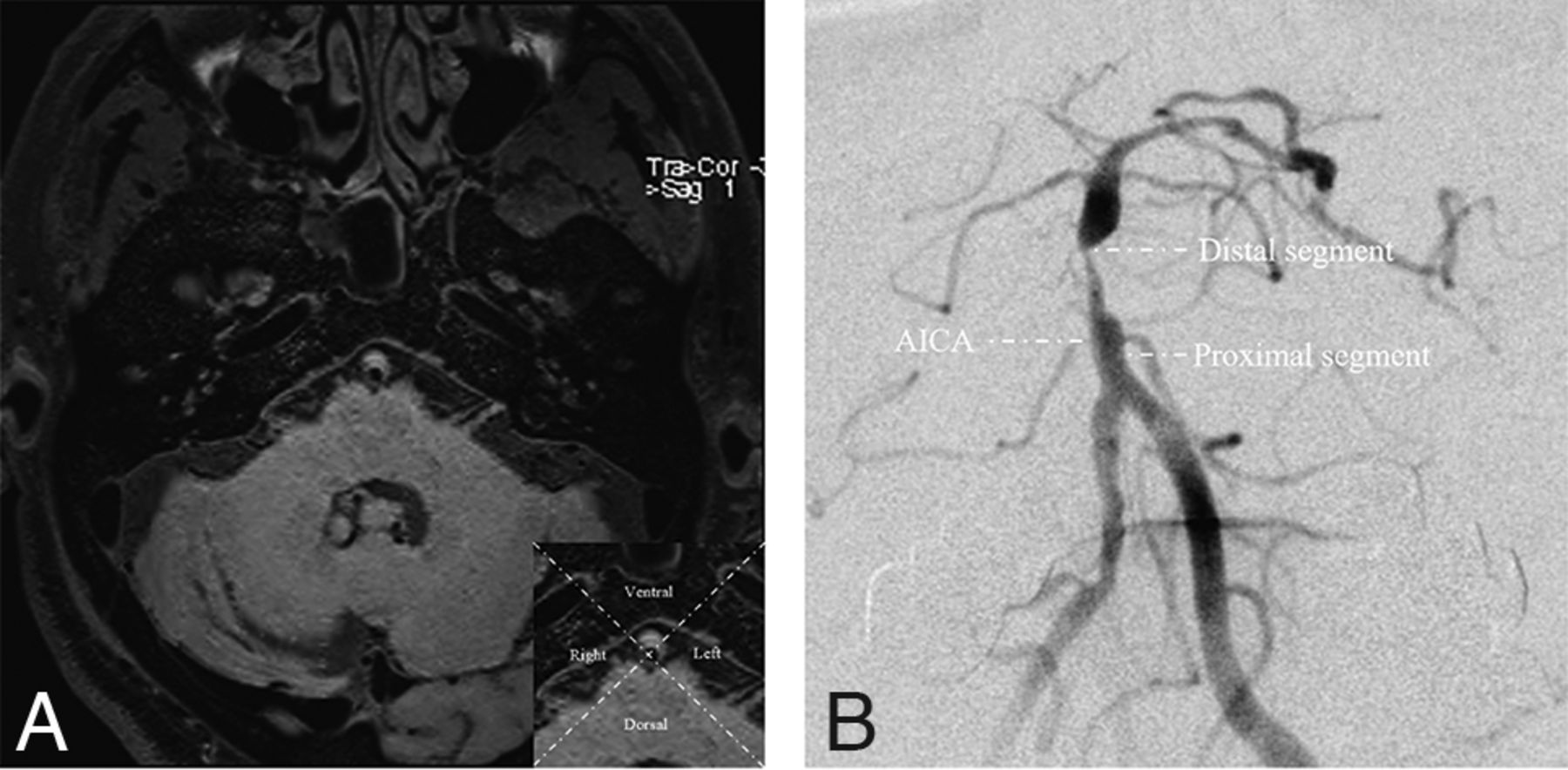
Using 2 perpendicular lines, we divided the cross-sectional plaque into 4 quadrants at the MLN: ventral, dorsal, left, and right.17 The left and right sites were classified as lateral sites. If the plaque was large and its thickest aspect spanned more than 2 quadrants, it was considered distributed between >2 quadrants (Fig 1A). In the coronal position, DSA was used to divide the basilar artery into 2 segments on the basis of the branches of the anterior-inferior cerebellar artery.18 Lesions crossing the anterior-inferior cerebellar artery were categorized as junctional segment lesions (Fig 1B).

FIG 1.
  • Download figure
  • Open in new tab
  • Download powerpoint
FIG 1.

A, The basilar artery is divided into 4 quadrants at the axial position based on high-resolution MR imaging. B, The basilar artery is divided into 2 segments referring to the AICA on the basis of DSA, the proximal and distal segments. The junctional segment is defined when the plaque involves both segments across the anterior-inferior cerebellar artery.

Interventional Procedure and Assessment of Outcome

All patients were treated by a team of neurosurgeons and neuroradiologists with extensive experience in endovascular treatment. Experienced operators determined the therapeutic strategy according to the lesion characteristics. Aspirin, 100 mg daily, was combined with clopidogrel, 75 mg daily for 5 days before, or a loading dose of aspirin and clopidogrel, 300 mg each, was used 1 day before endovascular treatment. Standard protocols for the procedure were followed as described previously.14 Three-month dual-antiplatelet therapy, comprising aspirin, 100 mg daily, and clopidogrel, 75 mg daily, was initiated following the intervention.

An experienced team of neurosurgeons and neuroradiologists investigated the postoperative ischemic events, including TIA and ischemic stroke. Ischemic stroke was defined as a neurologic deficit lasting >24 hours. TIA was defined as a neurologic deficit lasting <24 hours.19 Depending on whether postoperative outcome events occurred after endovascular treatment, patients were classified into an either ischemic or nonischemic events group.

Statistical Analysis

Data were analyzed using the SAS software (Version 9.4; SAS Institute). Quantitative variables are presented as means. Qualitative variables are presented as numbers and percentages. Descriptive analyses were performed for participants in the postoperative ischemic and nonischemic events group. The χ2 test or Fisher exact test was used to compare the categoric variables, as appropriate. The Student t test or Wilcoxon test was used to compare the quantitative variables. Univariate and multivariate regression analyses were performed to investigate the factors influencing the postoperative ischemic events. Variables with P < .1, along with sex, age, and treatment type, were included in the multivariate regression analysis. P < .05 was considered as statistically significant.

RESULTS

In total, 281 consecutive patients with severe symptomatic basilar artery stenosis underwent endovascular treatment. Among them, 140 patients having complete HR-MR imaging and DSA data were finally included in the analysis (Online Supplemental Data). The average age of these patients was 61.9 (SD, 7.7) years. Twenty-six (18.5%) patients underwent primary angioplasty, and 114 (81.5%) underwent stent placement, which included 30 balloon-mounted stents and 84 self-expansion stents. Furthermore, 16 patients with postoperative ischemic events, including 3 TIAs, 4 perforating infarctions, 7 artery-to-artery embolisms, and 2 mixed mechanisms were regarded as the ischemic events group, and 124 patients without postoperative ischemic events, as the nonischemic events group (Online Supplemental Data). Table 1 summarizes the clinical and lesion characteristics, with the exception of plaque burden (mean, 19.3% [SD, 18.3%] versus 3.7% [SD, 23.6%]; P = .047) between the ischemic events group and the nonischemic events group.

View this table:
  • View inline
  • View popup
Table 1:

Clinical and lesion characteristics

A total of 140 basilar artery plaques were reviewed from the axial and coronal positions. In the axial view at the MLN site, 12.9% of plaques were distributed ventral to the basilar artery wall, and 22.1% in the lateral, 34.3% in the dorsal, and 30.7% in ≥2 quadrants. The plaques distributed in the lateral wall were more common in the ischemic events group than in the nonischemic events group (43.8% versus 19.3%, P = .049). In the coronal view, plaque distribution was most common at the segment distal to the anterior-inferior cerebellar artery (60.7%), followed by the segment proximal to the anterior-inferior cerebellar artery (35.0%), and least in the junctional segment (4.3%). Plaques distributed at the junctional segment in the ischemic events group were more frequent compared with the nonischemic events group (18.8% versus 2.4%, P = .020). The additional details of plaque distribution are presented in Table 2. The risk of ischemic events was the highest in patients with plaques located at the lateral wall (22.5%) and junctional segment (50.0%) of the basilar artery (Online Supplemental Data). Additionally, the high risk of ischemic events in patients with plaques located at the junctional segment of the basilar artery was associated with a large remodeling index compared with that in the nonischemic events group (mean, 1.38 [SD, 0.20] versus 0.82 [SD, 0.29]; 95% CI, 0.05–1.13; P = .049) (Online Supplemental Data).

View this table:
  • View inline
  • View popup
Table 2:

Plaque distribution of the basilar artery

The multivariate logistic regression analysis showed that plaques distributed at the lateral wall (OR = 4.00; 95% CI, 1.21–13.23; P = .023) and junctional segments (OR = 8.75; 95% CI, 1.16–66.22; P = .036) as well as plaque burden (OR = 1.03; 95% CI, 1.01–1.06; P = .042) were associated with a higher risk of postoperative ischemic events (Table 3, Fig 2).

View this table:
  • View inline
  • View popup
Table 3:

Relationship between plaque distribution and ischemic events by multivariate analysis

FIG 2.
  • Download figure
  • Open in new tab
  • Download powerpoint
FIG 2.

An adult patient presented with recurrent stroke for 1 month. A, Preoperative DSA revealed 79.3% stenosis at the distal segment of the basilar artery. The middle column shows cross-sectional T1-weighted basilar artery images at the MLN (B) and reference (REF) (E) sites. The VA and LA at the MLN (B, VA = 0.157 cm2, LA = 0.014 cm2) and REF (E, VA = 0.237 cm2, LA = 0.075 cm2) sites were manually traced for measuring after zooming in ×400. The plaque burden was calculated as 16.6% using [(WAMLN-WAREF) / VA MLN] × 100%. D, A 2.5 × 8 mm Apollo balloon-mounted stent (MicroPort NeuroTech) was positioned at the stenotic segment. DWI (C) and ADC imaging (F) confirmed a new mixed type of infarction (white arrows) after the intervention.

DISCUSSION

In this study, symptomatic patients with severe basilar artery stenosis were enrolled to investigate the correlation of atherosclerotic plaque distribution with postoperative complications. The risk of postoperative ischemic events in patients with severe symptomatic basilar artery stenosis was 11.4% (16/140). Although plaque distribution of the basilar artery was most commonly observed at the dorsal wall of the basilar artery in the axial (34.3%) and distal segments in the coronal view (60.7%), the risk of postoperative complications was highest when the plaques were situated in the lateral wall and junctional segment of the basilar artery. In addition to the large plaque burden, plaques located at the lateral wall and junctional segment of the basilar artery were independent risk factors for postoperative ischemic events.

The high risk of postoperative complications is a substantial limitation for endovascular treatment of intracranial atherosclerotic stenosis, especially in the posterior circulation.4,20 The basilar artery is the main artery for the posterior circulation, which has the highest risk of postoperative complications for endovascular treatment among the intracranial arteries.3 The posterior circulation appears to be more capable of plaque burden because it has a lower blood flow and less sympathetic innervation compared with the anterior circulation.21⇓⇓-24 Additionally, the number of perforating arteries in the basilar artery is significantly higher than that of the other intracranial arteries.25 These reasons explain the high risk associated with endovascular treatment. Previous studies have suggested that SAMMPRIS, strict patient selection, technical development, and incremental experience of the operators reduces the risk of postoperative complications for endovascular treatment.26 Our study showed a relatively lower risk compared with SAMMPRIS (21.6%) and is comparable with several studies (11.2%–14.2%).27,28 However, the prevention of postoperative complications is necessary to maximize the benefits of endovascular treatment.

It is highly likely that postoperative complications can be reduced by assessing the plaque distribution and features.18 Plaque distribution has been abundantly indicated to be a significant indicator of postoperative complications, thus revealing that postoperative complications of intracranial atherosclerotic plaques located in the posterior circulation were higher than those in the anterior circulation (12.1% versus 6.6, P < .01).29⇓-31 However, the association of local distribution of basilar artery atherosclerotic plaques with postoperative complications is unclear. Several researchers have hypothesized that plaques near the perforating orifices might be a determinant of postoperative complications through narrative analysis in plaque microanatomy studies.6,18,32,33

In this study, plaques present in the lateral wall were associated with a higher risk of postoperative complications.34 Penetrating arteries from the lateral and dorsal sides of the basilar artery have been observed in previous studies.10 According to the anatomic studies, 65.3% of penetrating arteries originate from the lateral side of the basilar artery, whereas 34.7% originate from the dorsal side.35 Additionally, the anastomosis rate of penetrating arteries emerging from the basilar artery is high (range, 41.6%–66.6%).9 From an axial perspective, the anastomosis rate of penetrations emerging from the dorsal side was higher than that on the lateral side.36 Moreover, 99% of the anastomoses of penetrating arteries from the dorsal side of basilar artery are located on the ventral surface of the pontocerebrum surface, 57.5% of the anastomoses occur between the penetrating arteries of the pontocerebrum, and 21.3%, between penetrations originating from the basilar artery and anterior-inferior cerebellar artery.36 As a result, plaques involving the lateral side of the basilar artery may be a risk factor for endovascular treatment for patients with severe basilar artery stenosis.

In addition, a large plaque burden is associated with postoperative complications, which may be due to a large plaque burden being more vulnerable during the expansion process of the balloon or stent.37 Furthermore, a large plaque burden has a high risk of initial and recurrent ischemic symptoms in patients with intracranial stenosis.38⇓-40 Thus, an evaluation of both plaque burden and distribution characteristics may be helpful in excluding high-risk patients from endovascular treatment of basilar artery stenosis.

This study has some limitations. First, although the study cohort had a larger sample size compared with other HR-MR imaging studies, the incidence of postoperative complications was low, comprising only 16 cases.17, 41 Therefore, the impact of plaque location on different stroke mechanisms (eg, artery-to-artery and perforating stroke) in symptomatic basilar artery stenosis is currently unavailable and warrants further analysis. Second, plaque characteristics from the axial position based on HR-MR imaging were assessed at the site of the MLN and not in other planes of plaque. The site of MLN was considered as the evaluation level because stress is most pronounced during endovascular therapy and may be associated with postoperative complications. Third, plaque burden was not analyzed separately at various plaque distributions from the axial and coronal positions to determine the potential mechanism of plaque distribution, which was constrained by the small number of cases of postoperative complications. This issue warrants further analysis with a larger sample size.

CONCLUSIONS

The current study showed that a large burden of plaques located at the lateral wall and junctional segment of the basilar artery may serve as predictive factors for postoperative complications during endovascular treatment. This finding may be helpful in selecting appropriate patients for endovascular treatment among symptomatic patients with basilar artery stenosis.

Acknowledgments

We would like to thank Editage (www.editage.com) for English language editing.

Footnotes

  • This study was funded by the National Key Research and Development Program of China (2016YFC1301703), Beijing Science and Technology Planning Project (Z201100005520019), and Beijing Hospitals Authority's Ascent Plan (DFL20220702).

  • Disclosure forms provided by the authors are available with the full text and PDF of this article at www.ajnr.org.

Indicates open access to non-subscribers at www.ajnr.org

References

  1. 1.↵
    Prognosis of patients with symptomatic vertebral or basilar artery stenosis: the Warfarin-Aspirin Symptomatic Intracranial Disease (WASID) Study Group. Stroke 1998;29:1389–92 doi:10.1161/01.str.29.7.1389 pmid:9660392
    Abstract/FREE Full Text
  2. 2.↵
    1. Chow MM,
    2. Masaryk TJ,
    3. Woo HH, et al
    . Stent-assisted angioplasty of intracranial vertebrobasilar atherosclerosis: midterm analysis of clinical and radiologic predictors of neurological morbidity and mortality. AJNR Am J Neuroradiol 2005;26:869–74 pmid:15814936
    PubMed
  3. 3.↵
    1. Fiorella D,
    2. Derdeyn CP,
    3. Lynn MJ
    ; et al. SAMMPRIS Trial Investigators. Detailed analysis of periprocedural strokes in patients undergoing intracranial stenting in Stenting and Aggressive Medical Management for Preventing Recurrent Stroke in Intracranial Stenosis (SAMMPRIS). Stroke 2012;43:2682–88 doi:10.1161/STROKEAHA.112.661173 pmid:22984008
    Abstract/FREE Full Text
  4. 4.↵
    1. Almallouhi E,
    2. Al Kasab S,
    3. Yamada L, et al
    . Relationship between vascular risk factors and location of intracranial atherosclerosis in the SAMMPRIS Trial. J Stroke Cerebrovasc Dis 2020;29:104713 doi:10.1016/j.jstrokecerebrovasdis.2020.104713 pmid:32089436
    CrossRefPubMed
  5. 5.↵
    1. Ryu CW,
    2. Kwak HS,
    3. Jahng GH, et al
    . High-resolution MRI of intracranial atherosclerotic disease. Neurointervention 2014;9:9–20 doi:10.5469/neuroint.2014.9.1.9 pmid:24644529
    CrossRefPubMed
  6. 6.↵
    1. Deng S,
    2. Zheng J,
    3. Wu Y, et al
    . Geometrical characteristics associated with atherosclerotic disease in the basilar artery: a magnetic resonance vessel wall imaging study. Quant Imaging Med Surg 2021;11:2711–20 doi:10.21037/qims-20-1291 pmid:34079735
    CrossRefPubMed
  7. 7.↵
    1. Jia B,
    2. Liebeskind DS,
    3. Ma N, et al
    . Factors associated with perforator stroke after selective basilar artery angioplasty or stenting. J Neurointerv Surg 2017;9:738–42 doi:10.1136/neurintsurg-2016-012329 pmid:27387711
    Abstract/FREE Full Text
  8. 8.↵
    1. Xu WH,
    2. Li ML,
    3. Gao S, et al
    . Plaque distribution of stenotic middle cerebral artery and its clinical relevance. Stroke 2011;42:2957–59 doi:10.1161/STROKEAHA.111.618132 pmid:21799160
    Abstract/FREE Full Text
  9. 9.↵
    1. Marinković SV,
    2. Gibo H
    . The surgical anatomy of the perforating branches of the basilar artery. Neurosurgery 1993;33:80–87 doi:10.1227/00006123-199307000-00012 pmid:8355851
    CrossRefPubMed
  10. 10.↵
    1. Saeki N,
    2. Rhoton AL Jr.
    Microsurgical anatomy of the upper basilar artery and the posterior circle of Willis. J Neurosurg 1977;46:563–78 doi:10.3171/jns.1977.46.5.0563 pmid:845644
    CrossRefPubMed
  11. 11.↵
    1. Abou-Chebl A,
    2. Steinmetz H
    . Critique of “Stenting versus aggressive medical therapy for intracranial arterial stenosis” by Chimowitz et al in the New England Journal of Medicine. Stroke 2012;43:616–20 doi:10.1161/STROKEAHA.111.641563 pmid:22246688
    Abstract/FREE Full Text
  12. 12.↵
    1. Wang M,
    2. Wu F,
    3. Yang Y, et al
    . Quantitative assessment of symptomatic intracranial atherosclerosis and lenticulostriate arteries in recent stroke patients using whole-brain high-resolution cardiovascular magnetic resonance imaging. J Cardiovasc Magn Reson 2018;20:35 doi:10.1186/s12968-018-0465-8 pmid:29880054
    CrossRefPubMed
  13. 13.↵
    1. Mandell DM,
    2. Mossa-Basha M,
    3. Qiao Y, et al
    ; Vessel Wall Imaging Study Group of the American Society of Neuroradiology. Intracranial vessel wall MRI: principles and expert consensus recommendations of the American Society of Neuroradiology. AJNR Am J Neuroradiol 2017;38:218–29 doi:10.3174/ajnr.A4893 pmid:27469212
    Abstract/FREE Full Text
  14. 14.↵
    1. Wang Y,
    2. Wang T,
    3. Dmytriw AA, et al
    . Safety of endovascular therapy for symptomatic intracranial artery stenosis: a national prospective registry. Stroke Vasc Neurol 2022;7:166–71 doi:10.1136/svn-2021-000979 pmid:34642253
    Abstract/FREE Full Text
  15. 15.↵
    1. Agha R,
    2. Abdall-Razak A,
    3. Crossley E, et al
    ; Group S. STROCSS 2019 Guideline. Strengthening the reporting of cohort studies in surgery. Int J Surg 2019;72:156–65 doi:10.1016/j.ijsu.2019.11.002 pmid:31704426
    CrossRefPubMed
  16. 16.↵
    1. Samuels OB,
    2. Joseph GJ,
    3. Lynn MJ, et al
    . A standardized method for measuring intracranial arterial stenosis. AJNR Am J Neuroradiol 2000;21:643–46 pmid:10782772
    Abstract/FREE Full Text
  17. 17.↵
    1. Yu J,
    2. Li ML,
    3. Xu YY, et al
    . Plaque distribution of low-grade basilar artery atherosclerosis and its clinical relevance. BMC Neurol 2017;17:8 doi:10.1186/s12883-016-0785-y pmid:28068949
    CrossRefPubMed
  18. 18.↵
    1. Chen Z,
    2. Liu AF,
    3. Chen H, et al
    . Evaluation of basilar artery atherosclerotic plaque distribution by 3D MR vessel wall imaging. J Magn Reson Imaging 2016;44:1592–99 doi:10.1002/jmri.25296 pmid:27249041
    CrossRefPubMed
  19. 19.↵
    1. Sacco RL,
    2. Kasner SE,
    3. Broderick JP, et al
    ; Council on Nutrition, Physical Activity and Metabolism. An updated definition of stroke for the 21st century: a statement for healthcare professionals from the American Heart Association/American Stroke Association. Stroke 2013;44:2064–89 doi:10.1161/STR.0b013e318296aeca pmid:23652265
    Abstract/FREE Full Text
  20. 20.↵
    1. Derdeyn CP,
    2. Chimowitz MI,
    3. Lynn MJ, et al
    ; Stenting and Aggressive Medical Management for Preventing Recurrent Stroke in Intracranial Stenosis Trial Investigators. Aggressive medical treatment with or without stenting in high-risk patients with intracranial artery stenosis (SAMMPRIS): the final results of a randomised trial. Lancet 2014;383:333–41 doi:10.1016/S0140-6736(13)62038-3 pmid:24168957
    CrossRefPubMed
  21. 21.↵
    1. Buijs PC,
    2. Krabbe-Hartkamp MJ,
    3. Bakker C, et al
    . Effect of age on cerebral blood flow: measurement with ungated two-dimensional phase-contrast MR angiography in 250 adults. Radiology 1998;209:667–74 doi:10.1148/radiology.209.3.9844657 pmid:9844657
    CrossRefPubMed
  22. 22.↵
    1. Beausang‐Linder M,
    2. Bill A
    . Cerebral circulation in acute arterial hypertension: protective effects of sympathetic nervous activity. Acta Physiol Scand 1981;111:193–99 pmid:7282395
    CrossRefPubMed
  23. 23.↵
    1. Roth W,
    2. Morgello S,
    3. Goldman J, et al
    . Histopathological differences between the anterior and posterior brain arteries as a function of aging. Stroke 2017;48:638–44 doi:10.1161/STROKEAHA.116.015630 pmid:28196941
    Abstract/FREE Full Text
  24. 24.↵
    1. Ma N,
    2. Jiang WJ,
    3. Lou X, et al
    . Arterial remodeling of advanced basilar atherosclerosis: a 3-Tesla MRI study. Neurology 2010;75:253–58 doi:10.1212/WNL.0b013e3181e8e714 pmid:20644151
    CrossRefPubMed
  25. 25.↵
    1. Kim BJ,
    2. Lee KM,
    3. Kim HY, et al
    . Basilar artery plaque and pontine infarction location and vascular geometry. J Stroke 2018;20:92–98 doi:10.5853/jos.2017.00829 pmid:29402062
    CrossRefPubMed
  26. 26.↵
    1. Luo J,
    2. Wang T,
    3. Gao P, et al
    . Endovascular treatment of intracranial atherosclerotic stenosis: current debates and future prospects. Front Neurol 2018;9:666 doi:10.3389/fneur.2018.00666 pmid:30186219
    CrossRefPubMed
  27. 27.↵
    1. Bai WX,
    2. Gao BL,
    3. Li TX, et al
    . Wingspan stenting can effectively prevent long-term strokes for patients with severe symptomatic atherosclerotic basilar stenosis. Interv Neuroradiol 2016;22:318–24 doi:10.1177/1591019915623797 pmid:26823331
    CrossRefPubMed
  28. 28.↵
    1. Luo J,
    2. Li L,
    3. Wang T, et al
    . Risk factors of new cerebral infarctions after endovascular treatment for basilar artery stenosis based on high-resolution magnetic resonance imaging. Front Neurol 2020;11:620031 doi:10.3389/fneur.2020.620031 pmid:33551976
    CrossRefPubMed
  29. 29.↵
    1. Nordmeyer H,
    2. Chapot R,
    3. Aycil A, et al
    . Angioplasty and stenting of intracranial arterial stenosis in perforator-bearing segments: a comparison between the anterior and the posterior circulation. Front Neurol 2018;9:533 doi:10.3389/fneur.2018.00533 pmid:30038595
    CrossRefPubMed
  30. 30.↵
    1. Gröschel K,
    2. Schnaudigel S,
    3. Pilgram SM, et al
    . A systematic review on outcome after stenting for intracranial atherosclerosis. Stroke 2009;40:e340–47 doi:10.1161/STROKEAHA.108.532713 pmid:19182081
    Abstract/FREE Full Text
  31. 31.↵
    1. Nordmeyer H,
    2. Chapot R,
    3. Aycil A, et al
    . Angioplasty and stenting of intracranial arterial stenosis in perforator-bearing segments: a comparison between the anterior and the posterior circulation. Front Neurol 2018;9:533 doi:10.3389/fneur.2018.00533 pmid:30038595
    CrossRefPubMed
  32. 32.↵
    1. Guo R,
    2. Zhang X,
    3. Zhu X, et al
    . Morphologic characteristics of severe basilar artery atherosclerotic stenosis on 3D high-resolution MRI. BMC Neurol 2018;18:206 doi:10.1186/s12883-018-1214-1 pmid:30553271
    CrossRefPubMed
  33. 33.↵
    1. Lim SH,
    2. Choi H,
    3. Kim HT, et al
    . Basilar plaque on high-resolution MRI predicts progressive motor deficits after pontine infarction. Atherosclerosis 2015;240:278–83 doi:10.1016/j.atherosclerosis.2015.03.029 pmid:25818854
    CrossRefPubMed
  34. 34.↵
    1. Huang B,
    2. Yang WQ,
    3. Liu XT, et al
    . Basilar artery atherosclerotic plaques distribution in symptomatic patients: a 3.0T high-resolution MRI study. Eur J Radiol 2013;82:e199–203 doi:10.1016/j.ejrad.2012.10.031 pmid:23228280
    CrossRefPubMed
  35. 35.↵
    1. Torche M,
    2. Mahmood A,
    3. Araujo R, et al
    . Microsurgical anatomy of the lower basilar artery. Neurol Res 1992;14:259–62 doi:10.1080/01616412.1992.11740066 pmid:1355281
    CrossRefPubMed
  36. 36.↵
    1. Nishijima Y
    . Anatomical analysis of the basilar artery and its branches with special reference to the arterial anastomosis, and its course and distribution on the pontine ventral surface [in Japanese]. Nihon Ika Daigaku Zasshi 1994;61:529–47 doi:10.1272/jnms1923.61.529 pmid:7829648
    CrossRefPubMed
  37. 37.↵
    1. Sun B,
    2. Wang L,
    3. Li X, et al
    . Intracranial atherosclerotic plaque characteristics and burden associated with recurrent acute stroke: a 3D quantitative vessel wall MRI study. Front Aging Neurosci 2021;13:706544 doi:10.3389/fnagi.2021.706544 pmid:34393761
    CrossRefPubMed
  38. 38.↵
    1. Li F,
    2. Chen QX,
    3. Chen ZB, et al
    . Magnetic resonance imaging of plaque burden in vascular walls of the middle cerebral artery correlates with cerebral infarction. Curr Neurovasc Res 2016;13:263–70 doi:10.2174/1567202613666160829095324] pmid:27573442
    CrossRefPubMed
  39. 39.↵
    1. Cao Y,
    2. Sun Y,
    3. Zhou B, et al
    . Atherosclerotic plaque burden of middle cerebral artery and extracranial carotid artery characterized by MRI in patients with acute ischemic stroke in China: association and clinical relevance. Neurol Res 2017;39:344–50 doi:10.1080/01616412.2017.1281196 pmid:28136710
    CrossRefPubMed
  40. 40.↵
    1. Ran Y,
    2. Wang Y,
    3. Zhu M, et al
    . Higher plaque burden of middle cerebral artery is associated with recurrent ischemic stroke: a quantitative magnetic resonance imaging study. Stroke 2020;51:659–62 doi:10.1161/STROKEAHA.119.028405 pmid:31856694
    CrossRefPubMed
  41. 41.↵
    1. Kim BJ,
    2. Kim HY,
    3. Jho W, et al
    . Asymptomatic basilar artery plaque distribution and vascular geometry. J Atheroscler Thromb 2019;26:1007–14 doi:10.5551/jat.47365 pmid:30918163
    CrossRefPubMed
  • Received November 20, 2022.
  • Accepted after revision March 1, 2023.
  • © 2023 by American Journal of Neuroradiology
PreviousNext
Back to top
Advertisement
Print
Download PDF
Email Article

Thank you for your interest in spreading the word on American Journal of Neuroradiology.

NOTE: We only request your email address so that the person you are recommending the page to knows that you wanted them to see it, and that it is not junk mail. We do not capture any email address.

Enter multiple addresses on separate lines or separate them with commas.
Clinical Relevance of Plaque Distribution for Basilar Artery Stenosis
(Your Name) has sent you a message from American Journal of Neuroradiology
(Your Name) thought you would like to see the American Journal of Neuroradiology web site.
CAPTCHA
This question is for testing whether or not you are a human visitor and to prevent automated spam submissions.
Cite this article
J. Luo, X. Bai, K. Huang, T. Wang, R. Yang, L. Li, Q. Tian, R. Xu, T. Li, Y. Wang, Y. Chen, P. Gao, J. Chen, B. Yang, Y. Ma, L. Jiao
Clinical Relevance of Plaque Distribution for Basilar Artery Stenosis
American Journal of Neuroradiology Apr 2023, DOI: 10.3174/ajnr.A7839

Citation Manager Formats

  • BibTeX
  • Bookends
  • EasyBib
  • EndNote (tagged)
  • EndNote 8 (xml)
  • Medlars
  • Mendeley
  • Papers
  • RefWorks Tagged
  • Ref Manager
  • RIS
  • Zotero
0 Responses
Respond to this article
Share
Bookmark this article
Clinical Relevance of Plaque Distribution for Basilar Artery Stenosis
J. Luo, X. Bai, K. Huang, T. Wang, R. Yang, L. Li, Q. Tian, R. Xu, T. Li, Y. Wang, Y. Chen, P. Gao, J. Chen, B. Yang, Y. Ma, L. Jiao
American Journal of Neuroradiology Apr 2023, DOI: 10.3174/ajnr.A7839
del.icio.us logo Twitter logo Facebook logo Mendeley logo
  • Tweet Widget
  • Facebook Like
  • Google Plus One
Purchase

Jump to section

  • Article
    • Abstract
    • ABBREVIATIONS:
    • MATERIALS AND METHODS
    • RESULTS
    • DISCUSSION
    • CONCLUSIONS
    • Acknowledgments
    • Footnotes
    • References
  • Figures & Data
  • Info & Metrics
  • Responses
  • References
  • PDF

Related Articles

  • No related articles found.
  • PubMed
  • Google Scholar

Cited By...

  • No citing articles found.
  • Crossref (2)
  • Google Scholar

This article has been cited by the following articles in journals that are participating in Crossref Cited-by Linking.

  • Endovascular Treatment of Symptomatic Basilar Artery Stenosis
    Jae Ho Kim, Kwang-Chun Cho, Taemin Kim, Sang Woo Ha, Sang Hyun Suh
    Neurointervention 2023 18 3
  • Plaque features of the middle cerebral artery are associated with periprocedural complications of intracranial angioplasty and stenting
    Yao Meng, Miyengi Cosmas Magigi, Yun Song, Wei Zhao, Meimei Zheng, Lili Sun, Hao Yin, Wei Wang, Jun Zhang, Ju Han
    Neuroradiology 2024 66 1

More in this TOC Section

  • Factors Associated with Major Re-recanalization
  • A Key Factor Shapes LS-DAVFs EVT Outcome
  • Optimizing Voxel Size in 3D Rotational Angiography
Show more Neurointervention

Similar Articles

Advertisement

Indexed Content

  • Current Issue
  • Accepted Manuscripts
  • Article Preview
  • Past Issues
  • Editorials
  • Editor's Choice
  • Fellows' Journal Club
  • Letters to the Editor
  • Video Articles

Cases

  • Case Collection
  • Archive - Case of the Week
  • Archive - Case of the Month
  • Archive - Classic Case

More from AJNR

  • Trainee Corner
  • Imaging Protocols
  • MRI Safety Corner
  • Book Reviews

Multimedia

  • AJNR Podcasts
  • AJNR Scantastics

Resources

  • Turnaround Time
  • Submit a Manuscript
  • Submit a Video Article
  • Submit an eLetter to the Editor/Response
  • Manuscript Submission Guidelines
  • Statistical Tips
  • Fast Publishing of Accepted Manuscripts
  • Graphical Abstract Preparation
  • Imaging Protocol Submission
  • Evidence-Based Medicine Level Guide
  • Publishing Checklists
  • Author Policies
  • Become a Reviewer/Academy of Reviewers
  • News and Updates

About Us

  • About AJNR
  • Editorial Board
  • Editorial Board Alumni
  • Alerts
  • Permissions
  • Not an AJNR Subscriber? Join Now
  • Advertise with Us
  • Librarian Resources
  • Feedback
  • Terms and Conditions
  • AJNR Editorial Board Alumni

American Society of Neuroradiology

  • Not an ASNR Member? Join Now

© 2025 by the American Society of Neuroradiology All rights, including for text and data mining, AI training, and similar technologies, are reserved.
Print ISSN: 0195-6108 Online ISSN: 1936-959X

Powered by HighWire