Advertisement

Main menu

  • Home
  • Content
    • Current Issue
    • Accepted Manuscripts
    • Article Preview
    • Past Issue Archive
    • Video Articles
    • AJNR Case Collection
    • Case of the Week Archive
    • Case of the Month Archive
    • Classic Case Archive
  • Special Collections
    • Advancing NeuroMRI with High-Relaxivity Contrast Agents
    • AJNR Awards
    • Low-Field MRI
    • Alzheimer Disease
    • ASNR Foundation Special Collection
    • Photon-Counting CT
    • View All
  • Multimedia
    • AJNR Podcasts
    • AJNR SCANtastic
    • Trainee Corner
    • MRI Safety Corner
    • Imaging Protocols
  • For Authors
    • Submit a Manuscript
    • Submit a Video Article
    • Submit an eLetter to the Editor/Response
    • Manuscript Submission Guidelines
    • Statistical Tips
    • Fast Publishing of Accepted Manuscripts
    • Graphical Abstract Preparation
    • Imaging Protocol Submission
    • Author Policies
  • About Us
    • About AJNR
    • Editorial Board
    • Editorial Board Alumni
  • More
    • Become a Reviewer/Academy of Reviewers
    • Subscribers
    • Permissions
    • Alerts
    • Feedback
    • Advertisers
    • ASNR Home

User menu

  • Alerts
  • Log in

Search

  • Advanced search
American Journal of Neuroradiology

American Journal of Neuroradiology

ASHNR American Society of Functional Neuroradiology ASHNR American Society of Pediatric Neuroradiology ASSR
  • Alerts
  • Log in

Advanced Search

  • Home
  • Content
    • Current Issue
    • Accepted Manuscripts
    • Article Preview
    • Past Issue Archive
    • Video Articles
    • AJNR Case Collection
    • Case of the Week Archive
    • Case of the Month Archive
    • Classic Case Archive
  • Special Collections
    • Advancing NeuroMRI with High-Relaxivity Contrast Agents
    • AJNR Awards
    • Low-Field MRI
    • Alzheimer Disease
    • ASNR Foundation Special Collection
    • Photon-Counting CT
    • View All
  • Multimedia
    • AJNR Podcasts
    • AJNR SCANtastic
    • Trainee Corner
    • MRI Safety Corner
    • Imaging Protocols
  • For Authors
    • Submit a Manuscript
    • Submit a Video Article
    • Submit an eLetter to the Editor/Response
    • Manuscript Submission Guidelines
    • Statistical Tips
    • Fast Publishing of Accepted Manuscripts
    • Graphical Abstract Preparation
    • Imaging Protocol Submission
    • Author Policies
  • About Us
    • About AJNR
    • Editorial Board
    • Editorial Board Alumni
  • More
    • Become a Reviewer/Academy of Reviewers
    • Subscribers
    • Permissions
    • Alerts
    • Feedback
    • Advertisers
    • ASNR Home
  • Follow AJNR on Twitter
  • Visit AJNR on Facebook
  • Follow AJNR on Instagram
  • Join AJNR on LinkedIn
  • RSS Feeds

AJNR Awards, New Junior Editors, and more. Read the latest AJNR updates


Improved Turnaround Times | Median time to first decision: 12 days

Research ArticleSpine Imaging and Spine Image-Guided Interventions
Open Access

Fully Automatic Method for Reliable Spinal Cord Compartment Segmentation in Multiple Sclerosis

C. Tsagkas, A. Horvath-Huck, T. Haas, M. Amann, A. Todea, A. Altermatt, J. Müller, A. Cagol, M. Leimbacher, M. Barakovic, M. Weigel, S. Pezold, T. Sprenger, L. Kappos, O. Bieri, C. Granziera, P. Cattin and K. Parmar
American Journal of Neuroradiology January 2023, DOI: https://doi.org/10.3174/ajnr.A7756
C. Tsagkas
aFrom the Neurologic Clinic and Policlinic, Departments of Medicine (C.T., M.A., J.M., M.W., T.S., L.K., C.G., K.P.), Clinical Research and Biomedical Engineering
bTranslational Imaging in Neurology Basel (C.T., A.T., J.M., A.C., M.B., M.W., C.G., K.P.), Department of Medicine and Biomedical Engineering
  • Find this author on Google Scholar
  • Find this author on PubMed
  • Search for this author on this site
  • ORCID record for C. Tsagkas
A. Horvath-Huck
fDepartment of Biomedical Engineering (A.H.-H., M.A., A.C., M.B., M.W., S.P., O.B., C.G., P.C.), University of Basel, Allschwil, Switzerland
  • Find this author on Google Scholar
  • Find this author on PubMed
  • Search for this author on this site
  • ORCID record for A. Horvath-Huck
T. Haas
cDivision of Radiological Physics (T.H., M.W., O.B.), Department of Radiology
  • Find this author on Google Scholar
  • Find this author on PubMed
  • Search for this author on this site
  • ORCID record for T. Haas
M. Amann
aFrom the Neurologic Clinic and Policlinic, Departments of Medicine (C.T., M.A., J.M., M.W., T.S., L.K., C.G., K.P.), Clinical Research and Biomedical Engineering
fDepartment of Biomedical Engineering (A.H.-H., M.A., A.C., M.B., M.W., S.P., O.B., C.G., P.C.), University of Basel, Allschwil, Switzerland
gMedical Image Analysis Center AG (M.A., A.A.), Basel, Switzerland
  • Find this author on Google Scholar
  • Find this author on PubMed
  • Search for this author on this site
  • ORCID record for M. Amann
A. Todea
bTranslational Imaging in Neurology Basel (C.T., A.T., J.M., A.C., M.B., M.W., C.G., K.P.), Department of Medicine and Biomedical Engineering
dDepartment of Neuroradiology (A.T.), Clinic for Radiology & Nuclear Medicine
  • Find this author on Google Scholar
  • Find this author on PubMed
  • Search for this author on this site
  • ORCID record for A. Todea
A. Altermatt
gMedical Image Analysis Center AG (M.A., A.A.), Basel, Switzerland
  • Find this author on Google Scholar
  • Find this author on PubMed
  • Search for this author on this site
  • ORCID record for A. Altermatt
J. Müller
aFrom the Neurologic Clinic and Policlinic, Departments of Medicine (C.T., M.A., J.M., M.W., T.S., L.K., C.G., K.P.), Clinical Research and Biomedical Engineering
bTranslational Imaging in Neurology Basel (C.T., A.T., J.M., A.C., M.B., M.W., C.G., K.P.), Department of Medicine and Biomedical Engineering
  • Find this author on Google Scholar
  • Find this author on PubMed
  • Search for this author on this site
  • ORCID record for J. Müller
A. Cagol
bTranslational Imaging in Neurology Basel (C.T., A.T., J.M., A.C., M.B., M.W., C.G., K.P.), Department of Medicine and Biomedical Engineering
fDepartment of Biomedical Engineering (A.H.-H., M.A., A.C., M.B., M.W., S.P., O.B., C.G., P.C.), University of Basel, Allschwil, Switzerland
  • Find this author on Google Scholar
  • Find this author on PubMed
  • Search for this author on this site
  • ORCID record for A. Cagol
M. Leimbacher
hMedical Faculty (M.L., P.C.), University of Basel, Basel, Switzerland
  • Find this author on Google Scholar
  • Find this author on PubMed
  • Search for this author on this site
  • ORCID record for M. Leimbacher
M. Barakovic
bTranslational Imaging in Neurology Basel (C.T., A.T., J.M., A.C., M.B., M.W., C.G., K.P.), Department of Medicine and Biomedical Engineering
fDepartment of Biomedical Engineering (A.H.-H., M.A., A.C., M.B., M.W., S.P., O.B., C.G., P.C.), University of Basel, Allschwil, Switzerland
  • Find this author on Google Scholar
  • Find this author on PubMed
  • Search for this author on this site
  • ORCID record for M. Barakovic
M. Weigel
aFrom the Neurologic Clinic and Policlinic, Departments of Medicine (C.T., M.A., J.M., M.W., T.S., L.K., C.G., K.P.), Clinical Research and Biomedical Engineering
bTranslational Imaging in Neurology Basel (C.T., A.T., J.M., A.C., M.B., M.W., C.G., K.P.), Department of Medicine and Biomedical Engineering
cDivision of Radiological Physics (T.H., M.W., O.B.), Department of Radiology
fDepartment of Biomedical Engineering (A.H.-H., M.A., A.C., M.B., M.W., S.P., O.B., C.G., P.C.), University of Basel, Allschwil, Switzerland
  • Find this author on Google Scholar
  • Find this author on PubMed
  • Search for this author on this site
  • ORCID record for M. Weigel
S. Pezold
fDepartment of Biomedical Engineering (A.H.-H., M.A., A.C., M.B., M.W., S.P., O.B., C.G., P.C.), University of Basel, Allschwil, Switzerland
  • Find this author on Google Scholar
  • Find this author on PubMed
  • Search for this author on this site
  • ORCID record for S. Pezold
T. Sprenger
aFrom the Neurologic Clinic and Policlinic, Departments of Medicine (C.T., M.A., J.M., M.W., T.S., L.K., C.G., K.P.), Clinical Research and Biomedical Engineering
iDepartment of Neurology (T.S.), DKD Helios Klinik Wiesbaden, Wiesbaden, Germany
  • Find this author on Google Scholar
  • Find this author on PubMed
  • Search for this author on this site
  • ORCID record for T. Sprenger
L. Kappos
aFrom the Neurologic Clinic and Policlinic, Departments of Medicine (C.T., M.A., J.M., M.W., T.S., L.K., C.G., K.P.), Clinical Research and Biomedical Engineering
eResearch Center for Clinical Neuroimmunology and Neuroscience Basel (RC2NB) (L.K.), Departments of Medicine, Clinical Research, and Biomedical Imaging, University Hospital Basel and University of Basel, Basel, Switzerland
  • Find this author on Google Scholar
  • Find this author on PubMed
  • Search for this author on this site
  • ORCID record for L. Kappos
O. Bieri
cDivision of Radiological Physics (T.H., M.W., O.B.), Department of Radiology
fDepartment of Biomedical Engineering (A.H.-H., M.A., A.C., M.B., M.W., S.P., O.B., C.G., P.C.), University of Basel, Allschwil, Switzerland
  • Find this author on Google Scholar
  • Find this author on PubMed
  • Search for this author on this site
  • ORCID record for O. Bieri
C. Granziera
aFrom the Neurologic Clinic and Policlinic, Departments of Medicine (C.T., M.A., J.M., M.W., T.S., L.K., C.G., K.P.), Clinical Research and Biomedical Engineering
bTranslational Imaging in Neurology Basel (C.T., A.T., J.M., A.C., M.B., M.W., C.G., K.P.), Department of Medicine and Biomedical Engineering
fDepartment of Biomedical Engineering (A.H.-H., M.A., A.C., M.B., M.W., S.P., O.B., C.G., P.C.), University of Basel, Allschwil, Switzerland
  • Find this author on Google Scholar
  • Find this author on PubMed
  • Search for this author on this site
  • ORCID record for C. Granziera
P. Cattin
fDepartment of Biomedical Engineering (A.H.-H., M.A., A.C., M.B., M.W., S.P., O.B., C.G., P.C.), University of Basel, Allschwil, Switzerland
hMedical Faculty (M.L., P.C.), University of Basel, Basel, Switzerland
  • Find this author on Google Scholar
  • Find this author on PubMed
  • Search for this author on this site
  • ORCID record for P. Cattin
K. Parmar
aFrom the Neurologic Clinic and Policlinic, Departments of Medicine (C.T., M.A., J.M., M.W., T.S., L.K., C.G., K.P.), Clinical Research and Biomedical Engineering
bTranslational Imaging in Neurology Basel (C.T., A.T., J.M., A.C., M.B., M.W., C.G., K.P.), Department of Medicine and Biomedical Engineering
jReha Rheinfelden (K.P.), Rheinfelden, Switzerland
  • Find this author on Google Scholar
  • Find this author on PubMed
  • Search for this author on this site
  • ORCID record for K. Parmar
  • Article
  • Figures & Data
  • Info & Metrics
  • Responses
  • References
  • PDF
Loading

Article Figures & Data

Figures

  • FIG 1.
    • Download figure
    • Open in new tab
    • Download powerpoint
    FIG 1.

    Schematic illustration of the automatic SCORE method using AMIRA images and subsequent segmentation of all SC compartments using MDGRU in HCs and patients with MS. A, Median slice of a TSE sequence with indications of the acquired AMIRA slices in green. B, Stack of AMIRA slices; each slice constitutes an average of all inversion images. C, AMIRA inversion images of 1 representative slice acquired at different inversion times. D, Different averages of selected inversion images. Manual segmentations were performed using the 1–5 AMIRA average for the SC GM/WM and MS lesion borders, and the 6–8 AMIRA average for the SC/CSF borders. These manual segmentations were used to train MDGRU neural networks for automatic segmentation of the SC/CSF borders (gold), SC GM/WM (dark brown), SCGM subdivisions (anterior horns, red; posterior horns, dark blue; gray commissure, dark green) and SC MS lesion borders (purple). MDGRU always used all 8 inversion images simultaneously without averaging.

  • FIG 2.
    • Download figure
    • Open in new tab
    • Download powerpoint
    FIG 2.

    Cross-sectional areas of all SC compartments in HCs and patients with MS. Representative SCORE segmentations of all AMIRA slices acquired in one HC and one patient with MS. The SC/CSF borders are depicted in gold; the SC GM/WM borders, in dark brown; the anterior horn borders, in red; the posterior horn borders, in dark blue; the gray commissure borders, in dark green; and the lesion borders, in purple.

  • FIG 3.
    • Download figure
    • Open in new tab
    • Download powerpoint
    FIG 3.

    Reproducibility of SCORE and manual segmentations in all SC compartments of HCs. A, Representative SCORE segmentations of respective AMIRA slices acquired in a scan-rescan fashion on a single HC. The SC/CSF borders (gold) are shown on the average of the last 3 TI AMIRA images of each slice, whereas the SC GM/WM (dark brown) and SC GM subdivision borders (anterior horns, red; posterior horns, dark blue; gray commissure, dark green) are shown on the average of the first 5 TI AMIRA images. The first 2 MR images were obtained back-to-back without repositioning to allow intrasession comparisons. The third scan was obtained after patient repositioning to allow intersession comparisons. Note the high agreement of SCORE segmentations between scans. Intra- and intersession reproducibility of manual (blue) and SCORE (red) was measured by DSC (B).
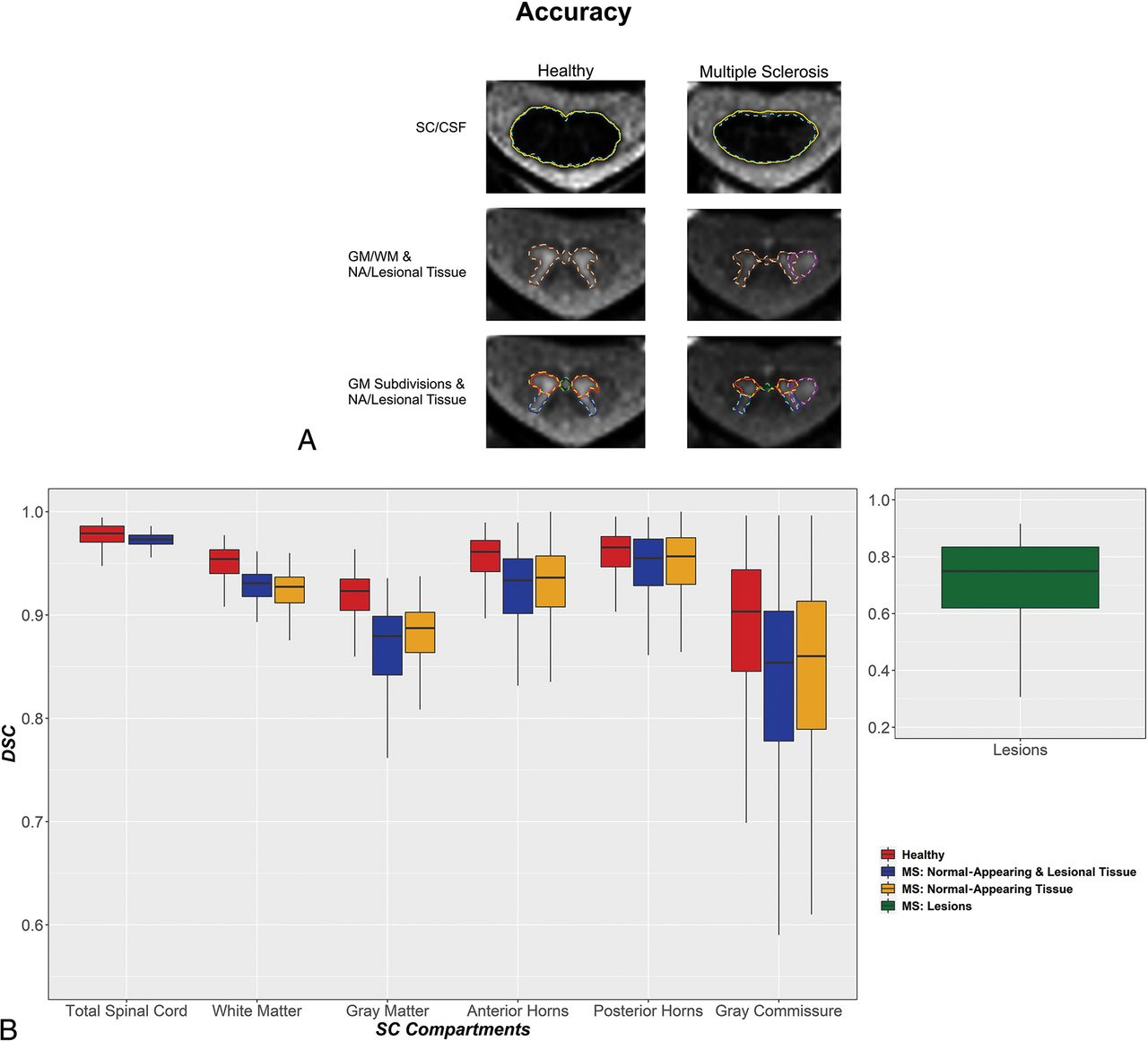
  • FIG 4.
    • Download figure
    • Open in new tab
    • Download powerpoint
    FIG 4.

    Accuracy of SCORE versus manual segmentations in all SC compartments of HCs and patients with MS. A, Representative SCORE and manual segmentations of AMIRA slices acquired in 1 HC and 1 patient with MS. The SC/CSF borders of manual and SCORE segmentations are shown in aqua (dashed line) and gold, respectively. The SC GM/WM borders of manual and SCORE segmentations are shown in coral (dashed line) and dark brown, respectively. The SC anterior horns, posterior horns, and gray commissure of manual and SCORE segmentations are shown in orange, turquoise, lime (dashed lines), red, navy blue, and dark green, respectively. Finally, the lesion borders of manual and SCORE segmentations are shown in violet (dashed line) and dark purple, respectively. Note the high agreement between manual and SCORE segmentations. The accuracy of SCORE versus manual segmentations was measured by DSC (B) and was generally high. Healthy, normal-appearing and lesional tissue and normal-appearing tissue are shown separately for each SC compartment in red, blue, and yellow, respectively. SC MS lesions are shown separately in green. Note the high accuracy of SCORE compared with manual segmentations in all SC compartments except for the gray commissure and lesions in both HCs and patients with MS.

PreviousNext
Back to top
Advertisement
Print
Download PDF
Email Article

Thank you for your interest in spreading the word on American Journal of Neuroradiology.

NOTE: We only request your email address so that the person you are recommending the page to knows that you wanted them to see it, and that it is not junk mail. We do not capture any email address.

Enter multiple addresses on separate lines or separate them with commas.
Fully Automatic Method for Reliable Spinal Cord Compartment Segmentation in Multiple Sclerosis
(Your Name) has sent you a message from American Journal of Neuroradiology
(Your Name) thought you would like to see the American Journal of Neuroradiology web site.
CAPTCHA
This question is for testing whether or not you are a human visitor and to prevent automated spam submissions.
Cite this article
C. Tsagkas, A. Horvath-Huck, T. Haas, M. Amann, A. Todea, A. Altermatt, J. Müller, A. Cagol, M. Leimbacher, M. Barakovic, M. Weigel, S. Pezold, T. Sprenger, L. Kappos, O. Bieri, C. Granziera, P. Cattin, K. Parmar
Fully Automatic Method for Reliable Spinal Cord Compartment Segmentation in Multiple Sclerosis
American Journal of Neuroradiology Jan 2023, DOI: 10.3174/ajnr.A7756

Citation Manager Formats

  • BibTeX
  • Bookends
  • EasyBib
  • EndNote (tagged)
  • EndNote 8 (xml)
  • Medlars
  • Mendeley
  • Papers
  • RefWorks Tagged
  • Ref Manager
  • RIS
  • Zotero
0 Responses
Respond to this article
Share
Bookmark this article
Fully Automatic Method for Reliable Spinal Cord Compartment Segmentation in Multiple Sclerosis
C. Tsagkas, A. Horvath-Huck, T. Haas, M. Amann, A. Todea, A. Altermatt, J. Müller, A. Cagol, M. Leimbacher, M. Barakovic, M. Weigel, S. Pezold, T. Sprenger, L. Kappos, O. Bieri, C. Granziera, P. Cattin, K. Parmar
American Journal of Neuroradiology Jan 2023, DOI: 10.3174/ajnr.A7756
del.icio.us logo Twitter logo Facebook logo Mendeley logo
  • Tweet Widget
  • Facebook Like
  • Google Plus One
Purchase

Jump to section

  • Article
    • Abstract
    • ABBREVIATIONS:
    • MATERIALS AND METHODS
    • RESULTS
    • DISCUSSION
    • CONCLUSIONS
    • Acknowledgments
    • Footnotes
    • References
  • Figures & Data
  • Info & Metrics
  • Responses
  • References
  • PDF

Related Articles

  • No related articles found.
  • PubMed
  • Google Scholar

Cited By...

  • EPISeg: Automated segmentation of the spinal cord on echo planar images using open-access multi-center data
  • Crossref
  • Google Scholar

This article has not yet been cited by articles in journals that are participating in Crossref Cited-by Linking.

More in this TOC Section

  • Dynamic CT Myelography: Patient Positioning
  • Evaluation of SIH MR Scoring Systems in Normals
  • Management Outcomes For VO Spine Biopsy
Show more SPINE IMAGING AND SPINE IMAGE-GUIDED INTERVENTIONS

Similar Articles

Advertisement

Indexed Content

  • Current Issue
  • Accepted Manuscripts
  • Article Preview
  • Past Issues
  • Editorials
  • Editor's Choice
  • Fellows' Journal Club
  • Letters to the Editor
  • Video Articles

Cases

  • Case Collection
  • Archive - Case of the Week
  • Archive - Case of the Month
  • Archive - Classic Case

More from AJNR

  • Trainee Corner
  • Imaging Protocols
  • MRI Safety Corner
  • Book Reviews

Multimedia

  • AJNR Podcasts
  • AJNR Scantastics

Resources

  • Turnaround Time
  • Submit a Manuscript
  • Submit a Video Article
  • Submit an eLetter to the Editor/Response
  • Manuscript Submission Guidelines
  • Statistical Tips
  • Fast Publishing of Accepted Manuscripts
  • Graphical Abstract Preparation
  • Imaging Protocol Submission
  • Evidence-Based Medicine Level Guide
  • Publishing Checklists
  • Author Policies
  • Become a Reviewer/Academy of Reviewers
  • News and Updates

About Us

  • About AJNR
  • Editorial Board
  • Editorial Board Alumni
  • Alerts
  • Permissions
  • Not an AJNR Subscriber? Join Now
  • Advertise with Us
  • Librarian Resources
  • Feedback
  • Terms and Conditions
  • AJNR Editorial Board Alumni

American Society of Neuroradiology

  • Not an ASNR Member? Join Now

© 2025 by the American Society of Neuroradiology All rights, including for text and data mining, AI training, and similar technologies, are reserved.
Print ISSN: 0195-6108 Online ISSN: 1936-959X

Powered by HighWire