
Article Figures & Data
Figures
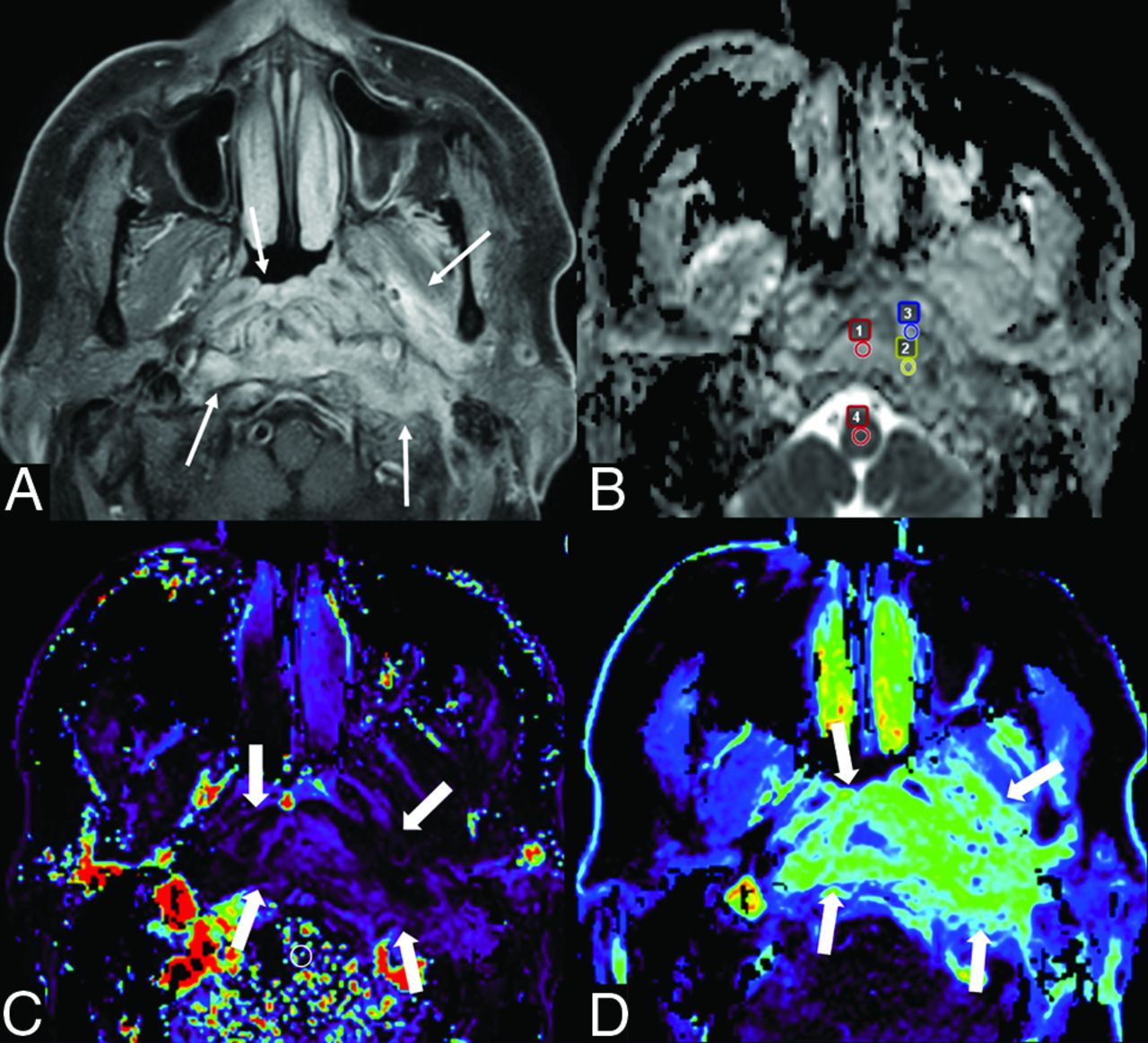
- FIG 1.
A case of SBO in a 68-year-old man. Postcontrast fat-suppressed T1-weighted image (A) shows ill-defined enhancement in the nasopharynx, prevertebral space, left parapharyngeal space, and left masticator space (arrows). The ADC map (B) shows 3 ROIs (Nos. 1 − 3, small circles) within high-signal lesion and 1 ROI (No. 4, small circle) for reference in the spinal cord, with an nADCmean of 1.86. DCE-MR imaging (C, Kep map; D, Ve map) shows the lesion (thick arrows) with a Kep of 0.42 minute−1 and Ve of 0.54.
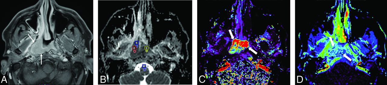
- FIG 2.
A case of nasopharyngeal cancer in a 37-year-old man. A mass lesion with enhancement (arrows) mainly on the right side of the nasopharynx is observed on the postcontrast fat-suppressed T1-weighted image (A). An ADC map (B) shows 3 ROIs (Nos. 1 − 3, small circles) within a low-signal lesion and 1 ROI (No. 4, small circle) for reference in the spinal cord, with an nADCmean of 0.86. DCE-MR imaging (C, Kep map; D, Ve map) shows the lesion (thick arrows) with a Kep of 3.14 minute−1 and a Ve of 0.36.
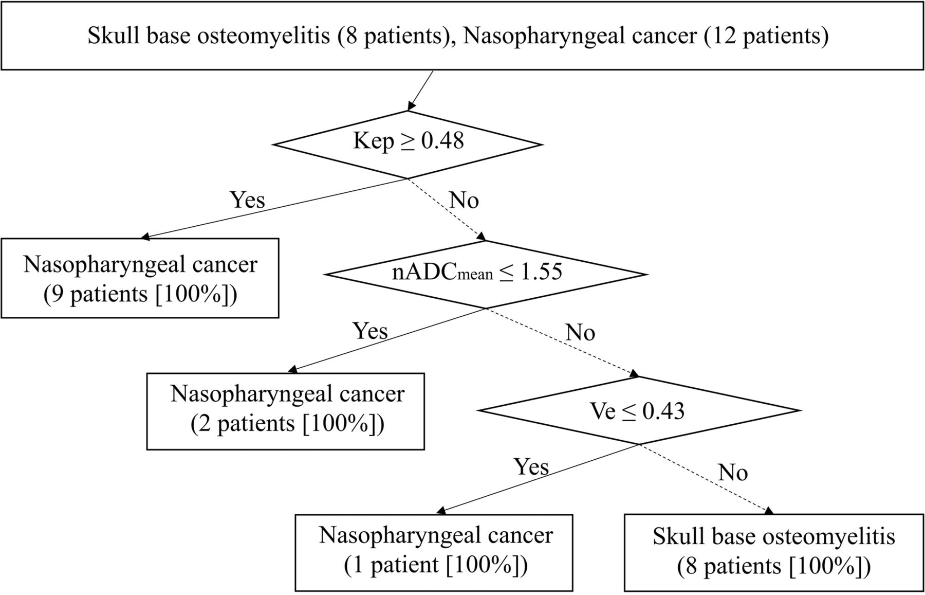
- FIG 3.
The decision tree model incorporating DCE parameters (Kep and Ve) and the nADCmean.


Tables
Clinical Information Demographic Sex Male = 8, female = 4 Median age (range) (yr) 52.5 (36–69) Clinical Pathology Squamous cell carcinoma 12/12 (100.0%) T classification T1 2/12 (16.7%) T2 5/12 (41.7%) T3 2/12 (16.7%) T4 3/12 (25.0%) DCE Parameters SBO Nasopharyngeal Cancer P Value Ve 0.48 (0.36–0.55) 0.38 (0.14–0.63) .15 Vp 0.14 (0.04–2.36) 0.12 (0.05–0.26) .99 Ktrans (min−1) 0.20 (0.15–0.39) 0.28 (0.06–1.42) .51 Kep (min−1) 0.43 (0.31–0.48) 0.57 (0.31–3.70) .04b ADC value 1.48 (1.14–1.84) 0.69 (0.55–1.39) <.001b Reference ADC 0.79 (0.54–0.94) 0.81 (0.73–0.90) .39 nADCmean 1.90 (0.61–2.48) 0.87 (0.61–1.90) <.001b


