
Article Figures & Data
Figures
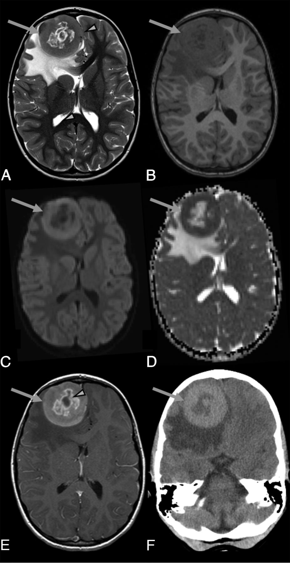
- FIG 1.
Brain images of a 6-year-old girl with pathology-proven unifocal JXG who presented with increased frequency of headaches, occasionally accompanied by vomiting. Brain MR imaging with an axial T2-weighted image (A), axial T1-weighted image (B), axial DWI (C), axial ADC map (D), axial T1+CE-weighted image (E), and axial noncontrast CT (F) shows a large, round, unifocal mass lesion located in the right frontal lobe (arrows in A–F) with central necrosis (arrowhead in E), perilesional edema, and right-to-left midline shift (arrowhead in A). Compared with cortical GM, the solid non-necrotic peripheral component of the lesion is T2-isointense (A) and T1-isointense (B) and shows contrast enhancement (E) and restricted diffusion (C and D). The axial CT image (F) demonstrates the hyperdense (solid portion), large mass lesion.
- FIG 2.
Brain MR images of a 3-year-old boy with pathology-proven multifocal JXG and new-onset strabismus. Axial T2-weighted (A–C), axial T1-weighted (D–F), and axial T1+CE-weighted (G–I) MR imaging sequences show exemplary 3 round-to-oval lesions located in the right (arrows in A, D, and G) and left paracentral (arrows in B, E, and H) regions and the pons (arrows in C, F, and I), respectively. Compared with cortical GM, the lesions are T2-iso- to hyperintense (A–C) and T1-hyperintense (D–F) and show homogeneous contrast enhancement (G–I) and mild perilesional edema.
- FIG 3.
MR images obtained in a 10-year-old boy with pathology-proven multifocal JXG with leptomeningeal brain and spine involvement. The patient presented with progressive right-ear pain and erythema and decreased hearing and right-sided facial nerve palsy along with headaches. Axial (A, B, and D) and coronal T1+CE-weighted (C) MR images of the brain. E, Sagittal T1+CE-weighted fat-suppressed MR image of the spine. For example, 2 centrally necrotic, peripherally contrast-enhancing lesions are noted within both cerebral hemispheres (arrows in A). In addition, nodular contrast enhancement involving the leptomeninges of the right temporal lobe (arrow in B) and contrast enhancement within both internal auditory canals (arrows in C and D) are shown. Sagittal T1+CE-weighted images of the spine depict increased contrast enhancement of the leptomeninges (“sugar coating”), partially with nodular components, most obvious at the C7 and T8 level (arrows in E).


Tables
Imaging features of CNS JXGa
No. T1-Weighted T2-Weighted T1+CE DWI/ADC 4/12 ↔ ↔ 4 Marked contrast enhancement 4 Diffusion restriction 3/12 ↔ ↔ - ↑ or ↑ 3 Marked contrast enhancement 1 Diffusion restriction, 2 no diffusion restriction 2/12 ↑ ↑ 2 Marked contrast enhancement 1 Diffusion restriction, 1 NA 2/12 ↑ ↔ or ↔ - ↑ 1 Marked contrast enhancement/1 with lesions with marked and lesions without contrast enhancement 1 No diffusion restriction, 1 NA 1/12 ↓ - ↔ ↓ - ↔ 1 Marked contrast enhancement 1 Diffusion restriction Note:—↔ indicates isointense; ↑, hyperintense; ↔ - ↑, iso- to hyperintense; ↓ - ↔, hypo- to isointense; NA, not applicable.
↵a Cases were included only when both T1WI and T2WI were available.


Jump to section
Related Articles
Cited By...
- No citing articles found.