Advertisement

Main menu

  • Home
  • Content
    • Current Issue
    • Accepted Manuscripts
    • Article Preview
    • Past Issue Archive
    • Video Articles
    • AJNR Case Collection
    • Case of the Week Archive
    • Case of the Month Archive
    • Classic Case Archive
  • Special Collections
    • AJNR Awards
    • Low-Field MRI
    • Alzheimer Disease
    • ASNR Foundation Special Collection
    • Photon-Counting CT
    • View All
  • Multimedia
    • AJNR Podcasts
    • AJNR SCANtastic
    • Trainee Corner
    • MRI Safety Corner
    • Imaging Protocols
  • For Authors
    • Submit a Manuscript
    • Submit a Video Article
    • Submit an eLetter to the Editor/Response
    • Manuscript Submission Guidelines
    • Statistical Tips
    • Fast Publishing of Accepted Manuscripts
    • Graphical Abstract Preparation
    • Imaging Protocol Submission
    • Author Policies
  • About Us
    • About AJNR
    • Editorial Board
    • Editorial Board Alumni
  • More
    • Become a Reviewer/Academy of Reviewers
    • Subscribers
    • Permissions
    • Alerts
    • Feedback
    • Advertisers
    • ASNR Home

User menu

  • Alerts
  • Log in

Search

  • Advanced search
American Journal of Neuroradiology

American Journal of Neuroradiology

ASHNR American Society of Functional Neuroradiology ASHNR American Society of Pediatric Neuroradiology ASSR
  • Alerts
  • Log in

Advanced Search

  • Home
  • Content
    • Current Issue
    • Accepted Manuscripts
    • Article Preview
    • Past Issue Archive
    • Video Articles
    • AJNR Case Collection
    • Case of the Week Archive
    • Case of the Month Archive
    • Classic Case Archive
  • Special Collections
    • AJNR Awards
    • Low-Field MRI
    • Alzheimer Disease
    • ASNR Foundation Special Collection
    • Photon-Counting CT
    • View All
  • Multimedia
    • AJNR Podcasts
    • AJNR SCANtastic
    • Trainee Corner
    • MRI Safety Corner
    • Imaging Protocols
  • For Authors
    • Submit a Manuscript
    • Submit a Video Article
    • Submit an eLetter to the Editor/Response
    • Manuscript Submission Guidelines
    • Statistical Tips
    • Fast Publishing of Accepted Manuscripts
    • Graphical Abstract Preparation
    • Imaging Protocol Submission
    • Author Policies
  • About Us
    • About AJNR
    • Editorial Board
    • Editorial Board Alumni
  • More
    • Become a Reviewer/Academy of Reviewers
    • Subscribers
    • Permissions
    • Alerts
    • Feedback
    • Advertisers
    • ASNR Home
  • Follow AJNR on Twitter
  • Visit AJNR on Facebook
  • Follow AJNR on Instagram
  • Join AJNR on LinkedIn
  • RSS Feeds

AJNR Awards, New Junior Editors, and more. Read the latest AJNR updates

Research ArticlePediatric Neuroimaging

MR Imaging of Pediatric Low-Grade Gliomas: Pretherapeutic Differentiation of BRAF V600E Mutation, BRAF Fusion, and Wild-Type Tumors in Patients without Neurofibromatosis-1

A. Trasolini, C. Erker, S. Cheng, C. Crowell, K. McFadden, R. Moineddin, M.A. Sargent and D. Mata-Mbemba
American Journal of Neuroradiology July 2022, DOI: https://doi.org/10.3174/ajnr.A7574
A. Trasolini
aFrom the IWK Health Centre (A.T., C.E., C.C., K.M., D.M.-M.), Halifax, Nova Scotia, Canada
bDalhousie University Medical School (A.T.), Halifax, Nova Scotia, Canada
  • Find this author on Google Scholar
  • Find this author on PubMed
  • Search for this author on this site
  • ORCID record for A. Trasolini
C. Erker
aFrom the IWK Health Centre (A.T., C.E., C.C., K.M., D.M.-M.), Halifax, Nova Scotia, Canada
cDepartments of Pediatrics (C.E.)
  • Find this author on Google Scholar
  • Find this author on PubMed
  • Search for this author on this site
  • ORCID record for C. Erker
S. Cheng
gDivision of Hematology, Oncology, and Bone Marrow Transplant (S.C.), Department of Pediatrics, University of British Columbia, Vancouver, British Columbia, Canada
  • Find this author on Google Scholar
  • Find this author on PubMed
  • Search for this author on this site
  • ORCID record for S. Cheng
C. Crowell
aFrom the IWK Health Centre (A.T., C.E., C.C., K.M., D.M.-M.), Halifax, Nova Scotia, Canada
fFaculty of Science (C.C.), Dalhousie University, Halifax, Nova Scotia, Canada
  • Find this author on Google Scholar
  • Find this author on PubMed
  • Search for this author on this site
  • ORCID record for C. Crowell
K. McFadden
aFrom the IWK Health Centre (A.T., C.E., C.C., K.M., D.M.-M.), Halifax, Nova Scotia, Canada
dPathology (K.M.)
  • Find this author on Google Scholar
  • Find this author on PubMed
  • Search for this author on this site
  • ORCID record for K. McFadden
R. Moineddin
hUniversity of Toronto Dalla Lana School of Public Health (R.M.), Toronto, Ontario, Canada
  • Find this author on Google Scholar
  • Find this author on PubMed
  • Search for this author on this site
  • ORCID record for R. Moineddin
M.A. Sargent
iDepartment of Radiology (M.A.S.), British Columbia Children’s Hospital and University of British Columbia, Vancouver, British Columbia, Canada
  • Find this author on Google Scholar
  • Find this author on PubMed
  • Search for this author on this site
  • ORCID record for M.A. Sargent
D. Mata-Mbemba
aFrom the IWK Health Centre (A.T., C.E., C.C., K.M., D.M.-M.), Halifax, Nova Scotia, Canada
eDiagnostic Radiology (D.M.-M.)
jDepartment of Diagnostic Imaging (D.M.-M.), IWK Health Centre, Halifax, Nova Scotia, Canada
  • Find this author on Google Scholar
  • Find this author on PubMed
  • Search for this author on this site
  • ORCID record for D. Mata-Mbemba
  • Article
  • Figures & Data
  • Supplemental
  • Info & Metrics
  • Responses
  • References
  • PDF
Loading

Article Figures & Data

Figures

  • Tables
  • FIG 1.
    • Download figure
    • Open in new tab
    • Download powerpoint
    FIG 1.

    A 3-year-old girl who presented with symptoms related to increased intracranial pressure. The brain MR imaging shows a large complex cystic/solid mass lesion arising from the vermis and anteriorly compressing the fourth ventricle, therefore causing supratentorial massive hydrocephalus. The solid component of the tumor shows slight hyperintense signal on T2WI (A) and diffuse enhancement (B) but not with diffusion restriction (C and D). The tissue diagnosis was pilocytic astrocytoma with KIAA1549:BRAF fusion.
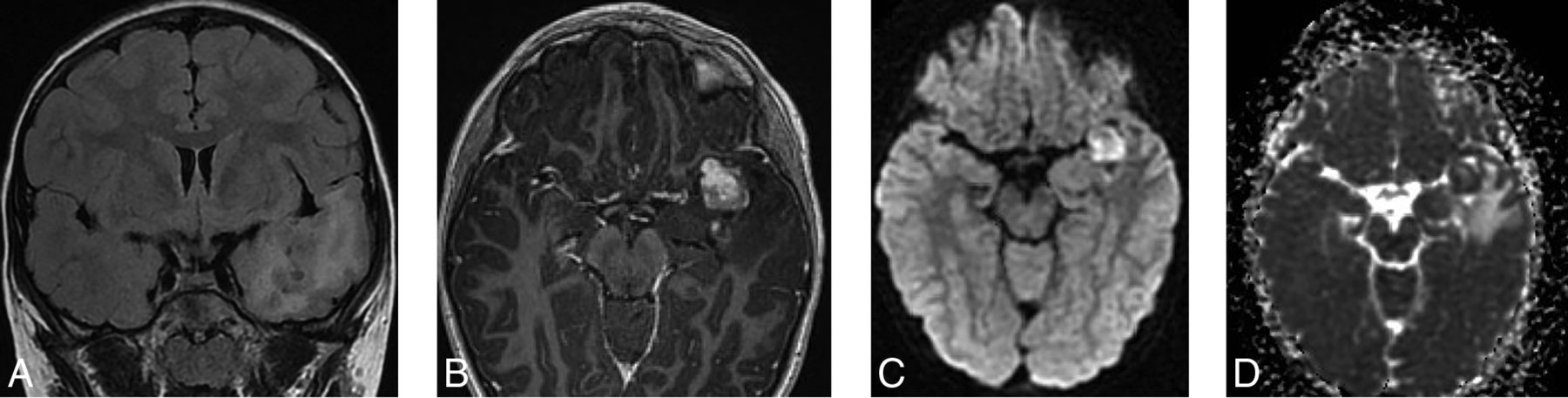
  • FIG 2.
    • Download figure
    • Open in new tab
    • Download powerpoint
    FIG 2.

    A 9-year-old boy with seizures. The brain MR imaging shows an ill-defined mass lesion with tiny internal cystic changes on T2 FLAIR (A) involving the mesial aspect of the left temporal lobe. The lesion shows moderate surrounding edema, mild mass effect against surrounding structures, as well as moderate enhancement (B). On DWI (C) and ADC (D), the mass shows diffusion restriction. The final tissue diagnosis was ganglioglioma with a BRAF V600E mutation.

Tables

  • Figures
    • View popup
    Table 1:

    Cohort information

    CharacteristicTotal, No. (%)
    Sex
     Male31 (44)
     Female39 (56)
    Age at diagnosis (yr)
     Median (IQR)6.3 (2.3–11.7)a
    WHO grade
     Grade I58 (85)
     Grade II10 (15)
    Molecular subtype
     KIAA1549:BRAF fusion30 (43)
     V600E19 (27)
     Wild-type/other21 (30)
    Disease progression
     Yes30 (43)
     No40 (57)
    Tumor location
     Brainstem4 (6)
     Cerebral hemisphere29 (41)
     OPHG14 (20)
     Posterior fossa21 (30)
    Spinal cord2 (3)
    Brain imaging completed
     Yes100
     No0
    Spine imaging completed
     Yes44 (63)
     No26 (37)
    Metastasis present
     Yes, only brain0 (0)
     Yes, only spine0 (0)
     Yes, brain and spine4 (6)
     No66 (94)
    • Note:—OPHG indicates optic pathway/hypothalamic glioma.

    • ↵a Median/IQR.

    • View popup
    Table 2:

    Univariate analysis of demographics and clinical characteristics by molecular group

    VariablesKIAA1549:BRAF FusionBRAF V600EWild-Type/OtherP1P2
    No. (%)No. (%)No. (%)(Fusion vs V600E vs WT)(Fusion vs V600E)
    Sex.3583.4436
     Male16 (53)8 (42)7 (33)
     Female14 (47)11 (58)14 (67)
    Age at time of MR imaging (yr).0012.0126
     Median3.3 (1.56–5.10)a9.7 (5.08–14.25)a10.5 (6.42–14.58)a
    Progression of disease.5769.3669
     Yes15 (50)12 (63)13 (62)
     No15 (50)7 (37)8 (38)
    WHO grade.0902.0724
     Grade I27 (90)13 (68)18 (95)
     Grade II3 (10)6 (32)1 (5)
    Tumor location<.0001<.0001
     Brainstem2 (7)1 (5)1 (5)
     Cerebral hemisphere3 (10)13 (68)13 (62)
     OPHG7 (23)4 (21)3 (14)
     Posterior fossa17 (57)0 (0)4 (19)
     Spinal cord1 (3)1 (5)0 (0)
    Metastatic status.81071.0000
     Yes1 (3)1 (5)2 (10)
     No29 (97)18 (95)19 (90)
    • Note:—WT indicates wild-type; OPHG, optic pathway/hypothalamic glioma.

    • ↵a Median and 95% confidence interval.

PreviousNext
Back to top
Advertisement
Print
Download PDF
Email Article

Thank you for your interest in spreading the word on American Journal of Neuroradiology.

NOTE: We only request your email address so that the person you are recommending the page to knows that you wanted them to see it, and that it is not junk mail. We do not capture any email address.

Enter multiple addresses on separate lines or separate them with commas.
MR Imaging of Pediatric Low-Grade Gliomas: Pretherapeutic Differentiation of BRAF V600E Mutation, BRAF Fusion, and Wild-Type Tumors in Patients without Neurofibromatosis-1
(Your Name) has sent you a message from American Journal of Neuroradiology
(Your Name) thought you would like to see the American Journal of Neuroradiology web site.
CAPTCHA
This question is for testing whether or not you are a human visitor and to prevent automated spam submissions.
Cite this article
A. Trasolini, C. Erker, S. Cheng, C. Crowell, K. McFadden, R. Moineddin, M.A. Sargent, D. Mata-Mbemba
MR Imaging of Pediatric Low-Grade Gliomas: Pretherapeutic Differentiation of BRAF V600E Mutation, BRAF Fusion, and Wild-Type Tumors in Patients without Neurofibromatosis-1
American Journal of Neuroradiology Jul 2022, DOI: 10.3174/ajnr.A7574

Citation Manager Formats

  • BibTeX
  • Bookends
  • EasyBib
  • EndNote (tagged)
  • EndNote 8 (xml)
  • Medlars
  • Mendeley
  • Papers
  • RefWorks Tagged
  • Ref Manager
  • RIS
  • Zotero
0 Responses
Respond to this article
Share
Bookmark this article
MR Imaging of Pediatric Low-Grade Gliomas: Pretherapeutic Differentiation of BRAF V600E Mutation, BRAF Fusion, and Wild-Type Tumors in Patients without Neurofibromatosis-1
A. Trasolini, C. Erker, S. Cheng, C. Crowell, K. McFadden, R. Moineddin, M.A. Sargent, D. Mata-Mbemba
American Journal of Neuroradiology Jul 2022, DOI: 10.3174/ajnr.A7574
del.icio.us logo Twitter logo Facebook logo Mendeley logo
  • Tweet Widget
  • Facebook Like
  • Google Plus One
Purchase

Jump to section

  • Article
    • Abstract
    • ABBREVIATIONS:
    • MATERIALS AND METHODS
    • RESULTS
    • DISCUSSION
    • CONCLUSIONS
    • Acknowledgments
    • Footnotes
    • References
  • Figures & Data
  • Supplemental
  • Info & Metrics
  • Responses
  • References
  • PDF

Related Articles

  • PubMed
  • Google Scholar

Cited By...

  • CNS Embryonal Tumor with PLAGL Amplification, a New Tumor in Children and Adolescents: Insights from a Comprehensive MRI Analysis
  • Multiparametric MRI Along with Machine Learning Informs on Molecular Underpinnings, Prognosis, and Treatment Response in Pediatric Low-Grade Glioma
  • Imaging Clusters of Pediatric Low-Grade Glioma are Associated with Distinct Molecular Characteristics
  • Identification of Multiclass Pediatric Low-Grade Neuroepithelial Tumor Molecular Subtype with ADC MR Imaging and Machine Learning
  • Crossref (6)
  • Google Scholar

This article has been cited by the following articles in journals that are participating in Crossref Cited-by Linking.

  • CNS Embryonal Tumor with PLAGL Amplification, a New Tumor in Children and Adolescents: Insights from a Comprehensive MRI Analysis
    A. Tietze, B. Bison, J. Engelhardt, T. Fenouil, D. Figarella-Branger, E. Goebell, J. Hakumäki, E. Koscielniak, L.E. Ludlow, D. Meyronet, P. Nyman, I. Øra, J. Pesola, T. Rauramaa, R.E. Reddingius, D. Samuel, A. Sexton-Oates, A. Vasiljevic, A.K. Wefers, J. Zamecnik, D.T.W. Jones, M.K. Keck, K. von Hoff
    American Journal of Neuroradiology 2025 46 3
  • Multiparametric MRI along with machine learning predicts prognosis and treatment response in pediatric low-grade glioma
    Anahita Fathi Kazerooni, Adam Kraya, Komal S. Rathi, Meen Chul Kim, Arastoo Vossough, Nastaran Khalili, Ariana M. Familiar, Deep Gandhi, Neda Khalili, Varun Kesherwani, Debanjan Haldar, Hannah Anderson, Run Jin, Aria Mahtabfar, Sina Bagheri, Yiran Guo, Qi Li, Xiaoyan Huang, Yuankun Zhu, Alex Sickler, Matthew R. Lueder, Saksham Phul, Mateusz Koptyra, Phillip B. Storm, Jeffrey B. Ware, Yuanquan Song, Christos Davatzikos, Jessica B. Foster, Sabine Mueller, Michael J. Fisher, Adam C. Resnick, Ali Nabavizadeh
    Nature Communications 2025 16 1
  • Identification of Multiclass Pediatric Low-Grade Neuroepithelial Tumor Molecular Subtype with ADC MR Imaging and Machine Learning
    Matheus D. Soldatelli, Khashayar Namdar, Uri Tabori, Cynthia Hawkins, Kristen Yeom, Farzad Khalvati, Birgit B. Ertl-Wagner, Matthias W. Wagner
    American Journal of Neuroradiology 2024 45 6
  • MRI characteristics predict BRAF V600E status in gangliogliomas and pleomorphic xanthoastrocytomas and provide survival prognostication
    Bingqing Jiang, Yingyan Zheng, Dejun She, Zhen Xing, Dairong Cao
    Acta Radiologica 2024 65 1
  • Multimodal contrastive learning for enhanced explainability in pediatric brain tumor molecular diagnosis
    Sara Ketabi, Matthias W. Wagner, Cynthia Hawkins, Uri Tabori, Birgit Betina Ertl-Wagner, Farzad Khalvati
    Scientific Reports 2025 15 1
  • Performance of Machine Learning Models in Predicting BRAF Alterations Using Imaging Data in Low-Grade Glioma: A Systematic Review and Meta-Analysis
    Shahryar Rajai Firouzabadi, Roozbeh Tavanaei, Ida Mohammadi, Alireza Alikhani, Ali Ansari, Mohammadhosein Akhlaghpasand, Bardia Hajikarimloo, Raymund L. Yong, Konstantinos Margetis
    World Neurosurgery 2025 195

More in this TOC Section

  • Neuroimaging Delineation and Progression of SLSMD
  • fetal brain development of 10 weeks gestation
  • CHARGE fetal MRI clival cleft
Show more Pediatric Neuroimaging

Similar Articles

Advertisement

Indexed Content

  • Current Issue
  • Accepted Manuscripts
  • Article Preview
  • Past Issues
  • Editorials
  • Editor's Choice
  • Fellows' Journal Club
  • Letters to the Editor
  • Video Articles

Cases

  • Case Collection
  • Archive - Case of the Week
  • Archive - Case of the Month
  • Archive - Classic Case

More from AJNR

  • Trainee Corner
  • Imaging Protocols
  • MRI Safety Corner
  • Book Reviews

Multimedia

  • AJNR Podcasts
  • AJNR Scantastics

Resources

  • Turnaround Time
  • Submit a Manuscript
  • Submit a Video Article
  • Submit an eLetter to the Editor/Response
  • Manuscript Submission Guidelines
  • Statistical Tips
  • Fast Publishing of Accepted Manuscripts
  • Graphical Abstract Preparation
  • Imaging Protocol Submission
  • Evidence-Based Medicine Level Guide
  • Publishing Checklists
  • Author Policies
  • Become a Reviewer/Academy of Reviewers
  • News and Updates

About Us

  • About AJNR
  • Editorial Board
  • Editorial Board Alumni
  • Alerts
  • Permissions
  • Not an AJNR Subscriber? Join Now
  • Advertise with Us
  • Librarian Resources
  • Feedback
  • Terms and Conditions
  • AJNR Editorial Board Alumni

American Society of Neuroradiology

  • Not an ASNR Member? Join Now

© 2025 by the American Society of Neuroradiology All rights, including for text and data mining, AI training, and similar technologies, are reserved.
Print ISSN: 0195-6108 Online ISSN: 1936-959X

Powered by HighWire