
Article Figures & Data
Figures
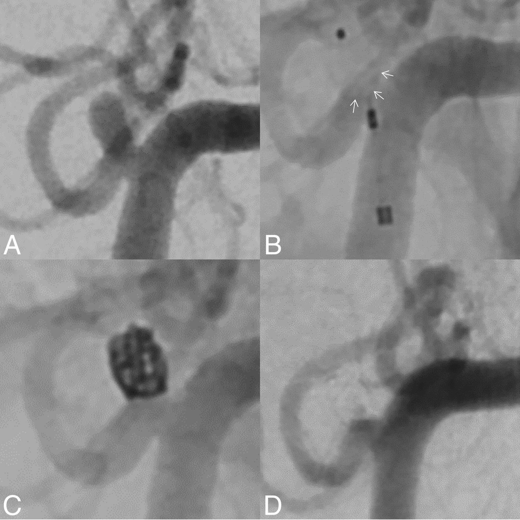
- FIG 1.
DSA shows a small, proximal PICA aneurysm (2 mm) in a patient with an SAH (A). Due to the broad neck of the aneurysm, the smallest available WEB in stock (SL, 3.5 × 2 mm) was deployed (B). However, the lower base of the WEB protruded markedly into the parent vessel (WEB contour highlighted by arrows). Implantation of an additional microstent seemed contraindicated because it might occlude the parent vessel (diameter, 1.3 mm) and would require permanent antiplatelet therapy. Hence, the WEB was removed before deployment, and the aneurysm was treated by implantation of a single coil (C). At final angiographic follow-up (21 months), the aneurysm was fully occluded and the PICA remained patent (D).
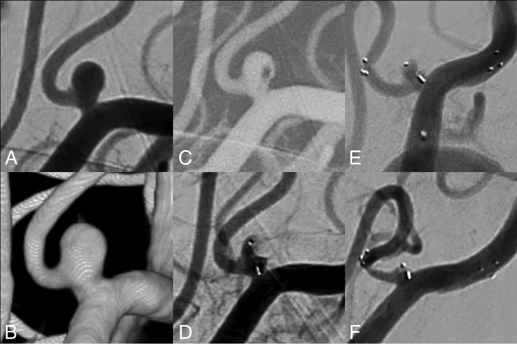
- FIG 2.
DSA shows a ruptured proximal PICA aneurysm (3.2 mm; neck width, 3.0 mm; dome-to-neck ratio, 1.1 mm) (A and B). Due to the broad-based geometry and the ruptured aneurysm status, intrasaccular flow disruption was envisaged. After probing the aneurysm with a low-profile VIA 17 microcatheter (C), a WEB 17 SL (4 × 2 mm) was placed within the aneurysm sac, achieving immediate contrast stasis to prevent aneurysm rerupture (D). After neurointensive care treatment of the patient, the aneurysm showed persistent residual filling. Hence, an Acclino microstent was placed across the aneurysm neck from the PICA into the distal vertebral artery to optimize WEB positioning (E). At 3-month angiographic follow-up, the aneurysm was completely occluded. There was a moderate in-stent stenosis, which was asymptomatic (F).
- FIG 3.
A 51-year-old female patient presented with a ruptured aneurysm at the proximal PICA (A and B). The aneurysm reruptured while the aneurysm sac was probed with the microcatheter. Extravasating contrast can be seen on the angiogram (C). However, directly after WEB deployment, the bleeding stopped due to intrasaccular stasis (D). Cerebellar infarction and herniation were excluded by a control CT (not shown). Although experiencing severe vasospasm, the patient survived and was transferred to a rehabilitation center with mild neurologic deficits (mRS 1). At 2 months, the WEB seemed to be fully thrombosed (E); however, the origin of the PICA, in particular the V4/PICA junction, appears to be dysplastic, warranting further angiographic control.
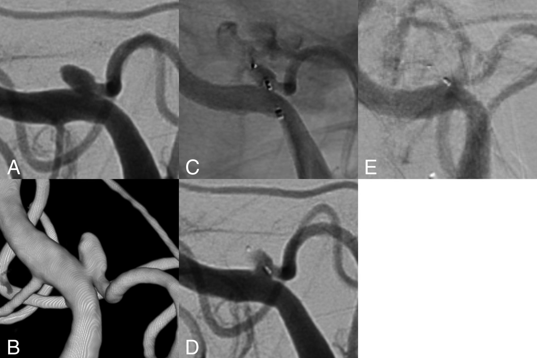
- FIG 4.
DSA (A) and 3D reconstructions of rotational data sets (B) show an unruptured wide-neck aneurysm at the branching of the PICA from the vertebral artery. Due to its very broad-based geometry, treatment by conventional coiling is not feasible. To avoid crossing-over stent-assisted coiling, WEB embolization was envisaged. After we probed the aneurysm sac with a low-profile VIA 17 microcatheter (C), a WEB SL 3.5 × 2 cm was implanted (D), which sealed the aneurysm at its neck level and maintained full patency of the PICA. Immediate angiographic control after WEB implantation shows contrast stasis within the WEB (E). Six-month DSA shows complete aneurysm occlusion (RROC I) and patency of the PICA (F).


Tables
Case Age/Sex Unruptured/Ruptured (WFNS) Size (mm) Neck Width (mm) D/N Ratio 1 65/F UR 3.4 2.7 1.3 2 57/F R (I) 3.2 3.0 1.1 3 48/F UR 2.9 2.6 1.1 4 51/F R (IV) 2.2 2.0 1.1 5 64/F R (II) 4.4 4.4 1.1 6 78/F R (V) 5.4 3.8 1.4 7 71/F UR 8.6 4.6 1.9 8 63/F UR 3.4 3.8 0.9 9 56/F R (V) 12.0 4.8 2.6 10 51/F UR 3.0 2.4 1.3 11 70/F UR 9.2 7.4 1.2 12 58/F R (V) 2.0 1.7 1.2 13 52/F R (V) 11.0 6.0 1.8 14 53/F UR 3.4 2.3 1.5 15 63/F UR 9.0 4.6 2.0 16 64/F UR 4.6 2.7 1.7 Note:—F indicates female; UR, unruptured; R, ruptured; WFNS, World Federation of Neurosurgical Societies grading scale; D/N, dome-to-neck ratio.
Case Treatment Complications Immediate RROC RROC at
FU (months)mRS at
FU (months)1 WEB SLS 4 mm Apposition thrombus, tirofiban, no neurologic deficit I I (6) 0 (6) 2 WEB SL 4 × 2 mm + Acclino stent 3.5 × 20 mm (staged) II I (3) 0 (20) 3 WEB SL 3.5 × 2 mm I I (6) 0 (6) 4 WEB SL 3 × 2 mm Aneurysm rerupture during WEB deployment, intrasaccular stasis, no deficit I I (1) 1 (1) 5 WEB SL 5 × 3 mm Transient hemianopsia partial posterior infarction, probably due to thromboembolism II I (7) 1 (7) 6 WEB SL 6 × 4 mm III II (1) 5 (1) 7 WEB DL 7 × 6 mm, 5 coils II I (17) 1 (17) 8 WEB SL 3 × 2 mm III I (8) 0 (8) 9 WEB SL 6 × 4 mm, 11 coils III I (10) 2 (10) 10 WEB SL 3 × 2 mm III I (5) 0 (5) 11 WEB SL 9 × 4 mm, LEO Babya stent 4.5 × 25 mm WEB protrusion into the parent artery, adjunctive stent implantation, no ischemic complications III II (6) 0 (6) 12 WEB SL 3.5 × 2 mm, coiling WEB implantation failed due to WEB protrusion, subsequent coil embolization II I (21) 0 (21) 13 WEB SL 11 × 6 mm III I (1) 6 (1) 14 WEB SL 4 × 3 mm III I (1) 0 (1) 15 WEB SL 9 × 4 mm III II (1) 1 (1) 16 WEB SL 3.5 × 2 mm III 0 (6) Note:—SLS indicates single-layer sphere; SL, single-layer; FU, follow-up; DL, dual-layer; RROC, Raymond-Roy occlusion classification.
↵a Balt Extrusion.


Jump to section
Related Articles
Cited By...
- No citing articles found.