
Article Figures & Data
Figures
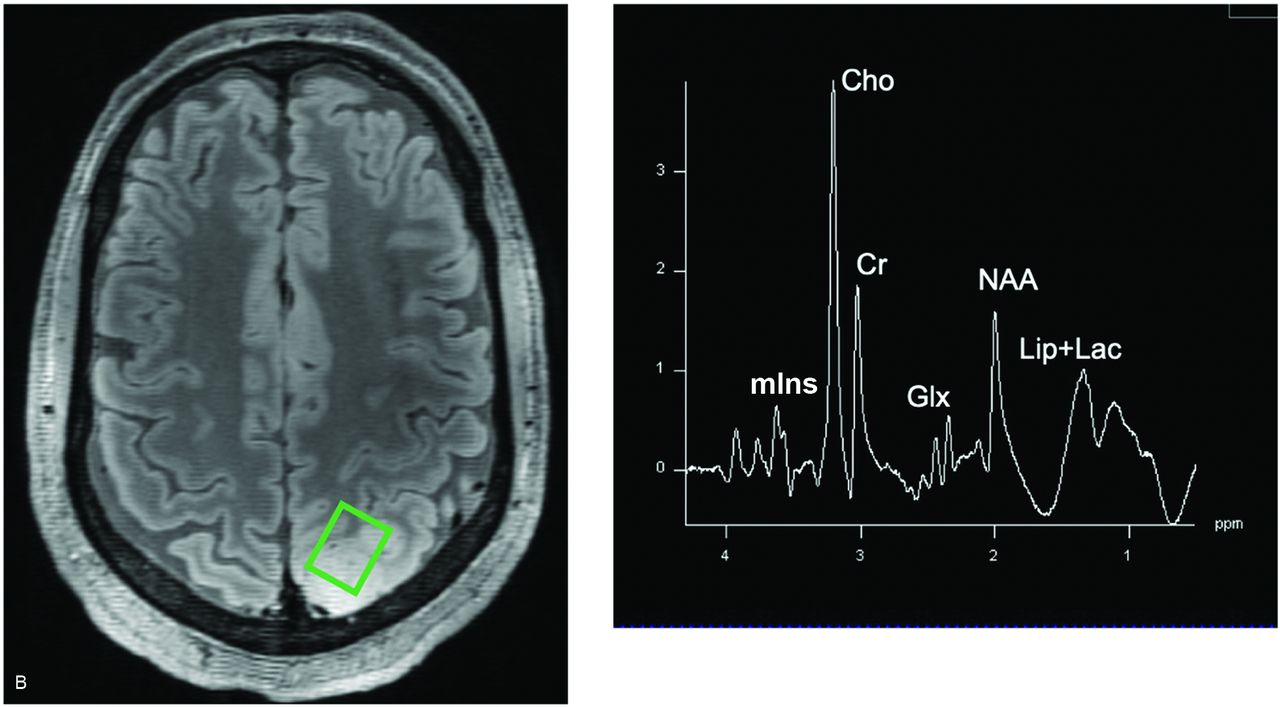
- FIG 1.
A, An MR spectrum generated at 7T from the occipital lobe (predominantly gray matter) of a healthy volunteer is shown (TR/TE = 2000/30.5 ms; number of averages = 32). The real part appears in blue and includes metabolite labels. The same image processed with LCModel is shown below and contains quantified metabolite values. Instead of capturing the increased resolution attainable at higher field strengths, these data are intended to primarily show a very clear spectrum from a normal brain. This was a 3 × 3 × 3 SVS acquisition in 1 minute obtained using an SASSI sequence. In contrast, note a single-voxel 1H-MRS spectrum (TR/TE = 3000/23 ms; number of averages = 8) from a patient with a grade III astrocytoma located in the left parietal region and having an IDH-mutant genotype showing various metabolites (B). The patient was scanned on a 7T whole‐body MR imaging scanner equipped with a single transmit/32‐channel receiver array head coil. Tissue infiltrated with gliomas such as the astrocytoma in B results in spectra with different metabolic characteristics than the normal tissue in A. Lip indicates lipids. The material from B was obtained with permission and in collaboration with Sanjeev Chawla in the Department of Radiology at the Perelman School of Medicine at the University of Pennsylvania.
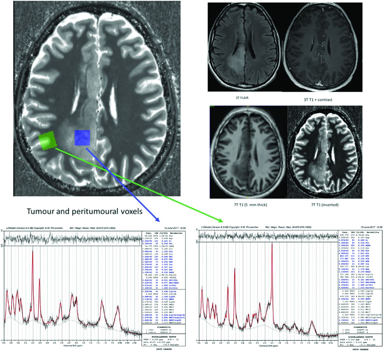
- FIG 2.
3T/7T MR imaging data, 7T SVS data, and LCModels from a patient with diffuse glioma are presented (TR/TE for the 7T SVS data = 8500/6 ms), with tumor data on the left (blue arrow) and peritumoral data on the right (green arrow) for each patient. Inspection of the MRS metabolic profiles shows that the peritumoral region has a relatively normal profile except for a highly elevated Glu concentration compared with both Gln and Cr. This is in contrast to the tumoral region, where there is a complete reversal of this profile with Gln and Cho being elevated compared with Glu, NAA, and Cr. These data provide further evidence of the dysregulation of the Glu-Gln shuffle, serving as a source of seizures in patients with gliomas. This material was obtained with permission and in collaboration with Andrew Neal and Bradford A. Moffatt at the Melbourne Brain Centre Imaging Unit (MBCIU), the University of Melbourne node of the Australian National Imaging Facility.
- FIG 3.
Major glutamine pathways in HGGs are featured. Gln undergoes a deamidation to Glu, which subsequently promotes tumorigenic growth either as a substrate in the tricarboxylic acid (TCA) cycle or as a precursor to GSH. The deamidation reaction is catalyzed by glutaminase. This enzyme is the target of glutaminase inhibitors, which may be useful therapeutic agents for patients with gliomas that involve high levels of glutamine detected by 7T MRS. PEP indicates phosphoenolpyruvate.
- FIG 4.
Exemplary 7T concentric ring trajectory FID-MRSI spectra of a patient with GBM. This method uses FID acquisition combined with concentric ring trajectories, bringing together multiple potential benefits: high resolution and acceleration while maintaining sufficient SNR, low SAR, B1 insensitivity, no selection box, and detection of J-coupled metabolites with 7T spectral separation. This measurement was acquired in 15 minutes with a nominal isotropic resolution of 3.4 mm, covering the cerebrum. Well-defined metabolic peaks are observed at 3 points: a tCho and Gly hotspot, a tCho and Gly cold spot, and normal-appearing white matter (NAWM). It is noticeable that even in the tumor tCho cold spot, tCho concentrations are higher than in NAWM but about 50% lower than in the tCho hotspot. Two of the metabolites that appear in these plots (Gly and Gln) have emerged as promising therapeutic targets and may serve as imaging biomarkers for patients with HGG. This material was obtained with permission from Gilbert Hangel at the High-field MR Center at the Medical University of Vienna. T1w indicates T1WI; tCr, total Cr.
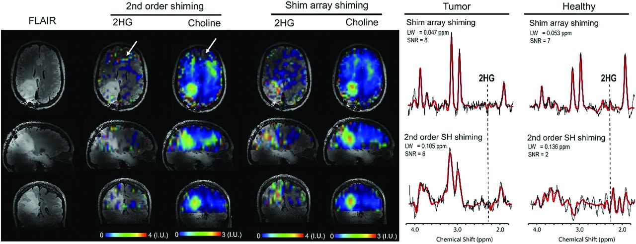
- FIG 5.
7T MRSI with standard second order shimming and higher order shimming with an AC/DC shim array was used to visualize Cho and 2HG in an IDH1 astrocytoma. A metabolic hallmark of IDH1 and IDH2 gliomas, 2HG generates a particularly strong signal at 7T, which may make it easier to diagnose patients with IDH-mutant profiles. The SNR of 2HG is lower in this example because this patient was treated with an operation and radiochemotherapy, which decrease 2HG levels. The primary clinical objective in this study was to determine whether there were residual mutant IDH tumor cells posttreatment. The white arrows indicate the frontal area where there are missing voxels in the Cho metabolic map and falsely increased values in the 2HG map due to a larger spectral linewidth obtained with second order shimming. The tumor has a higher contrast-to-noise ratio in the 2HG and Cho maps obtained with the AC/DC shim array. Examples of spectra are shown from tumor and healthy brain in the frontal region (corresponding to the white arrows). The position of the 2HG peak at 2.25 ppm is indicated in all spectra. For the adiabatic spin-echo excitation with TE = 78 ms, a negative peak should be obtained for 2HG, which is clearly visible in the tumor spectrum with AC/DC shim but not apparent in the tumor spectrum with standard second order shimming (2SH) shim. In particular, the frontal spectrum obtained with the 2SH shim is completely destroyed by the B0 inhomogeneity, while metabolite peaks are clearly visible with the AC/DC shim. 2HG is falsely fit in the frontal healthy spectrum with the 2SH shim due to negative spectral artifacts that appear at the 2.25-ppm 2HG peak position. The dashed vertical line indicates the location of the main 2HG peak at 2.25 ppm, which should be negative at 7T and TE = 78 ms. All the voxels in the MRSI were fit to create a metabolic map. A spectrum from the frontal voxel was chosen to show a false-positive fit of 2HG in healthy brain when the spectral quality is not adequate. Frontal brain regions are difficult to shim with standard methods, and the AC/DC coil can improve B0 homogeneity due to additional B0 shimming. Frontal loops in AC/DC are very close to frontal brain areas and create complex B0 field patterns to shim out the susceptibility induced by air tissue around the frontal sinus. The second order shimming was obtained with the manufacture’s software. This material was obtained with permission from and in collaboration with Jason P. Stockmann and Ovidiu C. Andronesi at the A. A. Martinos Center for Biomedical Imaging, Massachusetts General Hospital and Harvard Medical School. LW indicates linewidth.
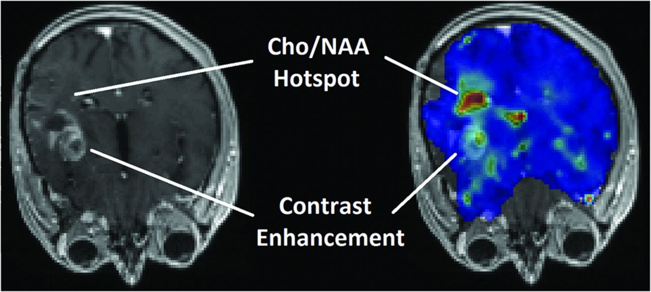
- FIG 6.
T1-weighted contrast-enhanced MR imaging (left) of a patient with a glioma subsequently diagnosed with recurrent tumor (true progression) and the same image with a superimposed map of the Cho/NAA ratio (right) acquired using echo-planar spectroscopic imaging. The map shows a hotspot of the elevated Cho/NAA ratio in a region that does not show contrast uptake on postcontrast T1 MR imaging (red depicts Cho/NAA > 1).
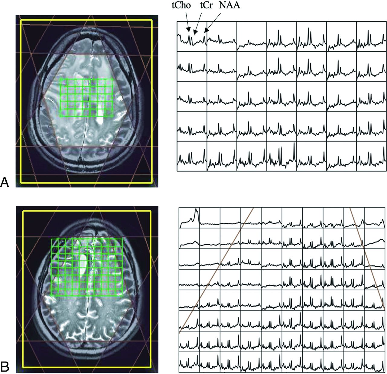
- FIG 7.
7T MRSI can capture gliomas in different stages, such as the recurrent grade II oligodendroglioma (progression) in the newly diagnosed, presymptomatic patient in A and the recurrent grade II oligodendroglioma (stable) in a treated patient in B. These spectra were acquired at 7T and show differences between progressing and nonprogressing tumors. The progressing tumor in the upper figure shows lower NAA in tumor regions and higher relative Cho, whereas the stable tumor in the lower figure demonstrates higher NAA with fewer metabolic abnormalities. NAA is on the right, and tCho and total Cr are in peaks just left of the center as labeled. Spectra data were processed after phasing/frequency corrections and coil combination (with baseline, no quantification). The metabolite range is from ∼1.8 to 4.2 ppm. This material was obtained with permission and in collaboration with Yan Li and Peder Larson in the Department of Radiology and Biomedical Imaging at the University of California, San Francisco. tCr indicates total Cr.


Tables
Comparison of 3T versus 7T MRS in visualization of metabolic markers in HGGsa
3T 7T SNR Lower (–) Higher (+) Spatial resolution Lower (–) Higher (+) Resolution of overlapping resonances (ie, PC vs GPC, Lac vs lipids, 2HG vs Glu, Gln, GABA, possibly Glu vs Gln) Poorer (–) Better (+) Range of metabolites Narrower (–) Wider (+) Uncertainty values of metabolite concentrations Greater (–) Smaller (+) Differentiation of IDH1/IDH2 gliomas vs wild-type gliomas Less specific (–) More specific (+) B0 inhomogeneity Lower (+) Higher (–) B1 inhomogeneity Lower (+) Higher (–) RF power deposition (SAR) Lower (+) Higher (–) CSL errors Less frequent (+) More frequent (–) Susceptibility artifacts Less frequent (+) More frequent (–) T1 relaxation time Shorter (+) Longer (–) T2 relaxation time Longer (+) Shorter (–) RF transmit body coils More accessible (+) Inaccessible (–) Metal hardware (ie, titanium plates placed during craniotomies) Safe (+) Contraindicated (–) Note:—CSL indicates chemical shift localization errors; (+), positive features; (–), negative features.
↵aThe assets and drawbacks of 7T MRS compared with 3T MRS are delineated.


