Advertisement

Main menu

  • Home
  • Content
    • Current Issue
    • Accepted Manuscripts
    • Article Preview
    • Past Issue Archive
    • Video Articles
    • AJNR Case Collection
    • Case of the Week Archive
    • Case of the Month Archive
    • Classic Case Archive
  • Special Collections
    • AJNR Awards
    • Low-Field MRI
    • Alzheimer Disease
    • ASNR Foundation Special Collection
    • Photon-Counting CT
    • View All
  • Multimedia
    • AJNR Podcasts
    • AJNR SCANtastic
    • Trainee Corner
    • MRI Safety Corner
    • Imaging Protocols
  • For Authors
    • Submit a Manuscript
    • Submit a Video Article
    • Submit an eLetter to the Editor/Response
    • Manuscript Submission Guidelines
    • Statistical Tips
    • Fast Publishing of Accepted Manuscripts
    • Graphical Abstract Preparation
    • Imaging Protocol Submission
    • Author Policies
  • About Us
    • About AJNR
    • Editorial Board
    • Editorial Board Alumni
  • More
    • Become a Reviewer/Academy of Reviewers
    • Subscribers
    • Permissions
    • Alerts
    • Feedback
    • Advertisers
    • ASNR Home

User menu

  • Alerts
  • Log in

Search

  • Advanced search
American Journal of Neuroradiology

American Journal of Neuroradiology

ASHNR American Society of Functional Neuroradiology ASHNR American Society of Pediatric Neuroradiology ASSR
  • Alerts
  • Log in

Advanced Search

  • Home
  • Content
    • Current Issue
    • Accepted Manuscripts
    • Article Preview
    • Past Issue Archive
    • Video Articles
    • AJNR Case Collection
    • Case of the Week Archive
    • Case of the Month Archive
    • Classic Case Archive
  • Special Collections
    • AJNR Awards
    • Low-Field MRI
    • Alzheimer Disease
    • ASNR Foundation Special Collection
    • Photon-Counting CT
    • View All
  • Multimedia
    • AJNR Podcasts
    • AJNR SCANtastic
    • Trainee Corner
    • MRI Safety Corner
    • Imaging Protocols
  • For Authors
    • Submit a Manuscript
    • Submit a Video Article
    • Submit an eLetter to the Editor/Response
    • Manuscript Submission Guidelines
    • Statistical Tips
    • Fast Publishing of Accepted Manuscripts
    • Graphical Abstract Preparation
    • Imaging Protocol Submission
    • Author Policies
  • About Us
    • About AJNR
    • Editorial Board
    • Editorial Board Alumni
  • More
    • Become a Reviewer/Academy of Reviewers
    • Subscribers
    • Permissions
    • Alerts
    • Feedback
    • Advertisers
    • ASNR Home
  • Follow AJNR on Twitter
  • Visit AJNR on Facebook
  • Follow AJNR on Instagram
  • Join AJNR on LinkedIn
  • RSS Feeds

AJNR Awards, New Junior Editors, and more. Read the latest AJNR updates

Research ArticlePediatric Neuroimaging
Open Access

Thalamus L-Sign: A Potential Biomarker of Neonatal Partial, Prolonged Hypoxic-Ischemic Brain Injury or Hypoglycemic Encephalopathy?

S.K. Misser, J.W. Lotz, R. van Toorn, N. Mchunu, M. Archary and A.J. Barkovich
American Journal of Neuroradiology May 2022, DOI: https://doi.org/10.3174/ajnr.A7511
S.K. Misser
aFrom the Departments of Radiology (S.K.M.)
cLake Smit and Partners Inc (S.K.M.), Durban, South Africa
  • Find this author on Google Scholar
  • Find this author on PubMed
  • Search for this author on this site
  • ORCID record for S.K. Misser
J.W. Lotz
dDepartments of Radiodiagnosis (J.W.L.)
  • Find this author on Google Scholar
  • Find this author on PubMed
  • Search for this author on this site
  • ORCID record for J.W. Lotz
R. van Toorn
ePaediatrics and Child Health (R.v.T.), Faculty of Medicine and Health Sciences, Stellenbosch University, Cape Town, South Africa
  • Find this author on Google Scholar
  • Find this author on PubMed
  • Search for this author on this site
  • ORCID record for R. van Toorn
N. Mchunu
fBiostatistics Research Unit (N.M.), South African Medical Research Council, Durban, South Africa
gSchool of Mathematics, Statistics and Computer Sciences, (N.M.), University of KwaZulu-Natal, Pietermaritzburg, South Africa
hCentre for the AIDS Programme of Research in South Africa (N.M.), Urban, South Africa
  • Find this author on Google Scholar
  • Find this author on PubMed
  • Search for this author on this site
  • ORCID record for N. Mchunu
M. Archary
bPediatrics (M.A.), Faculty of Health Sciences, University of KwaZulu-Natal, Nelson R Mandela School of Medicine, Durban, South Africa
  • Find this author on Google Scholar
  • Find this author on PubMed
  • Search for this author on this site
  • ORCID record for M. Archary
A.J. Barkovich
iSchool of Medicine (A.J.B.), University of California, San Francisco, San Francisco, California
  • Find this author on Google Scholar
  • Find this author on PubMed
  • Search for this author on this site
  • ORCID record for A.J. Barkovich
  • Article
  • Figures & Data
  • Supplemental
  • Info & Metrics
  • Responses
  • References
  • PDF
Loading

Article Figures & Data

Figures

  • Tables
  • FIG 1.
    • Download figure
    • Open in new tab
    • Download powerpoint
    FIG 1.

    Derivation of the 3 major study groups, the subgroups of HIBI, and the subtypes of watershed patterns of injury in patients who had partial, prolonged HIBI.
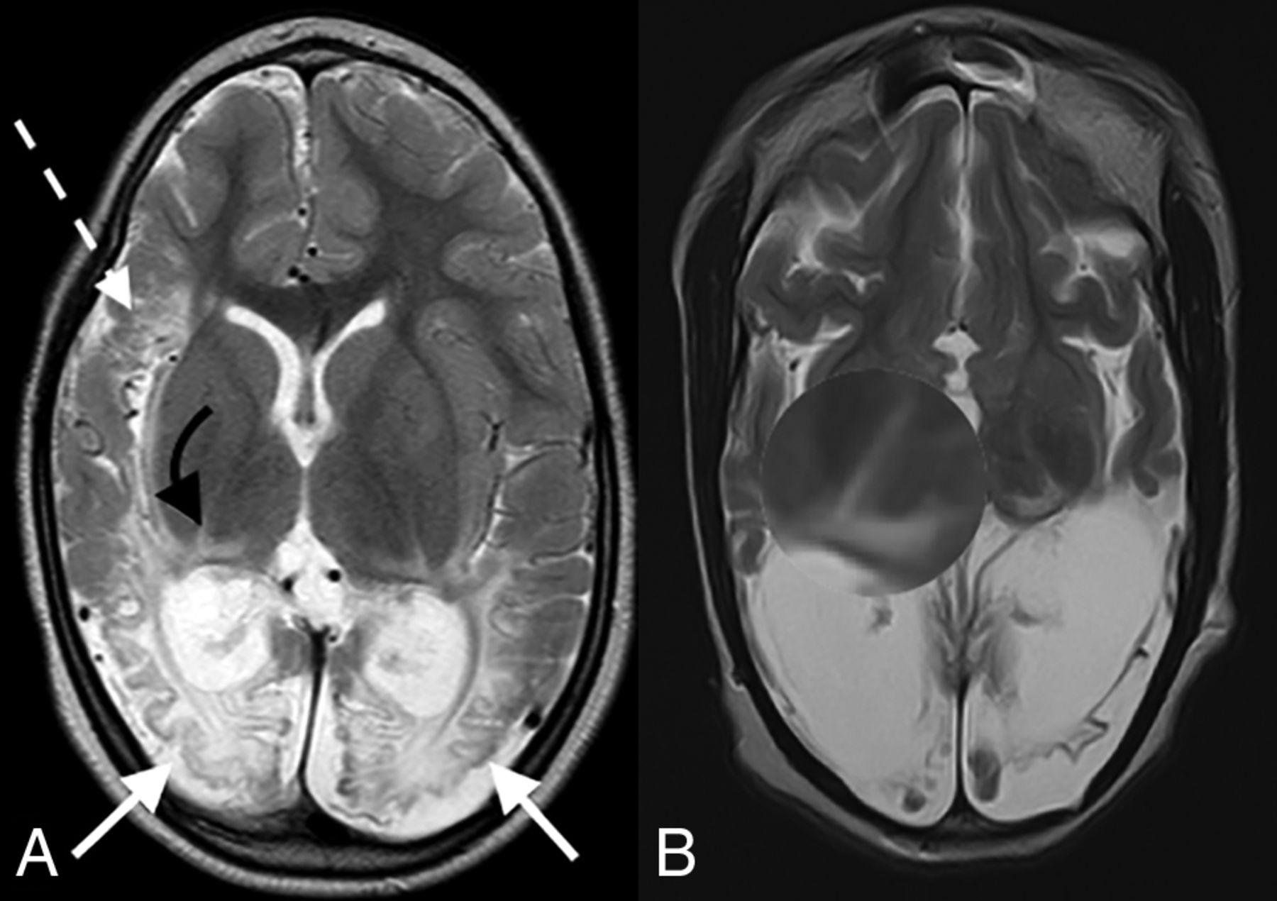
  • FIG 2.
    • Download figure
    • Open in new tab
    • Download powerpoint
    FIG 2.

    Axial T2-weighted images in a child with partial, prolonged HIBI demonstrating interarterial injuries at the peri-Sylvian (dashed white arrow) and posterior parieto-occipital (solid white arrows) watershed regions. Note the thalamus L-sign (curved arrows in A and highlighted by the loupe in a second patient in B).

  • FIG 3.
    • Download figure
    • Open in new tab
    • Download powerpoint
    FIG 3.

    Axial T2-weighted images in 2 children with proved neonatal hypoglycemia. There is bilateral occipital lobe encephalomalacia (arrows) related to hypoglycemic brain injury. Note the absence of any thalamic injury.
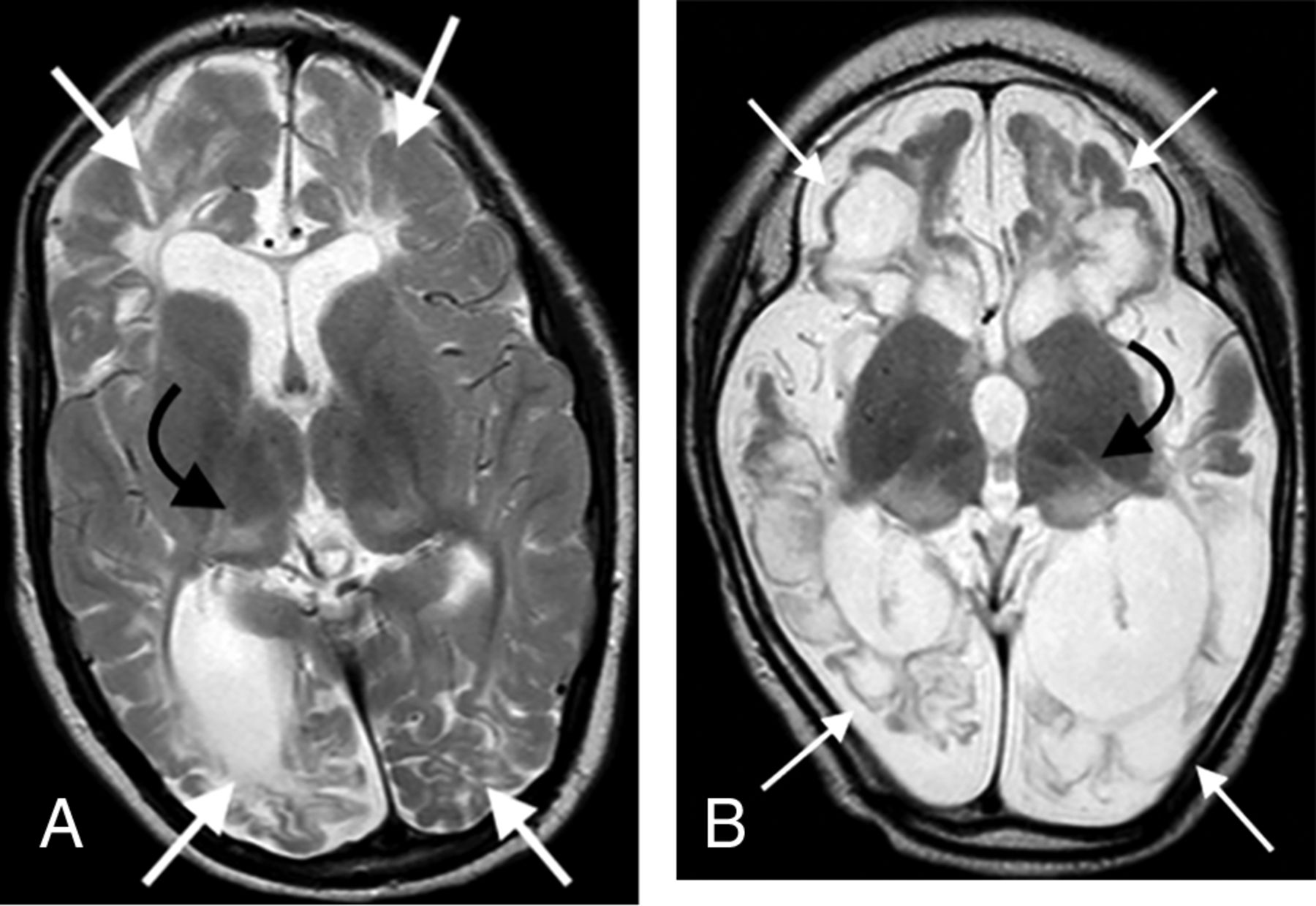
  • FIG 4.
    • Download figure
    • Open in new tab
    • Download powerpoint
    FIG 4.

    Combined hypoxic-ischemic and hypoglycemic brain injury in 2 children with documented neonatal encephalopathy. Note the exaggerated signal abnormality and thalamic volume loss (black arrows). There are multiple watershed areas (white arrows) demonstrating encephalomalacia change.

  • FIG 5.
    • Download figure
    • Open in new tab
    • Download powerpoint
    FIG 5.

    The key thalamic nuclei identified as components of the thalamus L-sign (dotted line) include the pulvinar, the lateral geniculate nucleus, and the reticular formation nuclei. Illustration by Neil Northey.

Tables

  • Figures
    • View popup
    Table 1:

    Key features of the 7 subtypes of partial prolonged/watershed HIBI

    Lobe/StructureFeaturesSubtype 1Subtype 2Subtype 3Subtype 4Subtype 5Subtype 6Subtype 7Overall
    Anterior (n = 10)Peri-Sylvian (n = 1)Posterior (n = 1)Anterior + Peri-Sylvian (n = 6)Peri-Sylvian + Posterior (n = 15)Anterior + Posterior (n = 7)All 3 Zones (n = 59)(n = 99)
    Thalamic injury location (No.) (%)Nil1 (16.7)3 (30.0)4 (4.0)
    Atypical2 (33.3)7 (70.0)1 (14.3)10 (10.1)
    Thalamus L-sign1 (100.0)1 (100.0)3 (50.0)15 (100.0)6 (85.7)59 (100.0)85 (85.9)
     Thalamus score (No.) (%)Not/less involved1 (100.0)1 (100.0)6 (100.0)14 (93.9)10 (100.0)6 (85.7)53 (89.8)91 (91.9)
    Markedly destroyed1 (6.7)1 (14.3)6 (10.2)8 (8.1)
     Parietal (No.) (%)Not involved3 (50.0)6 (60.0)1 (14.3)10 (10.1)
    Involved1 (100.0)1 (100.0)3 (50.0)15 (100.0)4 (40.0)6 (85.7)59 (100.0)89 (89.9)
     Occipital (No.) (%)Not involved1 (100.0)7 (70.0)8 (8.1)
    Involved1 (100.0)6 (100.0)15 (100.0)3 (30.0)7 (100.0)59 (100.0)91 (91.9)
     Frontal (No.) (%)Not involved1 (100.0)8 (53.3)1 (10.0)1 (1.7)11 (11.1)
    Involved1 (100.0)6 (100.0)7 (46.7)9 (90.0)7 (100.0)58 (98.3)88 (88.9)
     Temporal (No.) (%)Not involved3 (20.0)6 (60.0)3 (42.9)3 (5.1)15 (15.2)
    Involved1 (100.0)1 (100.0)6 (100.0)12 (80.0)4 (40.0)4 (57.1)56 (94.9)84 (84.8)
     Cerebellum (No.) (%)Not involved1 (100.0)4 (66.7)10 (66.7)7 (70.0)5 (71.4)40 (67.8)67 (67.7)
    Involved1 (100.0)2 (33.3)5 (33.3)3 (30.0)2 (28.6)19 (32.2)32 (32.3)
     Brainstem (No.) (%)Not involved1 (100.0)1 (100.0)6 (100.0)14 (93.3)10 (100.0)7 (100.0)49 (83.1)88 (88.9)
    Involved1 (6.7)10 (16.9)11 (11.1)
    • View popup
    Table 2:

    Key features involved in thalamus L-sign injury compared with other thalamic injuries (nil and atypical)

    Lobe/StructureFeaturesThalamus L-Sign (n = 85)Other (n = 14)Overall (n = 99)P Value
    Thalamus score (No.) (%)Not/less involved77 (90.6)14 (100.0)91 (91.9)<.001
    Markedly destroyed8 (9.4)
    Parietal (No.) (%)Not involved3 (3.5)7 (50.0)10 (10.1)<.001
    Involved82 (96.5)7 (50.0)89 (89.9)
    Occipital (No.) (%)Not involved1 (1.2)7 (50.0)8 (8.1)<.001
    Involved84 (98.8)7 (50.0)91 (91.9)
    Frontal (No.) (%)Not involved10 (11.8)1 (7.1)11 (11.1)1.000
    Involved75 (88.2)13 (92.9)88 (88.9)
    Temporal (No.) (%)Not involved9 (10.6)6 (42.9)15 (15.2).007
    Involved76 (89.4)8 (57.1)84 (84.8)
    Cerebellum (No.) (%)Not involved57 (67.1)10 (71.4)67 (67.7)1.000
    Involved28 (32.9)4 (28.6)32 (32.3)
    Brainstem (No.) (%)Not involved74 (87.1)14 (100.0)88 (88.9).355
    Involved11 (12.9)11 (11.1)
    • View popup
    Table 3:

    Factors associated with the thalamic L-sign injury

    Lobe/StructureRR95% CIP Value
    Parietal3.071.19−7.93.020
    Occipital7.381.18−46.23.033
    Frontal0.940.76−1.15.539
    Temporal1.510.99−2.29.055
    Cerebellum1.030.87−1.21.738
    Brainstem1.191.09−1.30<.001
    Parietal + occipital2.791.25−6.23.012
    • Note:—RR indicates relative risk.

PreviousNext
Back to top
Advertisement
Print
Download PDF
Email Article

Thank you for your interest in spreading the word on American Journal of Neuroradiology.

NOTE: We only request your email address so that the person you are recommending the page to knows that you wanted them to see it, and that it is not junk mail. We do not capture any email address.

Enter multiple addresses on separate lines or separate them with commas.
Thalamus L-Sign: A Potential Biomarker of Neonatal Partial, Prolonged Hypoxic-Ischemic Brain Injury or Hypoglycemic Encephalopathy?
(Your Name) has sent you a message from American Journal of Neuroradiology
(Your Name) thought you would like to see the American Journal of Neuroradiology web site.
CAPTCHA
This question is for testing whether or not you are a human visitor and to prevent automated spam submissions.
Cite this article
S.K. Misser, J.W. Lotz, R. van Toorn, N. Mchunu, M. Archary, A.J. Barkovich
Thalamus L-Sign: A Potential Biomarker of Neonatal Partial, Prolonged Hypoxic-Ischemic Brain Injury or Hypoglycemic Encephalopathy?
American Journal of Neuroradiology May 2022, DOI: 10.3174/ajnr.A7511

Citation Manager Formats

  • BibTeX
  • Bookends
  • EasyBib
  • EndNote (tagged)
  • EndNote 8 (xml)
  • Medlars
  • Mendeley
  • Papers
  • RefWorks Tagged
  • Ref Manager
  • RIS
  • Zotero
0 Responses
Respond to this article
Share
Bookmark this article
Thalamus L-Sign: A Potential Biomarker of Neonatal Partial, Prolonged Hypoxic-Ischemic Brain Injury or Hypoglycemic Encephalopathy?
S.K. Misser, J.W. Lotz, R. van Toorn, N. Mchunu, M. Archary, A.J. Barkovich
American Journal of Neuroradiology May 2022, DOI: 10.3174/ajnr.A7511
del.icio.us logo Twitter logo Facebook logo Mendeley logo
  • Tweet Widget
  • Facebook Like
  • Google Plus One
Purchase

Jump to section

  • Article
    • Abstract
    • ABBREVIATIONS:
    • MATERIALS AND METHODS
    • RESULTS
    • DISCUSSION
    • CONCLUSIONS
    • Footnotes
    • References
  • Figures & Data
  • Supplemental
  • Info & Metrics
  • Responses
  • References
  • PDF

Related Articles

  • PubMed
  • Google Scholar

Cited By...

  • No citing articles found.
  • Crossref (11)
  • Google Scholar

This article has been cited by the following articles in journals that are participating in Crossref Cited-by Linking.

  • Exploring the thalamus L-sign: initial findings and associations with white matter injury in premature infants
    Goni Merhav, Sarit Ravid, Mika Shapira Rootman
    Pediatric Radiology 2024 54 9
  • Use of Thalamus L-Sign to Differentiate Periventricular Leukomalacia From Neurometabolic Disorders
    Sabahattin Yuzkan, Merve Emecen Sanli, Merve Balci, Pakize Cennetoglu, Ihsan Kafadar, Burak Kocak
    Journal of Child Neurology 2023 38 6-7
  • Mimickers of hypoxic-ischaemic brain injury in term neonates: What the radiologist should know
    Shalendra K. Misser, Moherndran Archary
    South African Journal of Radiology 2024 28 1
  • The thalamus-L-sign in hypoxic-ischemic injury likely reflects secondary circuit injury and is only a feature on delayed magnetic resonance imaging in children with established cerebral palsy: reply to Merhav et al
    Shyam Sunder B. Venkatakrishna, Christelle Ackermann, Jelena Curic, Antrea Zouvani, Luis Octavio Tierradentro-Garcia, Savvas Andronikou
    Pediatric Radiology 2024 55 2
  • Developmental anatomy of the thalamus, perinatal lesions, and neurological development
    Paul Govaert, Roberta Arena, Jeroen Dudink, Sylke Steggerda, Thais Agut, Gertjan Marissens, Freek Hoebeek
    Developmental Medicine & Child Neurology 2025 67 1
  • Imaging of Cerebral Palsy: A Primer for the Radiologist
    M.S. Rootman, S. Shinnawi, G. Merhav, B.C. Friedman, L.-t. Pratt
    Neurographics 2025 15 2
  • L-Zeichen des Thalamus: spezifisch für eine perinatale Hypoxie-Ischämie
    Neuroradiologie Scan 2023 13 02
  • Neuroquantification enhances the radiological evaluation of term neonatal hypoxic-ischaemic cerebral injuries
    Shalendra K. Misser, Nobuhle Mchunu, Jan W. Lotz, Lisa Kjonigsen, Aziz Ulug, Moherndran Archary
    South African Journal of Radiology 2023 27 1
  • Periventricular Leukomalacia: Comparison of Parenchymal Signal and Volume Changes on Brain MRI in Paediatric Cases with Healthy Peers
    Hanife Gülden Düzkalır, İrem Sarı, Fulden Cantaş Türkiş, Safiye Güneş Sağer, Emine Çalışkan, Tamer Baysal
    Genel Tıp Dergisi 2024 34 4
  • Reply to Venkatakrishna SSB
    Goni Merhav, Mika Shapira Rootman
    Pediatric Radiology 2024 55 2

More in this TOC Section

  • Neuroimaging Delineation and Progression of SLSMD
  • fetal brain development of 10 weeks gestation
  • CHARGE fetal MRI clival cleft
Show more Pediatric Neuroimaging

Similar Articles

Advertisement

Indexed Content

  • Current Issue
  • Accepted Manuscripts
  • Article Preview
  • Past Issues
  • Editorials
  • Editor's Choice
  • Fellows' Journal Club
  • Letters to the Editor
  • Video Articles

Cases

  • Case Collection
  • Archive - Case of the Week
  • Archive - Case of the Month
  • Archive - Classic Case

More from AJNR

  • Trainee Corner
  • Imaging Protocols
  • MRI Safety Corner
  • Book Reviews

Multimedia

  • AJNR Podcasts
  • AJNR Scantastics

Resources

  • Turnaround Time
  • Submit a Manuscript
  • Submit a Video Article
  • Submit an eLetter to the Editor/Response
  • Manuscript Submission Guidelines
  • Statistical Tips
  • Fast Publishing of Accepted Manuscripts
  • Graphical Abstract Preparation
  • Imaging Protocol Submission
  • Evidence-Based Medicine Level Guide
  • Publishing Checklists
  • Author Policies
  • Become a Reviewer/Academy of Reviewers
  • News and Updates

About Us

  • About AJNR
  • Editorial Board
  • Editorial Board Alumni
  • Alerts
  • Permissions
  • Not an AJNR Subscriber? Join Now
  • Advertise with Us
  • Librarian Resources
  • Feedback
  • Terms and Conditions
  • AJNR Editorial Board Alumni

American Society of Neuroradiology

  • Not an ASNR Member? Join Now

© 2025 by the American Society of Neuroradiology All rights, including for text and data mining, AI training, and similar technologies, are reserved.
Print ISSN: 0195-6108 Online ISSN: 1936-959X

Powered by HighWire