Advertisement

Main menu

  • Home
  • Content
    • Current Issue
    • Accepted Manuscripts
    • Article Preview
    • Past Issue Archive
    • Video Articles
    • AJNR Case Collection
    • Case of the Week Archive
    • Case of the Month Archive
    • Classic Case Archive
  • Special Collections
    • AJNR Awards
    • Low-Field MRI
    • Alzheimer Disease
    • ASNR Foundation Special Collection
    • Photon-Counting CT
    • View All
  • Multimedia
    • AJNR Podcasts
    • AJNR SCANtastic
    • Trainee Corner
    • MRI Safety Corner
    • Imaging Protocols
  • For Authors
    • Submit a Manuscript
    • Submit a Video Article
    • Submit an eLetter to the Editor/Response
    • Manuscript Submission Guidelines
    • Statistical Tips
    • Fast Publishing of Accepted Manuscripts
    • Graphical Abstract Preparation
    • Imaging Protocol Submission
    • Author Policies
  • About Us
    • About AJNR
    • Editorial Board
    • Editorial Board Alumni
  • More
    • Become a Reviewer/Academy of Reviewers
    • Subscribers
    • Permissions
    • Alerts
    • Feedback
    • Advertisers
    • ASNR Home

User menu

  • Alerts
  • Log in

Search

  • Advanced search
American Journal of Neuroradiology

American Journal of Neuroradiology

ASHNR American Society of Functional Neuroradiology ASHNR American Society of Pediatric Neuroradiology ASSR
  • Alerts
  • Log in

Advanced Search

  • Home
  • Content
    • Current Issue
    • Accepted Manuscripts
    • Article Preview
    • Past Issue Archive
    • Video Articles
    • AJNR Case Collection
    • Case of the Week Archive
    • Case of the Month Archive
    • Classic Case Archive
  • Special Collections
    • AJNR Awards
    • Low-Field MRI
    • Alzheimer Disease
    • ASNR Foundation Special Collection
    • Photon-Counting CT
    • View All
  • Multimedia
    • AJNR Podcasts
    • AJNR SCANtastic
    • Trainee Corner
    • MRI Safety Corner
    • Imaging Protocols
  • For Authors
    • Submit a Manuscript
    • Submit a Video Article
    • Submit an eLetter to the Editor/Response
    • Manuscript Submission Guidelines
    • Statistical Tips
    • Fast Publishing of Accepted Manuscripts
    • Graphical Abstract Preparation
    • Imaging Protocol Submission
    • Author Policies
  • About Us
    • About AJNR
    • Editorial Board
    • Editorial Board Alumni
  • More
    • Become a Reviewer/Academy of Reviewers
    • Subscribers
    • Permissions
    • Alerts
    • Feedback
    • Advertisers
    • ASNR Home
  • Follow AJNR on Twitter
  • Visit AJNR on Facebook
  • Follow AJNR on Instagram
  • Join AJNR on LinkedIn
  • RSS Feeds

AJNR Awards, New Junior Editors, and more. Read the latest AJNR updates

Review ArticlePRACTICE PERSPECTIVES
Open Access

Implementation of a Low-Field Portable MRI Scanner in a Resource-Constrained Environment: Our Experience in Malawi

K. Chetcuti, C. Chilingulo, M.S. Goyal, L. Vidal, N.F. O’Brien, D.G. Postels, K.B. Seydel and T.E. Taylor
American Journal of Neuroradiology April 2022, DOI: https://doi.org/10.3174/ajnr.A7494
K. Chetcuti
aFrom the Department of Paediatrics and Child Health (K.C.)
  • Find this author on Google Scholar
  • Find this author on PubMed
  • Search for this author on this site
  • ORCID record for K. Chetcuti
C. Chilingulo
bBlantyre Malaria Project (C.C., N.F.O., D.G.P., K.B.S., T.E.T.), Kamuzu University of Health Sciences, Blantyre, Malawi
cDepartment of Radiology (C.C.), Queen Elizabeth Central Hospital, Blantyre, Malawi
  • Find this author on Google Scholar
  • Find this author on PubMed
  • Search for this author on this site
  • ORCID record for C. Chilingulo
M.S. Goyal
dMallinckrodt Institute of Radiology (M.S.G.), Washington University School of Medicine, St. Louis, Missouri
  • Find this author on Google Scholar
  • Find this author on PubMed
  • Search for this author on this site
  • ORCID record for M.S. Goyal
L. Vidal
eDivision of Neuroradiology (L.V.), Department of Radiology, Children’s Hospital of Philadelphia, Philadelphia, Pennsylvania
  • Find this author on Google Scholar
  • Find this author on PubMed
  • Search for this author on this site
  • ORCID record for L. Vidal
N.F. O’Brien
bBlantyre Malaria Project (C.C., N.F.O., D.G.P., K.B.S., T.E.T.), Kamuzu University of Health Sciences, Blantyre, Malawi
fDivision of Critical Care Medicine (N.F.O.), Department of Pediatrics, Nationwide Children’s Hospital, Ohio State University, Columbus, Ohio
  • Find this author on Google Scholar
  • Find this author on PubMed
  • Search for this author on this site
  • ORCID record for N.F. O’Brien
D.G. Postels
bBlantyre Malaria Project (C.C., N.F.O., D.G.P., K.B.S., T.E.T.), Kamuzu University of Health Sciences, Blantyre, Malawi
gDepartment of Neurology (D.G.P.), George Washington University/Children’s National Medical Center, Washington, DC
  • Find this author on Google Scholar
  • Find this author on PubMed
  • Search for this author on this site
  • ORCID record for D.G. Postels
K.B. Seydel
bBlantyre Malaria Project (C.C., N.F.O., D.G.P., K.B.S., T.E.T.), Kamuzu University of Health Sciences, Blantyre, Malawi
hDepartment of Osteopathic Medical Specialties (K.B.S., T.E.T.), College of Osteopathic Medicine, Michigan State University, East Lansing, Michigan
  • Find this author on Google Scholar
  • Find this author on PubMed
  • Search for this author on this site
  • ORCID record for K.B. Seydel
T.E. Taylor
bBlantyre Malaria Project (C.C., N.F.O., D.G.P., K.B.S., T.E.T.), Kamuzu University of Health Sciences, Blantyre, Malawi
hDepartment of Osteopathic Medical Specialties (K.B.S., T.E.T.), College of Osteopathic Medicine, Michigan State University, East Lansing, Michigan
  • Find this author on Google Scholar
  • Find this author on PubMed
  • Search for this author on this site
  • ORCID record for T.E. Taylor
  • Article
  • Figures & Data
  • Info & Metrics
  • Responses
  • References
  • PDF
Loading

Article Figures & Data

Figures

  • Tables
  • FIG 1.
    • Download figure
    • Open in new tab
    • Download powerpoint
    FIG 1.

    Hyperfine Swoop MR images of a 12-year-old child who was admitted to the pediatric ward at our institution with focal neurology, reduced consciousness, and septicemia when CT imaging was not available at our institution. The child’s MR imaging demonstrated a right-sided subdural empyema, which was confirmed at surgery. MR imaging findings were of a well-defined, septated T2-weighted hyperintense subdural fluid collection associated with mass effect (A), with a corresponding T1-weighted hyperintense subdural fluid collection (B).

  • FIG 2.
    • Download figure
    • Open in new tab
    • Download powerpoint
    FIG 2.

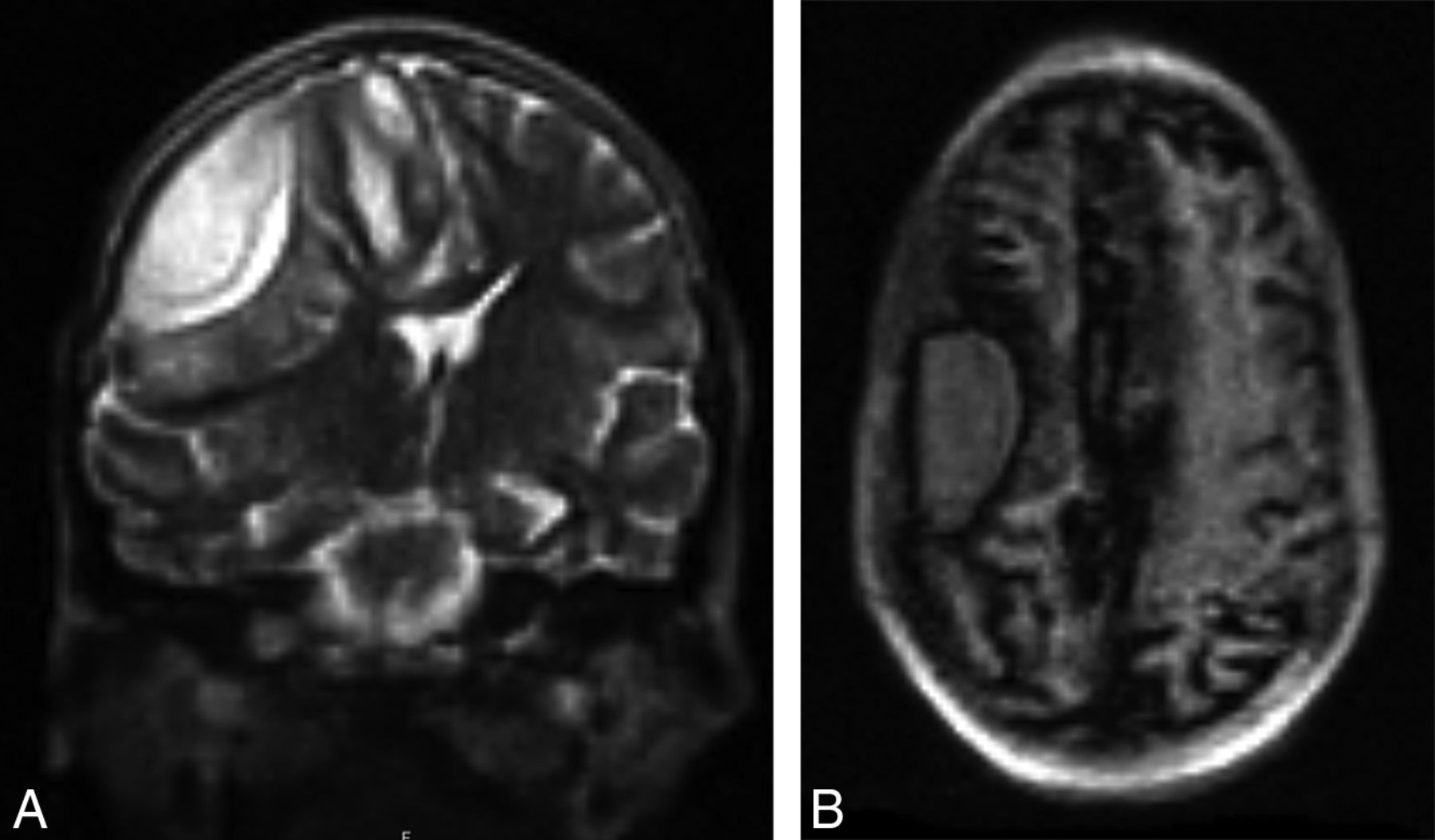
    Hyperfine Swoop MR images of a 6-year-old child who was admitted to our institution under the research study arm with a decreased level of consciousness and malaria parasitemia. The child’s MR imaging demonstrates typical findings associated with malarial encephalopathy of diffuse right-sided parietotemporal T2-weighted hyperintensity in the gray and subcortical white matter with an associated localized mass effect in the form of sulcal effacement on the coronal T2-weighted sequence (A), with corresponding restriction of diffusion on the DWI b=900 image and ADC images (B and C).

  • FIG 3.
    • Download figure
    • Open in new tab
    • Download powerpoint
    FIG 3.

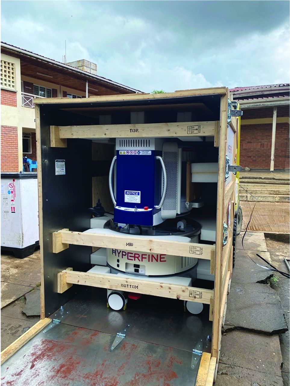
    The Hyperfine Swoop, still crated, at the time of receipt at our institution.

  • FIG 4.
    • Download figure
    • Open in new tab
    • Download powerpoint
    FIG 4.

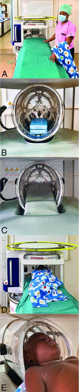
    The Hyperfine Swoop at our institution is set up in an appropriately optimized temperature- and humidity-controlled dedicated scanning room (A) with a patient scanning couch of an appropriate height adjacent to it. B and C, The blue padding block was sourced by our group and is applied within the head coil of the scanner with the gray manufacturer-provided padding applied over it, extending onto the patient’s scanning couch. The role of the blue padding block is to elevate the patient’s head a little further, thereby optimizing the patient’s head position further in the scanner head coil for more optimal head coverage during scanning. A patient model within the scanner (D) demonstrating the swaddling technique and a detailed view of the patient’s head position within the head coil (E).

Tables

  • Figures
  • Challenges encountered and proposed solutions for low-field MR imaging implementation in a resource-limited setting

    ChallengeProposed Solution/Troubleshooting Option
    Equipment delivery and receipt logisticsNeeds assessment of the most reliable and safe equipment transportation routeUse of a reputable carrier with tracking facilitiesInsurance coverageUncrating and setup training before equipment receiptAcquisition of tools needed for uncrating and setup before equipment receiptOn receipt, close inspection of external container gauges for warnings
    Equipment roadmapNeeds assessment of the expected routes of equipment portability within the health care facilityConstruction of low-incline, smooth ramps to mitigate steep and irregular terrain
    Equipment storageNeeds assessment of storage and operating conditions inclusive of temperature and humidity
    Equipment operationTraining of end user operators
    Equipment utilitiesNeeds assessment of hospital infrastructure including electricity supply and Internet speedAcquisition of a manufacturer-recommended compatible electrical surge protector
    Non-resource-constrained specific itemsPatient and equipment safetyPatient privacy and confidentiality
PreviousNext
Back to top
Advertisement
Print
Download PDF
Email Article

Thank you for your interest in spreading the word on American Journal of Neuroradiology.

NOTE: We only request your email address so that the person you are recommending the page to knows that you wanted them to see it, and that it is not junk mail. We do not capture any email address.

Enter multiple addresses on separate lines or separate them with commas.
Implementation of a Low-Field Portable MRI Scanner in a Resource-Constrained Environment: Our Experience in Malawi
(Your Name) has sent you a message from American Journal of Neuroradiology
(Your Name) thought you would like to see the American Journal of Neuroradiology web site.
CAPTCHA
This question is for testing whether or not you are a human visitor and to prevent automated spam submissions.
Cite this article
K. Chetcuti, C. Chilingulo, M.S. Goyal, L. Vidal, N.F. O’Brien, D.G. Postels, K.B. Seydel, T.E. Taylor
Implementation of a Low-Field Portable MRI Scanner in a Resource-Constrained Environment: Our Experience in Malawi
American Journal of Neuroradiology Apr 2022, DOI: 10.3174/ajnr.A7494

Citation Manager Formats

  • BibTeX
  • Bookends
  • EasyBib
  • EndNote (tagged)
  • EndNote 8 (xml)
  • Medlars
  • Mendeley
  • Papers
  • RefWorks Tagged
  • Ref Manager
  • RIS
  • Zotero
0 Responses
Respond to this article
Share
Bookmark this article
Implementation of a Low-Field Portable MRI Scanner in a Resource-Constrained Environment: Our Experience in Malawi
K. Chetcuti, C. Chilingulo, M.S. Goyal, L. Vidal, N.F. O’Brien, D.G. Postels, K.B. Seydel, T.E. Taylor
American Journal of Neuroradiology Apr 2022, DOI: 10.3174/ajnr.A7494
del.icio.us logo Twitter logo Facebook logo Mendeley logo
  • Tweet Widget
  • Facebook Like
  • Google Plus One
Purchase

Jump to section

  • Article
    • Abstract
    • Footnotes
    • References
  • Figures & Data
  • Info & Metrics
  • Responses
  • References
  • PDF

Related Articles

  • No related articles found.
  • PubMed
  • Google Scholar

Cited By...

  • Low-Field (64 mT) Portable MRI for Rapid Point-of-Care Diagnosis of Dissemination in Space in Patients Presenting with Optic Neuritis
  • Ultra-low-field brain MRI morphometry: test-retest reliability and correspondence to high-field MRI
  • MRI-Based Brain Volume Scoring in Cerebral Malaria Is Externally Valid and Applicable to Lower-Resolution Images
  • Bridging the gap: improving correspondence between low-field and high-field magnetic resonance images in young people
  • Crossref
  • Google Scholar

This article has not yet been cited by articles in journals that are participating in Crossref Cited-by Linking.

More in this TOC Section

  • Qualifying Certainty in Radiology Reports through Deep Learning–Based Natural Language Processing
  • Am I Ready to Be an Independent Neuroradiologist? Objective Trends in Neuroradiology Fellows' Performance during the Fellowship Year
  • Displaying Multiphase CT Angiography Using a Time-Variant Color Map: Practical Considerations and Potential Applications in Patients with Acute Stroke
Show more PRACTICE PERSPECTIVES

Similar Articles

Advertisement

Indexed Content

  • Current Issue
  • Accepted Manuscripts
  • Article Preview
  • Past Issues
  • Editorials
  • Editor's Choice
  • Fellows' Journal Club
  • Letters to the Editor
  • Video Articles

Cases

  • Case Collection
  • Archive - Case of the Week
  • Archive - Case of the Month
  • Archive - Classic Case

More from AJNR

  • Trainee Corner
  • Imaging Protocols
  • MRI Safety Corner
  • Book Reviews

Multimedia

  • AJNR Podcasts
  • AJNR Scantastics

Resources

  • Turnaround Time
  • Submit a Manuscript
  • Submit a Video Article
  • Submit an eLetter to the Editor/Response
  • Manuscript Submission Guidelines
  • Statistical Tips
  • Fast Publishing of Accepted Manuscripts
  • Graphical Abstract Preparation
  • Imaging Protocol Submission
  • Evidence-Based Medicine Level Guide
  • Publishing Checklists
  • Author Policies
  • Become a Reviewer/Academy of Reviewers
  • News and Updates

About Us

  • About AJNR
  • Editorial Board
  • Editorial Board Alumni
  • Alerts
  • Permissions
  • Not an AJNR Subscriber? Join Now
  • Advertise with Us
  • Librarian Resources
  • Feedback
  • Terms and Conditions
  • AJNR Editorial Board Alumni

American Society of Neuroradiology

  • Not an ASNR Member? Join Now

© 2025 by the American Society of Neuroradiology All rights, including for text and data mining, AI training, and similar technologies, are reserved.
Print ISSN: 0195-6108 Online ISSN: 1936-959X

Powered by HighWire