Advertisement

Main menu

  • Home
  • Content
    • Current Issue
    • Accepted Manuscripts
    • Article Preview
    • Past Issue Archive
    • Video Articles
    • AJNR Case Collection
    • Case of the Week Archive
    • Case of the Month Archive
    • Classic Case Archive
  • Special Collections
    • AJNR Awards
    • Low-Field MRI
    • Alzheimer Disease
    • ASNR Foundation Special Collection
    • Photon-Counting CT
    • View All
  • Multimedia
    • AJNR Podcasts
    • AJNR SCANtastic
    • Trainee Corner
    • MRI Safety Corner
    • Imaging Protocols
  • For Authors
    • Submit a Manuscript
    • Submit a Video Article
    • Submit an eLetter to the Editor/Response
    • Manuscript Submission Guidelines
    • Statistical Tips
    • Fast Publishing of Accepted Manuscripts
    • Graphical Abstract Preparation
    • Imaging Protocol Submission
    • Author Policies
  • About Us
    • About AJNR
    • Editorial Board
    • Editorial Board Alumni
  • More
    • Become a Reviewer/Academy of Reviewers
    • Subscribers
    • Permissions
    • Alerts
    • Feedback
    • Advertisers
    • ASNR Home

User menu

  • Alerts
  • Log in

Search

  • Advanced search
American Journal of Neuroradiology

American Journal of Neuroradiology

ASHNR American Society of Functional Neuroradiology ASHNR American Society of Pediatric Neuroradiology ASSR
  • Alerts
  • Log in

Advanced Search

  • Home
  • Content
    • Current Issue
    • Accepted Manuscripts
    • Article Preview
    • Past Issue Archive
    • Video Articles
    • AJNR Case Collection
    • Case of the Week Archive
    • Case of the Month Archive
    • Classic Case Archive
  • Special Collections
    • AJNR Awards
    • Low-Field MRI
    • Alzheimer Disease
    • ASNR Foundation Special Collection
    • Photon-Counting CT
    • View All
  • Multimedia
    • AJNR Podcasts
    • AJNR SCANtastic
    • Trainee Corner
    • MRI Safety Corner
    • Imaging Protocols
  • For Authors
    • Submit a Manuscript
    • Submit a Video Article
    • Submit an eLetter to the Editor/Response
    • Manuscript Submission Guidelines
    • Statistical Tips
    • Fast Publishing of Accepted Manuscripts
    • Graphical Abstract Preparation
    • Imaging Protocol Submission
    • Author Policies
  • About Us
    • About AJNR
    • Editorial Board
    • Editorial Board Alumni
  • More
    • Become a Reviewer/Academy of Reviewers
    • Subscribers
    • Permissions
    • Alerts
    • Feedback
    • Advertisers
    • ASNR Home
  • Follow AJNR on Twitter
  • Visit AJNR on Facebook
  • Follow AJNR on Instagram
  • Join AJNR on LinkedIn
  • RSS Feeds

AJNR Awards, New Junior Editors, and more. Read the latest AJNR updates

Review ArticleReview Articles
Open Access

Anatomy of Spinal Venous Drainage for the Neurointerventionalist: From Puncture Site to Intervertebral Foramen

N. Borg, J. Cutsforth-Gregory, S. Oushy, T. Huynh, L.E. Savastano, H.J. Cloft, G. Lanzino and W. Brinjikji
American Journal of Neuroradiology January 2022, DOI: https://doi.org/10.3174/ajnr.A7409
N. Borg
aFrom the Departments of Neurologic Surgery (N.B., S.O., L.E.S., G.L.)
  • Find this author on Google Scholar
  • Find this author on PubMed
  • Search for this author on this site
  • ORCID record for N. Borg
J. Cutsforth-Gregory
bNeurology (J.C.-G.)
  • Find this author on Google Scholar
  • Find this author on PubMed
  • Search for this author on this site
  • ORCID record for J. Cutsforth-Gregory
S. Oushy
aFrom the Departments of Neurologic Surgery (N.B., S.O., L.E.S., G.L.)
  • Find this author on Google Scholar
  • Find this author on PubMed
  • Search for this author on this site
  • ORCID record for S. Oushy
T. Huynh
dDepartment of Neurologic Surgery (T.H.), Mayo Clinic, Jacksonville, Florida
  • Find this author on Google Scholar
  • Find this author on PubMed
  • Search for this author on this site
  • ORCID record for T. Huynh
L.E. Savastano
aFrom the Departments of Neurologic Surgery (N.B., S.O., L.E.S., G.L.)
  • Find this author on Google Scholar
  • Find this author on PubMed
  • Search for this author on this site
  • ORCID record for L.E. Savastano
H.J. Cloft
cRadiology (H.J.C., W.B.), Mayo Clinic, Rochester, Minnesota
  • Find this author on Google Scholar
  • Find this author on PubMed
  • Search for this author on this site
  • ORCID record for H.J. Cloft
G. Lanzino
aFrom the Departments of Neurologic Surgery (N.B., S.O., L.E.S., G.L.)
  • Find this author on Google Scholar
  • Find this author on PubMed
  • Search for this author on this site
  • ORCID record for G. Lanzino
W. Brinjikji
cRadiology (H.J.C., W.B.), Mayo Clinic, Rochester, Minnesota
  • Find this author on Google Scholar
  • Find this author on PubMed
  • Search for this author on this site
  • ORCID record for W. Brinjikji
  • Article
  • Figures & Data
  • Info & Metrics
  • Responses
  • References
  • PDF
Loading

Article Figures & Data

Figures

  • FIG 1.
    • Download figure
    • Open in new tab
    • Download powerpoint
    FIG 1.

    The site of the CSF leak in the patients with CSF-venous fistula is the nerve root foramen. The nerve root is covered with a dural sleeve, which, in turn, is surrounded by a plexus of veins that forms the foraminal vein. This joins the segmental vein above to form the paraspinal vein. There may be anastomotic veins running perpendicular to the segmental veins that provide a direct connection between adjacent levels. v indicates vein; vv, veins.

  • FIG 2.
    • Download figure
    • Open in new tab
    • Download powerpoint
    FIG 2.

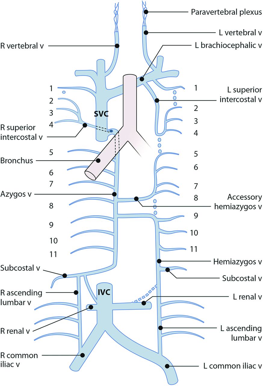
    The venous drainage of the spine can be separated into 3 compartments, from medial to lateral. The intradural compartment is further subdivided into intrinsic and extrinsic components. The extradural compartment refers to the epidural venous plexus, also referred to as the internal vertebral plexus. The paraspinal compartment is also called the external vertebral plexus. Efferent pathways are arranged in a segmental fashion, carrying venous blood toward the great veins and on to the right atrium.

  • FIG 3.
    • Download figure
    • Open in new tab
    • Download powerpoint
    FIG 3.

    A–C, The epidural plexus is richest in 2 anterolateral and 2 posterolateral columns. The 4 columns are joined by circumferential rings surrounding the thecal sac at the midlevel of each vertebral body. When viewed from anterior to posterior, the 4 columns, rings, and foraminal veins form a characteristic hexagonal shape. D, The composite view of the anterior and posterior columns on a frontal view is referred to as the lateral epidural plexus.

  • FIG 4.
    • Download figure
    • Open in new tab
    • Download powerpoint
    FIG 4.

    A, Coronal view in the cervical region showing that the rich plexus adjacent to the vertebral bodies is anatomically continuous and functionally analogous to the epidural plexus. Inferiorly, the paravertebral plexus condenses into the vertebral veins. B and C, Vertebral venograms rapidly opacify the paravertebral plexus, foraminal veins, and epidural plexus. Filling defects just adjacent to the vertebral bodies represent the pedicles. The more lateral filling defects represent the vertebral artery running through transverse foramina. Between adjacent pedicles is the foraminal vein. Lateral to the pedicle is the paravertebral plexus; medial to it is the epidural plexus, with its characteristic hexagonal shape. v indicates vein; vv, veins.
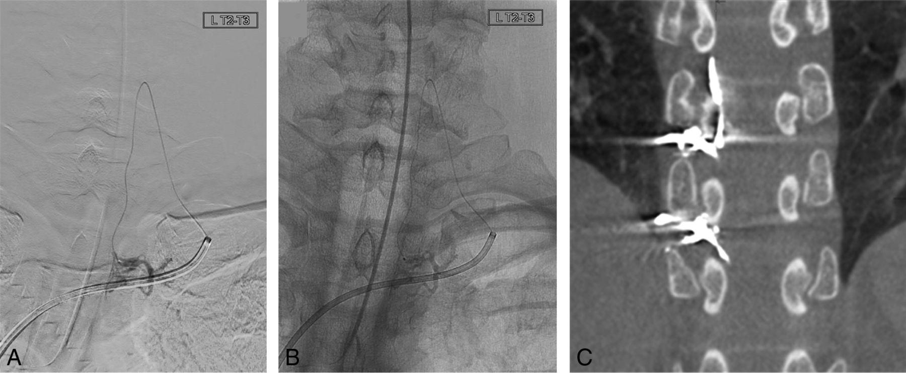
  • FIG 5.
    • Download figure
    • Open in new tab
    • Download powerpoint
    FIG 5.

    Examples of the appearance of the lateral epidural space at different levels. Subtracted (A) and unsubtracted (B) microcatheter venography performed at the left T2 lateral epidural plexus fills the foraminal veins. In this case, the guide catheter is in the left brachiocephalic vein. The microcatheter enters the left C6 foramen via the vertebral vein, before forming a hairpin turn and traveling caudally across 3 levels through the lateral epidural plexus. C, Postembolization conebeam CT in a different patient shows the embolic agent within the right T8 and T9 foraminal veins and the adjacent lateral epidural plexus.

  • FIG 6.
    • Download figure
    • Open in new tab
    • Download powerpoint
    FIG 6.

    Overview of the azygos system and its main tributaries. The right mainstem bronchus is included as a landmark for the arch of the azygos vein. R indicates right; L, left; v, vein.

  • FIG 7.
    • Download figure
    • Open in new tab
    • Download powerpoint
    FIG 7.

    Subtracted (A) and unsubtracted (B) views of azygos venography. To the right of the vertebral bodies, the guide catheter is seen to ascend from the IVC to the SVC, before coursing backward into the azygos vein. The trachea and carina are seen in relation to the arch of the azygos vein. Numerous venous stumps (black arrows) along the azygos vein represent reflux into paraspinal veins at each level. In this image, an embolic agent cast is seen filling the right superior intercostal vein (white arrow), which drains into the superior aspect of the arch of the azygos. There is also some opacification of 2 bronchial veins (asterisks).

  • FIG 8.
    • Download figure
    • Open in new tab
    • Download powerpoint
    FIG 8.

    Subtracted (A) and unsubtracted (B) images showing a 6F guide catheter crossing the midline from the azygos to the hemiazygos vein at the level of T9. Also apparent is the hexagonal shape of the epidural plexus, with a central filling defect at the level of the intervertebral disc. The subtracted image below shows the corresponding bony landmarks.

  • FIG 9.
    • Download figure
    • Open in new tab
    • Download powerpoint
    FIG 9.

    Venography performed with a catheter in the superior aspect of the left internal jugular vein (larger and more lateral, black arrow) shows contrast draining via the condylar vein (asterisk) into the vertebral vein (medial and smaller, white arrow), which inferiorly drains into the junction of the internal jugular vein with the left brachiocephalic vein. Along its course, multiple tributaries drain into the vertebral vein from the paravertebral plexus and foraminal veins. A catheter and wire are angled medially and superiorly from the junction of the brachiocephalic vein with the internal jugular vein to select the vertebral vein from below.

  • FIG 10.
    • Download figure
    • Open in new tab
    • Download powerpoint
    FIG 10.

    Roadmaps to each foramen, grouped by common venous drainage pathways. A, Right T5–12. B, Right lumbar. C, Left T9–12 and lumbar. D, Left T5–8. E, Right T1–4. F, Left T1–4. G, Cervical.

  • FIG 11.
    • Download figure
    • Open in new tab
    • Download powerpoint
    FIG 11.

    Postembolization image showing the liquid embolic agent in the accessory hemiazygos vein (white arrow) in a patient with multiple left-upper-thoracic CSF-venous fistulas. The catheter is seen to ascend in the SVC, course around the arch down the azygos vein (black arrow), and then cross the midline (asterisk) to enter the accessory hemiazygos vein.

PreviousNext
Back to top
Advertisement
Print
Download PDF
Email Article

Thank you for your interest in spreading the word on American Journal of Neuroradiology.

NOTE: We only request your email address so that the person you are recommending the page to knows that you wanted them to see it, and that it is not junk mail. We do not capture any email address.

Enter multiple addresses on separate lines or separate them with commas.
Anatomy of Spinal Venous Drainage for the Neurointerventionalist: From Puncture Site to Intervertebral Foramen
(Your Name) has sent you a message from American Journal of Neuroradiology
(Your Name) thought you would like to see the American Journal of Neuroradiology web site.
CAPTCHA
This question is for testing whether or not you are a human visitor and to prevent automated spam submissions.
Cite this article
N. Borg, J. Cutsforth-Gregory, S. Oushy, T. Huynh, L.E. Savastano, H.J. Cloft, G. Lanzino, W. Brinjikji
Anatomy of Spinal Venous Drainage for the Neurointerventionalist: From Puncture Site to Intervertebral Foramen
American Journal of Neuroradiology Jan 2022, DOI: 10.3174/ajnr.A7409

Citation Manager Formats

  • BibTeX
  • Bookends
  • EasyBib
  • EndNote (tagged)
  • EndNote 8 (xml)
  • Medlars
  • Mendeley
  • Papers
  • RefWorks Tagged
  • Ref Manager
  • RIS
  • Zotero
0 Responses
Respond to this article
Share
Bookmark this article
Anatomy of Spinal Venous Drainage for the Neurointerventionalist: From Puncture Site to Intervertebral Foramen
N. Borg, J. Cutsforth-Gregory, S. Oushy, T. Huynh, L.E. Savastano, H.J. Cloft, G. Lanzino, W. Brinjikji
American Journal of Neuroradiology Jan 2022, DOI: 10.3174/ajnr.A7409
del.icio.us logo Twitter logo Facebook logo Mendeley logo
  • Tweet Widget
  • Facebook Like
  • Google Plus One
Purchase

Jump to section

  • Article
    • Abstract
    • ABBREVIATIONS:
    • CONCLUSIONS
    • Footnotes
    • References
  • Figures & Data
  • Info & Metrics
  • Responses
  • References
  • PDF

Related Articles

  • No related articles found.
  • PubMed
  • Google Scholar

Cited By...

  • Azygos Vein Stenosis in Frontotemporal Dementia Sagging Brain Syndrome
  • Spinal CSF Leaks: The Neuroradiologist Transforming Care
  • Temporal Characteristics of CSF-Venous Fistulas on Dynamic Decubitus CT Myelography: A Retrospective Multi-Institution Cohort Study
  • Transvenous embolization of cerebrospinal fluid-venous fistulas: Independent validation and feasibility of upper-extremity approach and using dual-microcatheter and balloon pressure cooker technique
  • Resisted Inspiration Improves Visualization of CSF-Venous Fistulas in Spontaneous Intracranial Hypotension
  • Dual microcatheter and coil/balloon pressure cooker technique for transvenous embolization of cerebrospinal fluid-venous fistulas
  • Conebeam CT as an Adjunct to Digital Subtraction Myelography for Detection of CSF-Venous Fistulas
  • Dual microcatheter and coil/balloon pressure cooker technique for transvenous embolization of cerebrospinal fluid-venous fistulas
  • Crossref
  • Google Scholar

This article has not yet been cited by articles in journals that are participating in Crossref Cited-by Linking.

More in this TOC Section

  • An Atlas of Neonatal Neurovascular Imaging Anatomy as Depicted with Microvascular Imaging: The Intracranial Arteries
  • An Atlas of Neonatal Neurovascular Imaging Anatomy as Depicted with Microvascular Imaging: The Intracranial Veins
  • Clinical Translation of Hyperpolarized 13C Metabolic Probes for Glioma Imaging
Show more Review Articles

Similar Articles

Advertisement

Indexed Content

  • Current Issue
  • Accepted Manuscripts
  • Article Preview
  • Past Issues
  • Editorials
  • Editor's Choice
  • Fellows' Journal Club
  • Letters to the Editor
  • Video Articles

Cases

  • Case Collection
  • Archive - Case of the Week
  • Archive - Case of the Month
  • Archive - Classic Case

More from AJNR

  • Trainee Corner
  • Imaging Protocols
  • MRI Safety Corner
  • Book Reviews

Multimedia

  • AJNR Podcasts
  • AJNR Scantastics

Resources

  • Turnaround Time
  • Submit a Manuscript
  • Submit a Video Article
  • Submit an eLetter to the Editor/Response
  • Manuscript Submission Guidelines
  • Statistical Tips
  • Fast Publishing of Accepted Manuscripts
  • Graphical Abstract Preparation
  • Imaging Protocol Submission
  • Evidence-Based Medicine Level Guide
  • Publishing Checklists
  • Author Policies
  • Become a Reviewer/Academy of Reviewers
  • News and Updates

About Us

  • About AJNR
  • Editorial Board
  • Editorial Board Alumni
  • Alerts
  • Permissions
  • Not an AJNR Subscriber? Join Now
  • Advertise with Us
  • Librarian Resources
  • Feedback
  • Terms and Conditions
  • AJNR Editorial Board Alumni

American Society of Neuroradiology

  • Not an ASNR Member? Join Now

© 2025 by the American Society of Neuroradiology All rights, including for text and data mining, AI training, and similar technologies, are reserved.
Print ISSN: 0195-6108 Online ISSN: 1936-959X

Powered by HighWire