Advertisement

Main menu

  • Home
  • Content
    • Current Issue
    • Accepted Manuscripts
    • Article Preview
    • Past Issue Archive
    • Video Articles
    • AJNR Case Collection
    • Case of the Week Archive
    • Case of the Month Archive
    • Classic Case Archive
  • Special Collections
    • Advancing NeuroMRI with High-Relaxivity Contrast Agents
    • AJNR Awards
    • Low-Field MRI
    • Alzheimer Disease
    • ASNR Foundation Special Collection
    • Photon-Counting CT
    • View All
  • Multimedia
    • AJNR Podcasts
    • AJNR SCANtastic
    • Trainee Corner
    • MRI Safety Corner
    • Imaging Protocols
  • For Authors
    • Submit a Manuscript
    • Submit a Video Article
    • Submit an eLetter to the Editor/Response
    • Manuscript Submission Guidelines
    • Statistical Tips
    • Fast Publishing of Accepted Manuscripts
    • Graphical Abstract Preparation
    • Imaging Protocol Submission
    • Author Policies
  • About Us
    • About AJNR
    • Editorial Board
    • Editorial Board Alumni
  • More
    • Become a Reviewer/Academy of Reviewers
    • Subscribers
    • Permissions
    • Alerts
    • Feedback
    • Advertisers
    • ASNR Home

User menu

  • Alerts
  • Log in

Search

  • Advanced search
American Journal of Neuroradiology

American Journal of Neuroradiology

ASHNR American Society of Functional Neuroradiology ASHNR American Society of Pediatric Neuroradiology ASSR
  • Alerts
  • Log in

Advanced Search

  • Home
  • Content
    • Current Issue
    • Accepted Manuscripts
    • Article Preview
    • Past Issue Archive
    • Video Articles
    • AJNR Case Collection
    • Case of the Week Archive
    • Case of the Month Archive
    • Classic Case Archive
  • Special Collections
    • Advancing NeuroMRI with High-Relaxivity Contrast Agents
    • AJNR Awards
    • Low-Field MRI
    • Alzheimer Disease
    • ASNR Foundation Special Collection
    • Photon-Counting CT
    • View All
  • Multimedia
    • AJNR Podcasts
    • AJNR SCANtastic
    • Trainee Corner
    • MRI Safety Corner
    • Imaging Protocols
  • For Authors
    • Submit a Manuscript
    • Submit a Video Article
    • Submit an eLetter to the Editor/Response
    • Manuscript Submission Guidelines
    • Statistical Tips
    • Fast Publishing of Accepted Manuscripts
    • Graphical Abstract Preparation
    • Imaging Protocol Submission
    • Author Policies
  • About Us
    • About AJNR
    • Editorial Board
    • Editorial Board Alumni
  • More
    • Become a Reviewer/Academy of Reviewers
    • Subscribers
    • Permissions
    • Alerts
    • Feedback
    • Advertisers
    • ASNR Home
  • Follow AJNR on Twitter
  • Visit AJNR on Facebook
  • Follow AJNR on Instagram
  • Join AJNR on LinkedIn
  • RSS Feeds

AJNR Awards, New Junior Editors, and more. Read the latest AJNR updates


Improved Turnaround Times | Median time to first decision: 12 days

Research ArticleAdult Brain

Hypovascular Cellular Tumor in Primary Central Nervous System Lymphoma is Associated with Treatment Resistance: Tumor Habitat Analysis Using Physiologic MRI

S.Y. Jeong, J.E. Park, N. Kim and H.S. Kim
American Journal of Neuroradiology November 2021, DOI: https://doi.org/10.3174/ajnr.A7351
S.Y. Jeong
aFrom the Department of Radiology and Research Institute of Radiology (S.Y.J., J.E.P., H.S.K.), University of Ulsan College of Medicine, Asan Medical Center, Seoul, Republic of Korea
  • Find this author on Google Scholar
  • Find this author on PubMed
  • Search for this author on this site
  • ORCID record for S.Y. Jeong
J.E. Park
aFrom the Department of Radiology and Research Institute of Radiology (S.Y.J., J.E.P., H.S.K.), University of Ulsan College of Medicine, Asan Medical Center, Seoul, Republic of Korea
  • Find this author on Google Scholar
  • Find this author on PubMed
  • Search for this author on this site
  • ORCID record for J.E. Park
N. Kim
bJAPEX LLC (N.K.), Seoul, Korea.
  • Find this author on Google Scholar
  • Find this author on PubMed
  • Search for this author on this site
  • ORCID record for N. Kim
H.S. Kim
aFrom the Department of Radiology and Research Institute of Radiology (S.Y.J., J.E.P., H.S.K.), University of Ulsan College of Medicine, Asan Medical Center, Seoul, Republic of Korea
  • Find this author on Google Scholar
  • Find this author on PubMed
  • Search for this author on this site
  • ORCID record for H.S. Kim
  • Article
  • Figures & Data
  • Supplemental
  • Info & Metrics
  • Responses
  • References
  • PDF
Loading

Article Figures & Data

Figures

  • Tables
  • FIG 1.
    • Download figure
    • Open in new tab
    • Download powerpoint
    FIG 1.

    Flow chart of patient recruitment.

  • FIG 2.
    • Download figure
    • Open in new tab
    • Download powerpoint
    FIG 2.

    The overall process of the deep learning segmentation and tumor habitat analysis. A, Image acquisition, registration, and deep learning segmentation for a contrast-enhancing lesion (CEL). B, Extraction of ADC and nCBV values from the CEL and voxel classifications based on ADC and nCBV values. The individual voxels in each cluster are grouped according to their similarities and differences using a K-means clustering algorithm. C, The cluster number is set to 3 to depict 3 different habitats according to the combinations of ADC and nCBV parameters: hypervascular cellular, hypovascular cellular, and hypovascular hypocellular. D, Voxels are shown as spatial habitats in the original image space. Associations of pretreatment tumor habitats with TTP were analyzed.

  • FIG 3.
    • Download figure
    • Open in new tab
    • Download powerpoint
    FIG 3.

    A, Demonstration of the 3 spatial habitats defined by clustering of voxels using normalized ADC and nCBV maps in a 53-year-old male patient. The hypervascular cellular habitat (red) shows high nCBV and low ADC, the hypovascular cellular habitat (green) shows low nCBV and low ADC, and the hypovascular hypocellular habitat (blue) shows low nCBV and high ADC. The tumor exhibits a large hypovascular cellular habitat (green), and a persistent enhancing mass was associated with a short TTP after initial chemotherapy. B, Demonstration of the 3 spatial habitats defined by clustering of voxels using normalized ADC and nCBV maps in a 57-year-old male patient. The hypervascular cellular habitat (red) shows high nCBV and low ADC, the hypovascular cellular habitat (green) shows low nCBV and low ADC, and the hypovascular hypocellular habitat (blue) shows low nCBV and high ADC. The tumor has a small hypovascular cellular habitat (green) and showed a complete response at 53 days after initial chemotherapy.
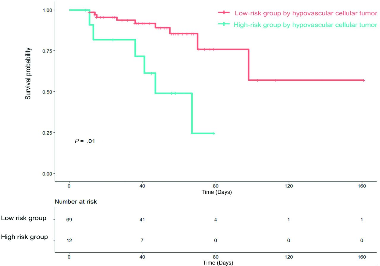
  • FIG 4.
    • Download figure
    • Open in new tab
    • Download powerpoint
    FIG 4.

    Kaplan-Meier analysis of TTP in patients with PCNSL stratified by hypovascular cellular habitat (log-rank, P = .01).

Tables

  • Figures
    • View popup
    Table 1:

    Patients and imaging characteristics of the PCNSLs included in this study

    Clinical Characteristics (n = 81)
    Mean age (yr)61.6 (SD, 11.8)
    Sex
     Male/female38/43 (46.9%/53.1%)
    ECOG performance status at diagnosis (case No) (%)
     171 (87.7%)
     22 (2.5%)
     36 (7.4%)
     42 (2.5%)
    Mean serum LDH level225.9 (SD, 59.9)
    Mean CSF-total protein107.0 (SD, 103.0)
    Initial treatment response (case No) (%)
     Treatment response (CR and PR)64 (79.0%)
     Treatment failure (SD and PD)17 (21.0%)
    Imaging characteristics (case No) (%)
     Location
      Deepa69 (85.2%)
      Hemisphere12 (14.8%)
     Atypical findingsb (case No) (%)
      Positive14 (17.3%)
      Negative67 (82.7%)
    • Note:—CR, complete response; PR, partial response; PD, progressive disease; SD, stable disease.

    • ↵a Deep location: thalamus, basal ganglia, corpus callosum, periventricular area, cerebellum.

    • ↵b Atypical findings: presence of hemorrhage or necrosis.

    • View popup
    Table 2:

    Clinical and imaging predictors associated with TTP in PCNSL

    VariableHazard Ratio95% CIP Value
    Age0.980.94–1.03.55
    Atypical findinga4.411.32–14.71.016
    CSF-total protein1.000.99–1.00.70
    ECOG
     2 (reference)
     3 or 45.821.07–31.66.04
    Serum LDH level1.011.00–1.02.013
    Deep locationb1.750.33–9.08.50
    • ↵a Atypical findings: presence of hemorrhage or necrosis.

    • ↵b Deep location: thalamus, basal ganglia, corpus callosum, periventricular area, cerebellum.

    • View popup
    Table 3:

    Exploratory analysis of spatial habitats for predicting TTP in patients with PCNSL

    Spatial Tumor HabitatsTTP
    Hazard Ratioa95% CIP Value
    No. of voxels (20,000 voxels)
     Hypervascular cellular habitat1.390.29–6.44.29
     Hypovascular cellular habitat2.831.20–6.65.017
     Hypovascular hypocelluar habitat2.030.58–7.15.27
    Voxel fraction (%)
     Hypervascular cellular habitat0.880.04–20.97.83
     Hypovascular cellular habitat2.070.29–14.83.46
     Hypovascular hypocelluar habitat0.460.05–4.01.48
    ADC0.980.95–1.01.16
    CBV0.800.45–1.32.38
    • ↵a Hazard ratios reported here indicate the relative change in hazard that a 10-unit (20,000 voxels) increase in each imaging parameter incurs.

    • View popup
    Table 4:

    Prediction of TTP in PCNSLs according to tumor habitat, ADC, and CBVa

    Combination of Clinical, Conventional Imaging Predictors, and Tumor HabitatsCombined Clinical and Conventional PredictorsTumor HabitatsClinical Predictors OnlyConventional Imaging Predictors Only
    C-index0.730.710.650.680.63
    95% CI0.67–0.800.54–0.780.52–0.780.54–0.820.50–0.76
    P valueReference.01.012.81.62
    • ↵a Combined clinical predictors were age, ECOG score, and mean serum LDH level; the conventional imaging predictor was the presence of atypical image findings. P value refers to the significance in the difference of the C-indices between the combined model and the single model assessed using “CompareC” (https://cran.r-project.org/web/packages/compareC/index.html) in the R statistical and computing software.

PreviousNext
Back to top
Advertisement
Print
Download PDF
Email Article

Thank you for your interest in spreading the word on American Journal of Neuroradiology.

NOTE: We only request your email address so that the person you are recommending the page to knows that you wanted them to see it, and that it is not junk mail. We do not capture any email address.

Enter multiple addresses on separate lines or separate them with commas.
Hypovascular Cellular Tumor in Primary Central Nervous System Lymphoma is Associated with Treatment Resistance: Tumor Habitat Analysis Using Physiologic MRI
(Your Name) has sent you a message from American Journal of Neuroradiology
(Your Name) thought you would like to see the American Journal of Neuroradiology web site.
CAPTCHA
This question is for testing whether or not you are a human visitor and to prevent automated spam submissions.
Cite this article
S.Y. Jeong, J.E. Park, N. Kim, H.S. Kim
Hypovascular Cellular Tumor in Primary Central Nervous System Lymphoma is Associated with Treatment Resistance: Tumor Habitat Analysis Using Physiologic MRI
American Journal of Neuroradiology Nov 2021, DOI: 10.3174/ajnr.A7351

Citation Manager Formats

  • BibTeX
  • Bookends
  • EasyBib
  • EndNote (tagged)
  • EndNote 8 (xml)
  • Medlars
  • Mendeley
  • Papers
  • RefWorks Tagged
  • Ref Manager
  • RIS
  • Zotero
0 Responses
Respond to this article
Share
Bookmark this article
Hypovascular Cellular Tumor in Primary Central Nervous System Lymphoma is Associated with Treatment Resistance: Tumor Habitat Analysis Using Physiologic MRI
S.Y. Jeong, J.E. Park, N. Kim, H.S. Kim
American Journal of Neuroradiology Nov 2021, DOI: 10.3174/ajnr.A7351
del.icio.us logo Twitter logo Facebook logo Mendeley logo
  • Tweet Widget
  • Facebook Like
  • Google Plus One
Purchase

Jump to section

  • Article
    • Abstract
    • ABBREVIATIONS:
    • MATERIALS AND METHODS
    • RESULTS
    • DISCUSSION
    • CONCLUSIONS
    • ACKNOWLEDGMENT
    • Footnotes
    • References
  • Figures & Data
  • Supplemental
  • Info & Metrics
  • Responses
  • References
  • PDF

Related Articles

  • PubMed
  • Google Scholar

Cited By...

  • No citing articles found.
  • Crossref
  • Google Scholar

This article has not yet been cited by articles in journals that are participating in Crossref Cited-by Linking.

More in this TOC Section

  • Diagnostic Neuroradiology of Monoclonal Antibodies
  • Cerebral ADC Changes in Fabry Disease
  • ML for Glioma Molecular Subtype Prediction
Show more Adult Brain

Similar Articles

Advertisement

Indexed Content

  • Current Issue
  • Accepted Manuscripts
  • Article Preview
  • Past Issues
  • Editorials
  • Editor's Choice
  • Fellows' Journal Club
  • Letters to the Editor
  • Video Articles

Cases

  • Case Collection
  • Archive - Case of the Week
  • Archive - Case of the Month
  • Archive - Classic Case

More from AJNR

  • Trainee Corner
  • Imaging Protocols
  • MRI Safety Corner
  • Book Reviews

Multimedia

  • AJNR Podcasts
  • AJNR Scantastics

Resources

  • Turnaround Time
  • Submit a Manuscript
  • Submit a Video Article
  • Submit an eLetter to the Editor/Response
  • Manuscript Submission Guidelines
  • Statistical Tips
  • Fast Publishing of Accepted Manuscripts
  • Graphical Abstract Preparation
  • Imaging Protocol Submission
  • Evidence-Based Medicine Level Guide
  • Publishing Checklists
  • Author Policies
  • Become a Reviewer/Academy of Reviewers
  • News and Updates

About Us

  • About AJNR
  • Editorial Board
  • Editorial Board Alumni
  • Alerts
  • Permissions
  • Not an AJNR Subscriber? Join Now
  • Advertise with Us
  • Librarian Resources
  • Feedback
  • Terms and Conditions
  • AJNR Editorial Board Alumni

American Society of Neuroradiology

  • Not an ASNR Member? Join Now

© 2025 by the American Society of Neuroradiology All rights, including for text and data mining, AI training, and similar technologies, are reserved.
Print ISSN: 0195-6108 Online ISSN: 1936-959X

Powered by HighWire