
Article Figures & Data
Figures
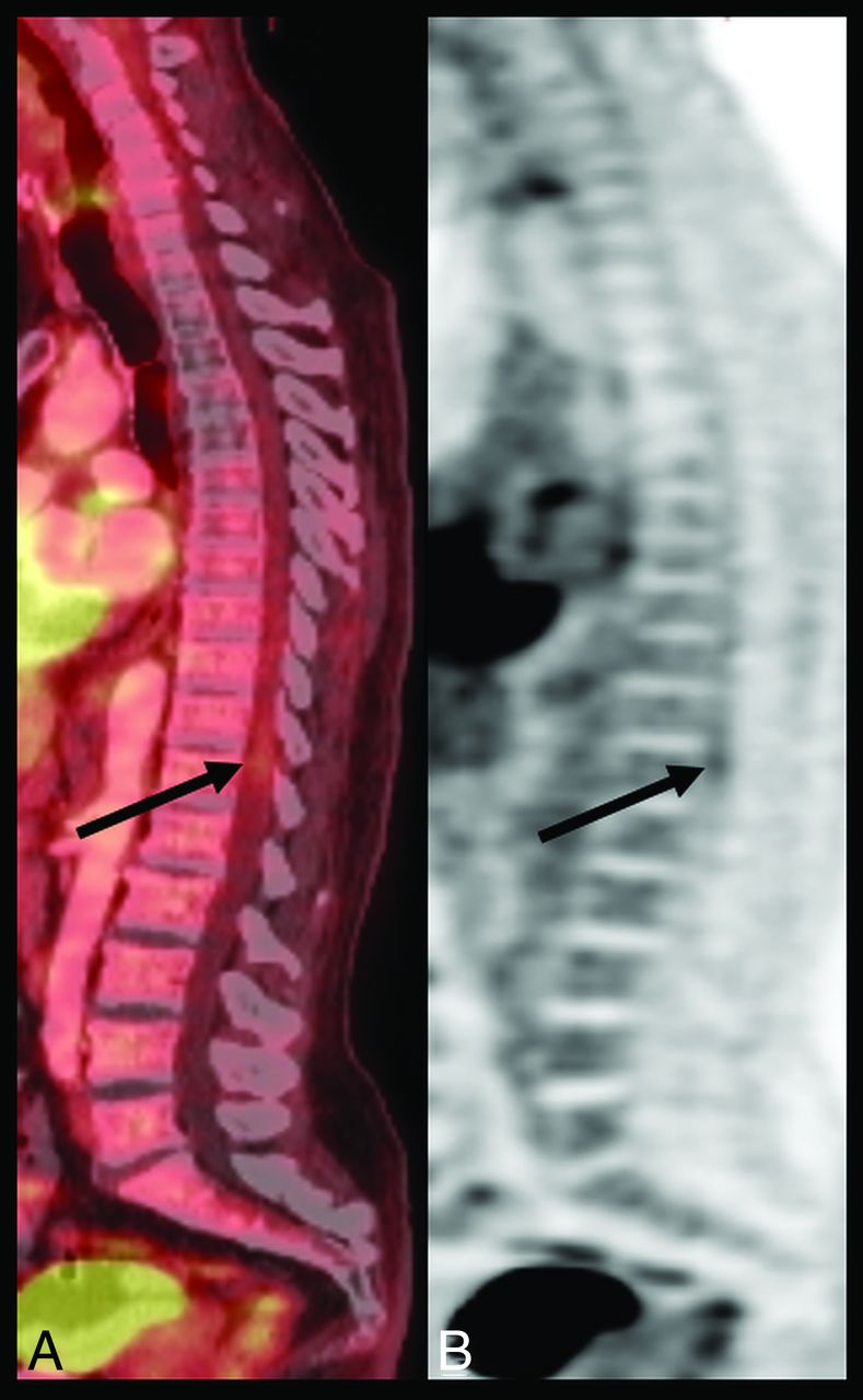
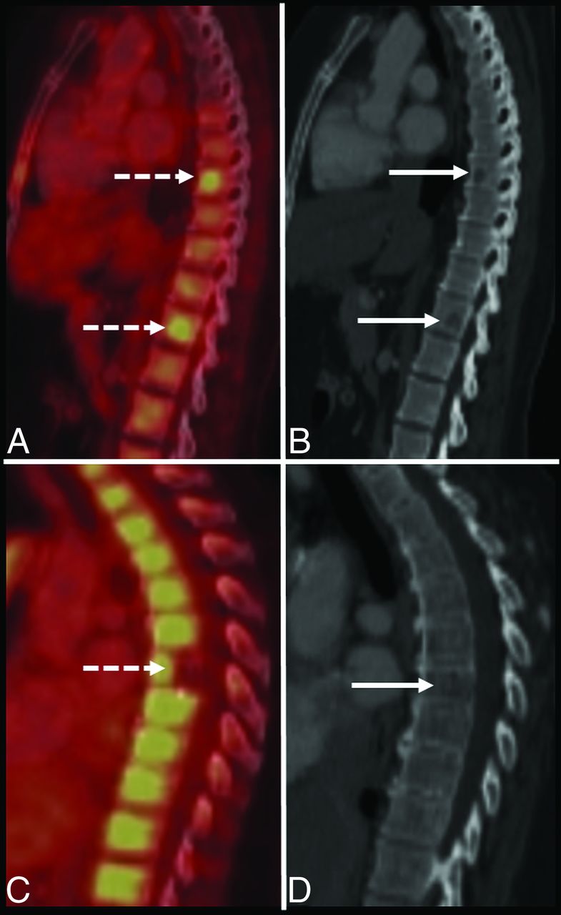
- FIG 1.
A 59-year-old man with lung cancer without metastatic disease. Sagittal fused (A) and AC PET (B) images demonstrate physiologic [18F]FDG uptake throughout the spine as well as focal physiologic [18F]FDG spinal cord uptake at T11–T12 (arrows). Absent uptake in the midthoracic spine is related to previous radiation therapy. Fused indicates fused PET and CT image; AC, attenuation-corrected.
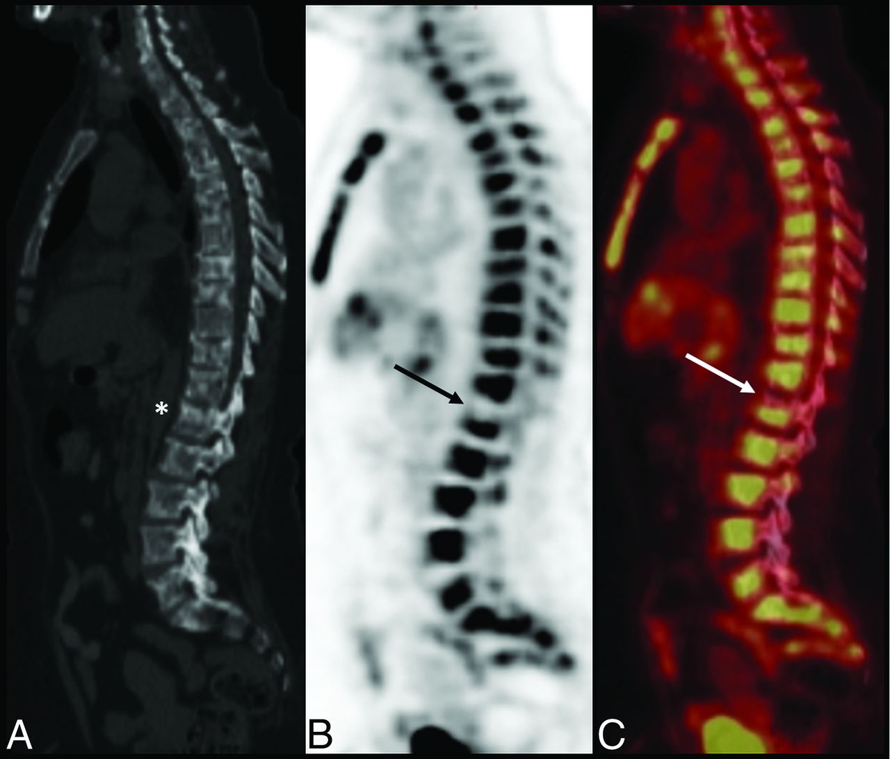
- FIG 2.
A 62-year-old woman with breast cancer. Sagittal CT (A), AC PET (B), and fused (C) images demonstrate a sclerotic (asterisk) and photopenic region (solid arrows) in the L1 vertebral body consistent with a site of treated metastasis. Note the diffusely increased radiotracer uptake through the remaining axial skeleton, which obscures multilevel osseous metastases seen on CT. Fused indicates fused PET and CT image; AC, attenuation-corrected.
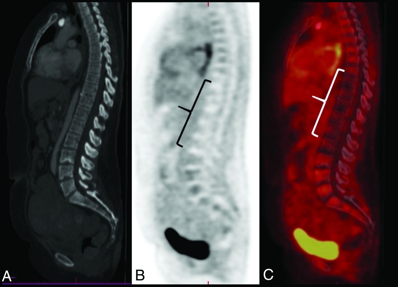
- FIG 3.
A 48-year-old man with B-cell lymphoma of the gastric fundus after radiation therapy. Sagittal CT (A), AC PET (B), and fused (C) images demonstrate a large photopenic segment (bracket) in the radiation field. Note the absence of a correlative abnormality on CT in this region. Fused indicates fused PET and CT image; AC, attenuation-corrected.
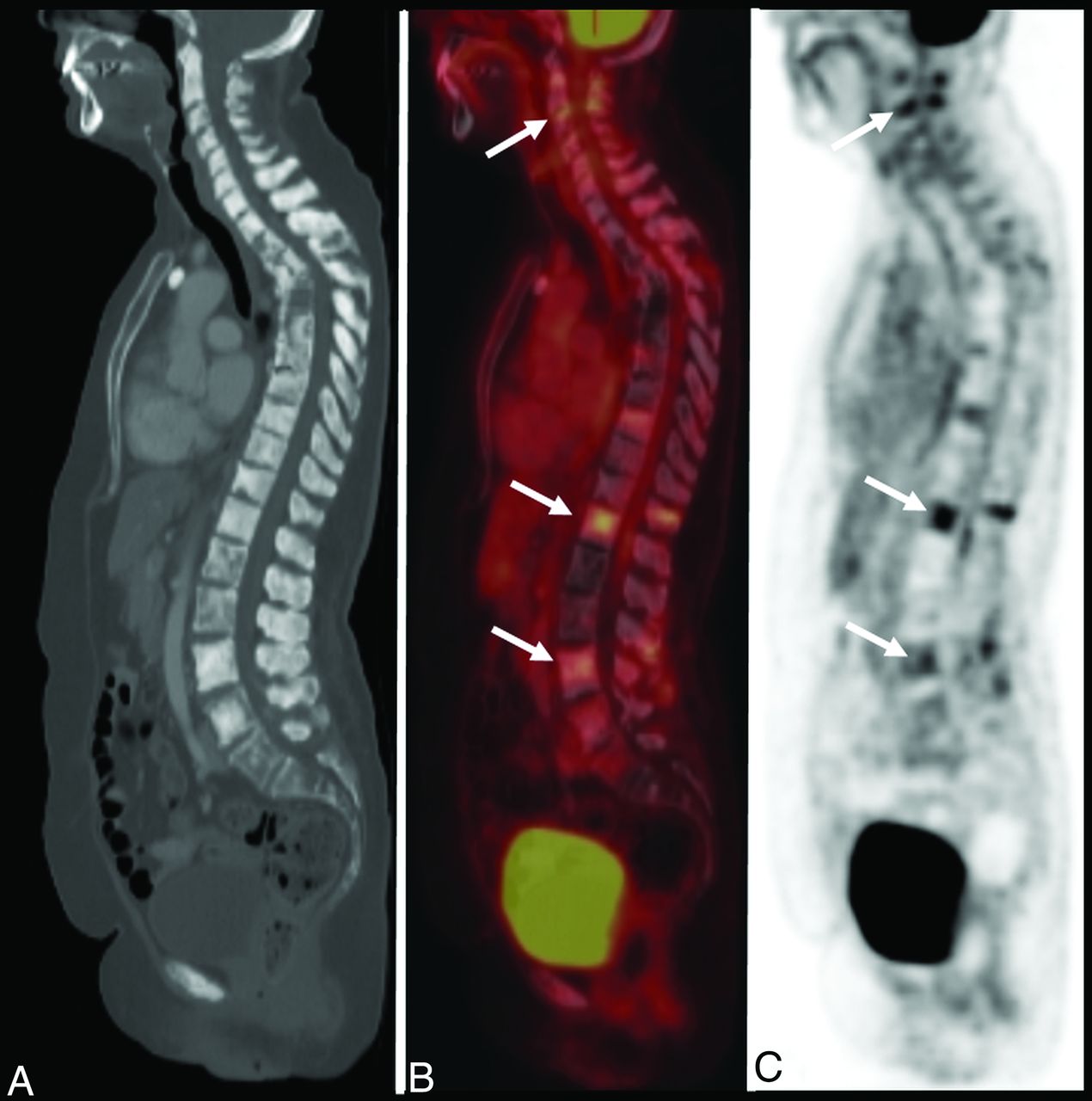
- FIG 4.
A 66-year-old woman with breast cancer. Sagittal CT (A), fused (B), and AC PET (C) images demonstrate extensive osseous metastases throughout the spine with a number of lesions demonstrating varying degrees of increased uptake (arrows). Heterogeneous radiotracer uptake is due to posttreatment changes. Absent radiotracer uptake suggests treated disease including in sclerotic vertebral bodies. This case demonstrates the superiority of PET/CT over conventional imaging in demonstrating a response to therapy. Fused indicates fused PET and CT image; AC, attenuation-corrected.
- FIG 5.
An 80-year-old woman with mucosa-associated lymphatic tissue lymphoma. Axial fused (A), sagittal fused (B), and sagittal T2-weighted MR images (C) demonstrate an intensely hypermetabolic focus within the spinal column at T7–T8 (dashed arrow), which corresponds to a T2-isointense posterior epidural mass (solid arrow), which was found to be biopsy-proved metastatic lymphoma. Fused indicates fused PET and CT image.
- FIG 6.
A 54-year-old woman with breast cancer. Sagittal AC (A) and fused (B) images show a linear segment of hypermetabolic activity (asterisk) in the thoracolumbar spinal column. This correlates with multiple enhancing intramedullary metastatic lesions (arrows) on corresponding sagittal T1-weighted postcontrast MR imaging (C). Fused indicates fused PET and CT image; AC, attenuation-corrected.
- FIG 7.
A 61-year-old man with chronic lymphocytic leukemia. Sagittal fused (A), AC (B), and postcontrast T1-weighted MR images (C) demonstrate a hypermetabolic focus within the anterior thoracic spinal canal (dashed white arrow) corresponding to a solid, enhancing intradural extramedullary lesion (solid white arrow), which was found to be a schwannoma. There is additional subtle hypermetabolic uptake predominantly along the inferior thoracic cord (dashed circle), which demonstrates a “sugar-coating” pattern of enhancement on MR imaging, consistent with leptomeningeal spread of disease. Fused indicates fused PET and CT image; AC, attenuation-corrected.
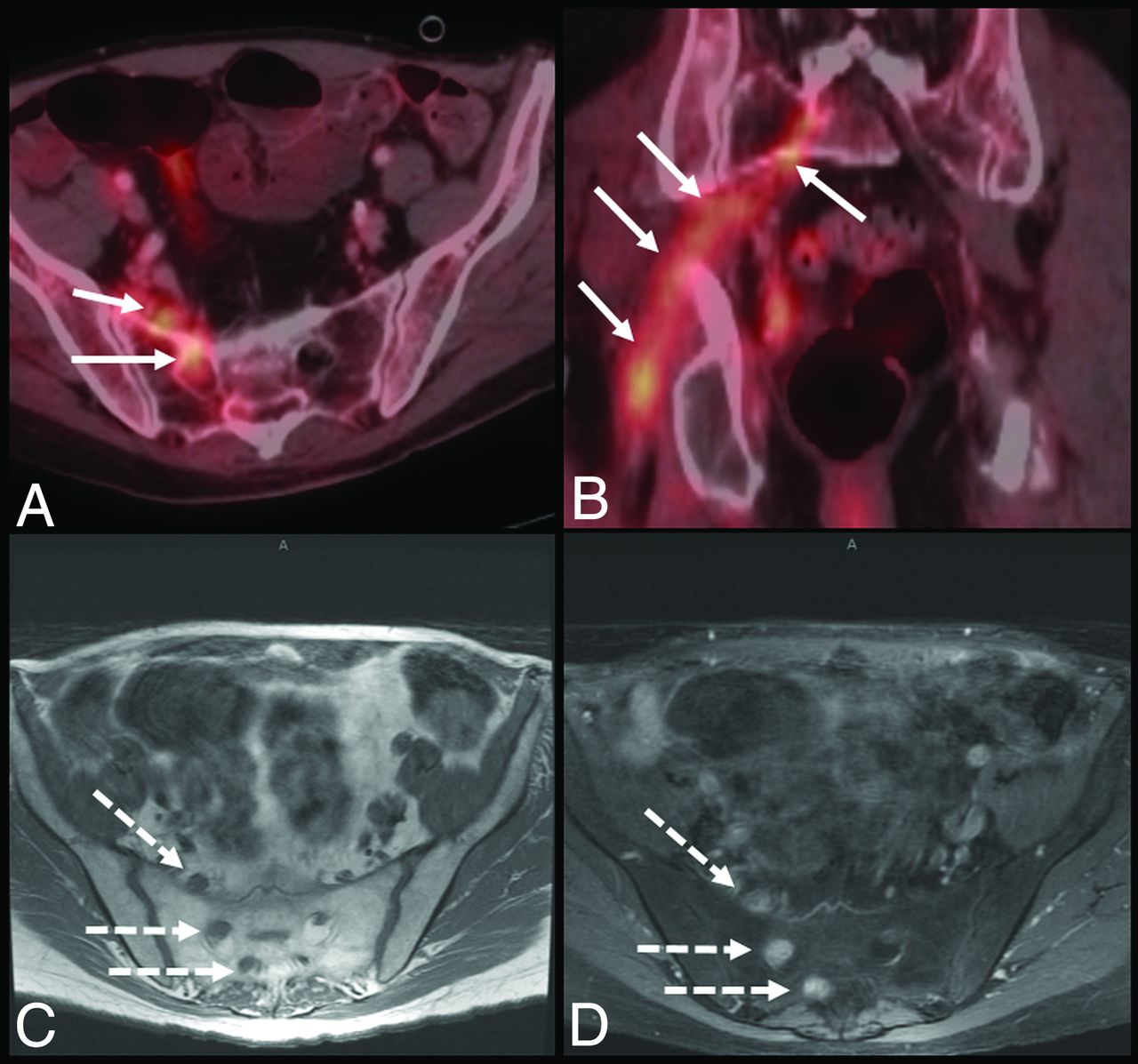
- FIG 8.
A 62-year-old man with penile cancer. Axial and coronal fused (A and B) images demonstrate a long, nodular segment of radiotracer uptake within the right pelvis suspicious for perineural spread of metastases (white arrows). Corresponding axial T1-weighted precontrast and T1-weighted fat-saturated postcontrast MR imaging (C and D) show nodular thickening and enhancement (dashed arrows) along the right sacral nerve roots. Fused indicates fused PET and CT image.
- FIG 9.
An 87-year-old woman with lung cancer. Coronal CT (A), [18F]FDG-PET (B), and PET/CT (C) images at the sacroiliac level demonstrate bilateral linear lucencies through the sacral ala (white arrows), with corresponding linear radiotracer uptake (black arrows) compatible with insufficiency fractures.
- FIG 10.
A 68-year-old woman with breast cancer. Axial (A) and coronal (C) CT and axial (B) and coronal (D) fused images demonstrate an intense focus of increased [18F]FDG uptake in the lumbar spine (dashed arrows) corresponding to a bulky osteophytic pseudoarthrosis on CT (solid arrows), which can mimic blastic osseous metastases. Fused indicates fused PET and CT image.
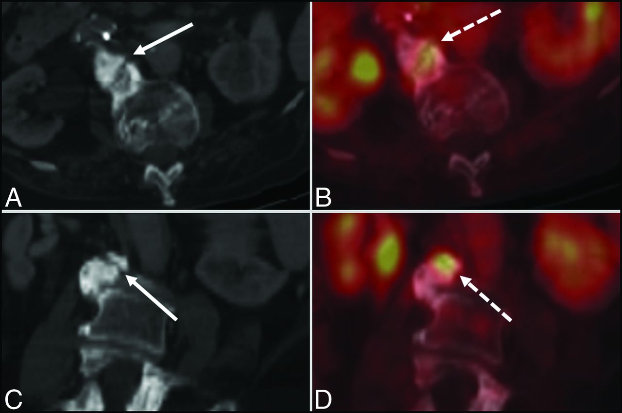
- FIG 11.
Upper row: A 50-year-old woman with multiple myeloma. Sagittal fused (A) and CT (B) images demonstrate foci of intensely increased FDG uptake (dashed arrows) in the thoracolumbar spine vertebral bodies, corresponding to lytic myelomatous lesions (solid arrows) on CT, which in the absence of a proper history, can appear as lytic osseous metastases. Lower row: A 62-year-old woman with breast cancer after recent chemotherapy. Sagittal fused image (C) demonstrates an incidental photopenic lesion (dashed arrow) in the posterior T6 vertebral body corresponding to a hemangioma on CT (solid arrow). Fused indicates fused PET and CT image.
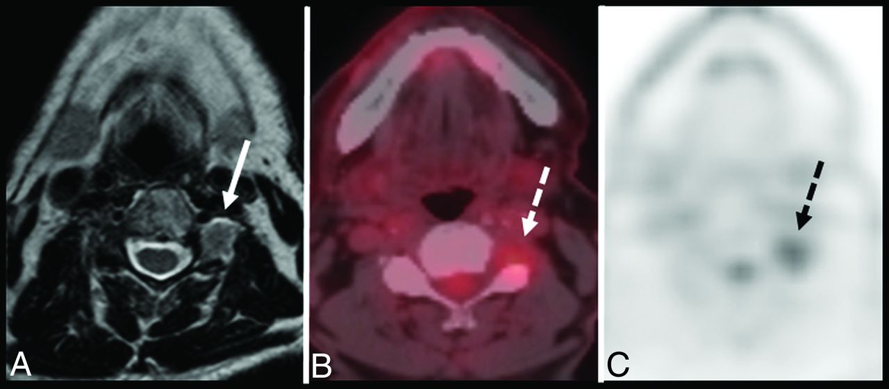
- FIG 12.
A 74-year-old man with thyroid cancer. Axial T2-weighted MR imaging (A), fused (B), and AC (C) images demonstrate mild increased [18F]FDG uptake (dashed arrows) within the paraspinal region at the C3–C4 level, appearing as a rounded soft-tissue density with neuroforaminal widening/remodeling and intermediate T2 signal on MR imaging (solid arrow). Findings corresponded to a schwannoma, which, in the setting of known primary malignancy, can mimic perineural metastasis. Fused indicates fused PET and CT image; AC, attenuation-corrected.
- FIG 13.
A 70-year-old man with rectal cancer. Axial CT (A), fused (B), and AC (C) images demonstrate intense hypermetabolic uptake (dashed arrows) in the paraspinal musculature of the mid and lower thoracic spine, which corresponds to a hypodense region of phlegmon/developing abscess on CT (solid arrow). Fluid cultures were positive for methicillin-resistant Staphylococcus aureus. Fused indicates fused PET and CT image; AC, attenuation-corrected.


Jump to section
Related Articles
Cited By...
- No citing articles found.