
Article Figures & Data
Figures
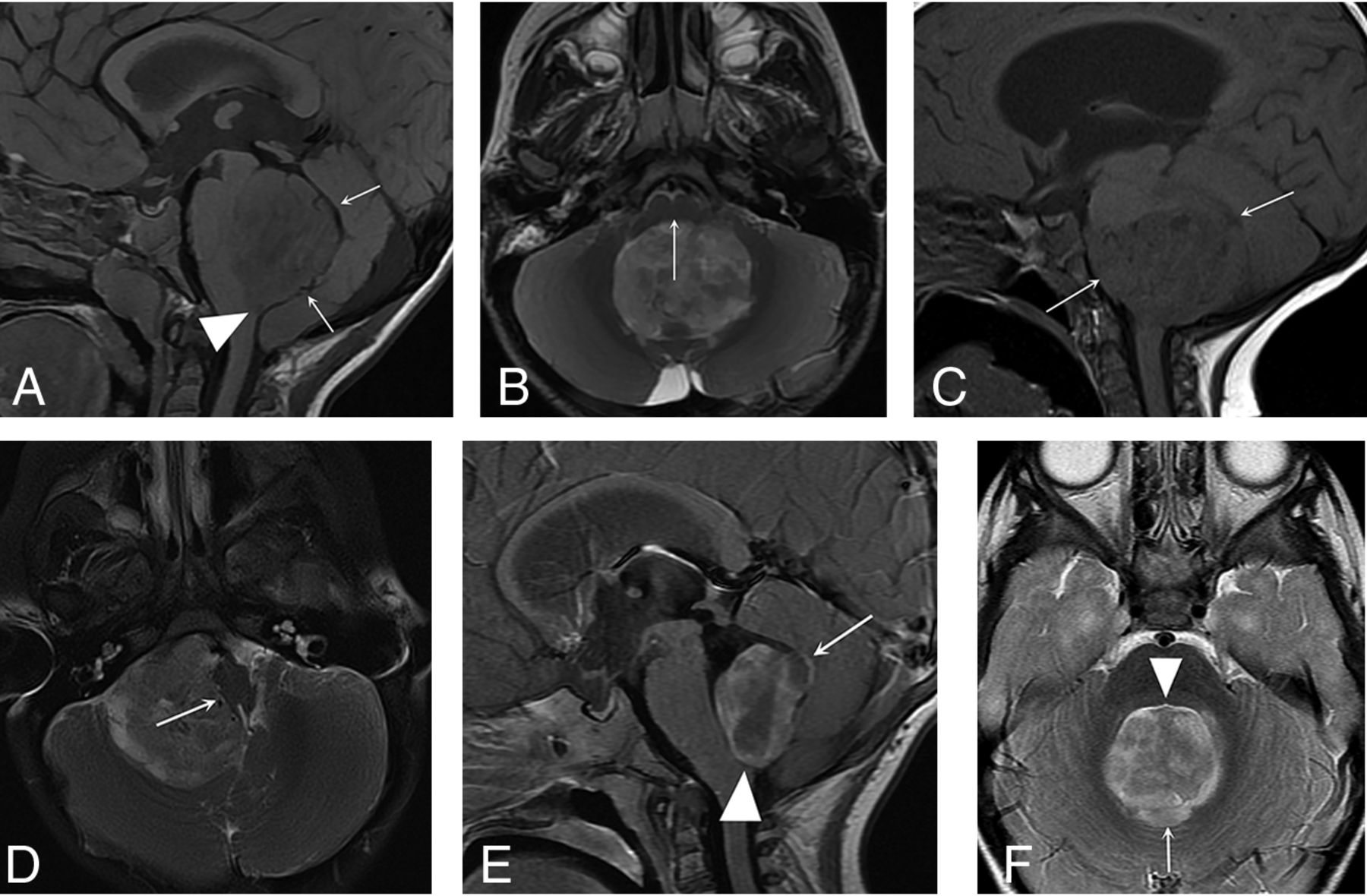
- FIG 1.
PFA ependymoma types based on location. Sagittal T1- (A) and axial T2-weighted images (B): Midfloor-type posterior fossa ependymomas fill the obex (arrowhead), and a gap may be seen between the roof of the fourth ventricle and the tumor (arrows, A). The brainstem is displaced anteriorly (arrow, B), and in the sagittal plane, it is seen in its entirety in a single sagittal image. Sagittal T1- (C) and axial T2-weighted images (D): Lateral-type posterior fossa ependymomas are centered in the lateral recess of the fourth ventricle or cerebellopontine angle (arrows, C) and displace the brainstem laterally (arrow, D). The brainstem usually cannot be seen in a single sagittal image. Postgadolinium sagittal T1- (E) and axial T2-weighted images (F): Roof-type posterior fossa ependymomas appear closely associated with the roof of the fourth ventricle (arrows, E and F) and do not fill the obex (arrowhead, E). A gap is usually seen between the tumor and the floor of the fourth ventricle (arrowhead, F).
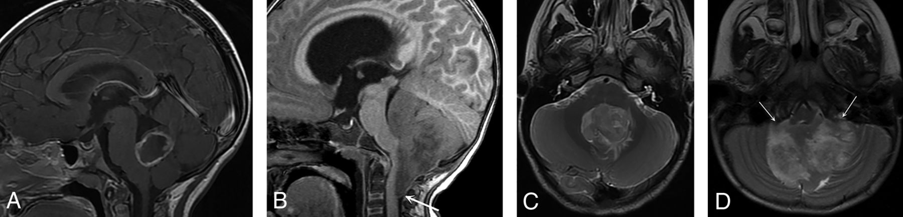
- FIG 2.
PFA-2c ependymoma versus another subtype. Postgadolinium sagittal T1- (A) and axial T2-weighted (C) MR images of a PFA-2c subtype ependymoma and sagittal T1- (B) and axial T2-weighted (D) MR images of another PFA ependymoma subtype (1b) show that the PFA-2c ependymoma has a more circumscribed appearance in that it does not wrap around the brainstem or extend below the foramen magnum in distinction from the PFA-1b subtype (arrow, B). The PFA-2c ependymoma in this case did not extend through the foramina of Luschka, unlike the PFA-1b tumor (arrows, D), or encase the basilar or vertebral arteries. This more circumscribed appearance was seen in the 5 PFA-2c tumors in our study, though evaluation of a larger number of this subtype is needed to confirm this appearance as a characteristic of PFA-2c ependymomas.


Tables
Yes (%) No (%) Extends below foramen magnum, P = .18 PFA-1 67 (89.3) 8 (10.7) PFA-2 37 (78.7) 10 (21.3) Total 104 (85.2) 18 (14.8) Involves foramina of Luschka, P = .55 PFA-1 61 (81.3) 14 (18.7) PFA-2 41 (87.2) 6 (12.8) Total 102 (83.6) 20 (16.4) Encases blood vessels, P = .73 PFA-1 33 (44.0) 42 (56.0) PFA-2 23 (48.9) 24 (51.1) Total 56 (45.9) 66 (54.1) Necrosis (P = .03)a PFA-1 59 (80.8) 14 (19.2) PFA-2 28 (60.9) 18 (39.1) Total 87 (73.1) 32 (26.9) Hydrocephalus, P = .36 PFA-1 65 (86.7) 10 (13.3) PFA-2 44 (93.6) 3 (6.4) Total 109 (89.3) 13 (10.7) ↵a Data for this variable were available for 119 subjects because 3 patients did not have preoperative postcontrast images available.
Lateral (%) Midfloor (%) Roof (%) PFA-1 33 (44.0) 32 (42.7) 10 (13.3) PFA-2 14 (29.8) 23 (48.9) 10 (21.3) Total 47 (38.5) 55 (45.1) 20 (16.4) - Table 3:
Imaging characteristics of PFA-2c ependymomas compared with other PFA-2 subtypes and all other PFA subtypesa
Yes No Extends below foramen magnum PFA-2c 0 (0%) 5 (100%) PFA-2a and PFA-2b 29 (87.9%) 4 (12.1%) All other PFA tumors 96 (88.9%) 12 (11.1%) Encases blood vessels PFA-2c 0 (0%) 5 (100%) PFA-2a and PFA-2b 17 (51.5%) 16 (48.5%) All other PFA tumors 50 (46.3%) 58 (53.7%) Involves foramina of Luschka PFA-2c 2 (40%) 3 (60%) PFA-2a and PFA-2b 31 (93.9%) 2 (6.1%) All other PFA tumors 92 (85.2%) 16 (14.8%) ↵a Ninety-eight subjects had subtype data available. “All other PFA tumors” includes the PFA-1 subgroup and the PFA-2a and 2b subtypes. The 15 subjects with PFA-1 subgroup ependymomas that could not be subtyped were included in the category of All other PFA tumors. The 8 patients with PFA-2 tumors that could not be subtyped were not included in this analysis.
- Table 4:
Imaging type of PFA-2c ependymomas compared with other PFA-2 subtypes and all other PFA subtypesa
Lateral Midfloor Roof PFA-2c 0 (0%) 5 (100%) 0 (0%) PFA-2a and PFA-2b 10 (30.3%) 14 (42.4%) 9 (27.3%) All other PFA tumors 43 (39.8%) 46 (42.6%) 19 (17.6%) ↵a Ninety-eight subjects had subtype data available. “All other PFA tumors” includes the PFA-1 subgroup and the PFA-2a and 2b subtypes. The 15 subjects with PFA-1 subgroup ependymomas that could not be subtyped were included in the category of All other PFA tumors. The 8 patients with PFA-2 tumors that could not be subtyped were not included in this analysis.
- Table 5:
PFA-2c ependymomas compared with other PFA-2 ependymoma subtypes and all other PFA subtypes with respect to flag variable 1a
Flag Variable 1b Yes No PFA-2c 5 (100%) 0 (0%) PFA-2a and PFA-2b 3 (9%) 30 (91%) All other PFA tumors 6 (5.6%) 102 (94.4%) ↵a Ninety-eight subjects had subtype data available. “All other PFA tumors” includes the PFA-1 subgroup and the PFA-2a and 2b subtypes. The 15 subjects with PFA-1 subgroup ependymomas that could not be subtyped were included in the category of All other PFA tumors. The 9 patients with PFA-2 tumors that could not be subtyped were not included in this analysis.
↵b The flag variable incorporates the following 3 variables: “no” for extension below the foramen magnum, “no” for encases blood vessels, and “midfloor-type” tumor.


Jump to section
Related Articles
Cited By...
- Arterial Spin-Labeling Perfusion Lightbulb Sign: An Imaging Biomarker of Pediatric Posterior Fossa Hemangioblastoma
- Ependymal Tumors: Overview of the Recent World Health Organization Histopathologic and Genetic Updates with an Imaging Characteristic
- Newly Recognized CNS Tumors in the 2021 World Health Organization Classification: Imaging Overview with Histopathologic and Genetic Correlation