
Article Figures & Data
Figures
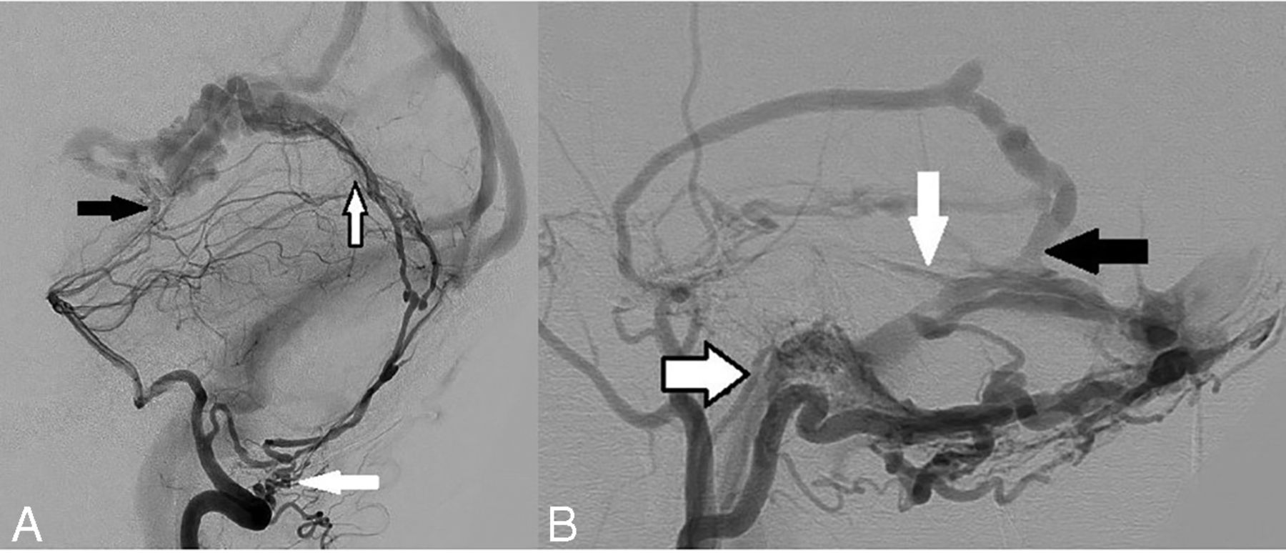
- FIG 1.
Arterial supply and venous drainage patterns in dAVFs. A, Arterial supply: Left vertebral artery angiogram in a lateral projection shows a falcotentorial dAVF with all 3 different forms of arterial supply: 1) extradural origin of a dural artery: left posterior meningeal artery arising from the extracranial distal V3 segment of the left vertebral artery (white arrow) and providing posterior supply to the fistula via the artery of the falx cerebelli; 2) intradural origin of a dural artery: Artery of Davidoff and Schechter arising from the P1–2 junction of the left posterior cerebral artery, traveling along the free edge of the tentorium cerebelli and supplying the AVF from its undersurface (black arrow); 3) secondary-induced pial artery supply: several small, irregular, induced pial branches from the distal aspect of the left superior cerebellar artery supplying the posterior aspect of the fistula (black border arrow). Reproduced from Bhatia K et al.7 B, Venous drainage: Left external carotid artery angiogram in a lateral projection in a patient with multiple intracranial dAVFs demonstrates different patterns of venous drainage. A large left transverse sinus fistula is present with supply from an enlarged squamous temporal branch of the middle meningeal artery and draining directly into the transverse sinus (black arrow). There is sinus reflux of contrast into the torcula (Cognard IIa) and cortical venous reflux into the vein of Labbe (white arrow, Cognard IIb). This is a Cognard IIa+b AVF. A left condylar fistula is also present with arterial supply from the jugular branch of the left ascending pharyngeal artery (black border arrow) and draining directly into a condylar vein (direct cortical venous drainage). This is a Cognard III AVF.
- FIG 2.
Noninvasive imaging characteristics of dAVFs. A, Axial bone window noncontrast CT image of a patient with a right transverse-sigmoid sinus dAVF demonstrates enlarged right retromastoid transosseous vascular channels from occipital artery feeders (white arrow). B, Axial TOF-MRA image in the same patient demonstrates abnormal hyperintense signal in the right transverse-sigmoid sinus junction (white arrow) due to arterialization of the sinus, with adjacent asymmetrically enlarged occipital artery feeders. C, Coronal contrast-enhanced CT image in a patient with a left transverse sinus dAVF demonstrates enlarged tortuous pial veins over the left cerebellum (white arrow), resulting from cortical venous reflux.
- FIG 3.
Angiographic assessment of a left petrous dAVF. A, Lateral projection DSA of a left ECA injection shows arterial supply to the AVF by the petrous branch of the middle meningeal artery (white arrow), a component of the facial nerve arterial arcade, by the accessory meningeal artery (black arrow) with an intervening nidus before the venous drainage and the fistula draining into the petrosal vein (black-border arrow) over the posterior superior surface of the left petrous temporal bone. B, 3D rotational DSA via a left ECA injection in the lateral projection reconstruction again shows the petrous branch of the left middle meningeal artery (white arrow) and the accessory meningeal artery (white border arrow) entering a nidal network before the fistulous point. C, Reconstructed axial MPR MIP image of a 3D rotational DSA via a left ECA injection demonstrates, in exquisite anatomic detail, the petrous branch of the left middle meningeal artery extending posteriorly from the foramen spinosum over the petrous temporal bone (white arrow), accessory meningeal artery entering via foramen ovale (black arrow), and the draining petrosal vein (black border arrow). This level of anatomic detail and spatial relationship to the skull base foramina is difficult to appreciate on standard DSA images, and this allows assessment of the potential risks of facial nerve ischemia that may be associated with a transarterial approach. D, Axial TOF-MRA image demonstrates pathologic hyperintense signal in the left petrosal vein (black border arrow) due to pre-excited protons entering via the high-flow left petrosal dAVF. A–C, Reproduced from the supplemental material of Bhatia et al.41


Tables
Venous drainage pattern classification in intracranial dAVFs
Classification of Borden et al3 Classification of Cognard et al2 Benign venous drainage patterns I Drains to dural sinus I Drains to dural sinus No CVR No sinus reflux No CVR IIa Drains to dural sinus Sinus reflux present No CVR Aggressive venous drainage patterns II Drains to dural sinus IIb Drains to dural sinus CVR present No sinus reflux CVR present IIa+b Drains to dural sinus Sinus reflux present CVR present III Direct drainage to cortical veins III Direct drainage to cortical veins Or No venous ectasia Drainage to an isolated segment of dural sinus IV Direct drainage to cortical veins Venous ectasia present V Spinal perimedullary venous drainage Note:—CVR indicates cortical venous reflux.


