Advertisement

Main menu

  • Home
  • Content
    • Current Issue
    • Accepted Manuscripts
    • Article Preview
    • Past Issue Archive
    • Video Articles
    • AJNR Case Collection
    • Case of the Week Archive
    • Case of the Month Archive
    • Classic Case Archive
  • Special Collections
    • AJNR Awards
    • Low-Field MRI
    • Alzheimer Disease
    • ASNR Foundation Special Collection
    • Photon-Counting CT
    • View All
  • Multimedia
    • AJNR Podcasts
    • AJNR SCANtastic
    • Trainee Corner
    • MRI Safety Corner
    • Imaging Protocols
  • For Authors
    • Submit a Manuscript
    • Submit a Video Article
    • Submit an eLetter to the Editor/Response
    • Manuscript Submission Guidelines
    • Statistical Tips
    • Fast Publishing of Accepted Manuscripts
    • Graphical Abstract Preparation
    • Imaging Protocol Submission
    • Author Policies
  • About Us
    • About AJNR
    • Editorial Board
    • Editorial Board Alumni
  • More
    • Become a Reviewer/Academy of Reviewers
    • Subscribers
    • Permissions
    • Alerts
    • Feedback
    • Advertisers
    • ASNR Home

User menu

  • Alerts
  • Log in

Search

  • Advanced search
American Journal of Neuroradiology

American Journal of Neuroradiology

ASHNR American Society of Functional Neuroradiology ASHNR American Society of Pediatric Neuroradiology ASSR
  • Alerts
  • Log in

Advanced Search

  • Home
  • Content
    • Current Issue
    • Accepted Manuscripts
    • Article Preview
    • Past Issue Archive
    • Video Articles
    • AJNR Case Collection
    • Case of the Week Archive
    • Case of the Month Archive
    • Classic Case Archive
  • Special Collections
    • AJNR Awards
    • Low-Field MRI
    • Alzheimer Disease
    • ASNR Foundation Special Collection
    • Photon-Counting CT
    • View All
  • Multimedia
    • AJNR Podcasts
    • AJNR SCANtastic
    • Trainee Corner
    • MRI Safety Corner
    • Imaging Protocols
  • For Authors
    • Submit a Manuscript
    • Submit a Video Article
    • Submit an eLetter to the Editor/Response
    • Manuscript Submission Guidelines
    • Statistical Tips
    • Fast Publishing of Accepted Manuscripts
    • Graphical Abstract Preparation
    • Imaging Protocol Submission
    • Author Policies
  • About Us
    • About AJNR
    • Editorial Board
    • Editorial Board Alumni
  • More
    • Become a Reviewer/Academy of Reviewers
    • Subscribers
    • Permissions
    • Alerts
    • Feedback
    • Advertisers
    • ASNR Home
  • Follow AJNR on Twitter
  • Visit AJNR on Facebook
  • Follow AJNR on Instagram
  • Join AJNR on LinkedIn
  • RSS Feeds

AJNR Awards, New Junior Editors, and more. Read the latest AJNR updates

Research ArticlePediatric Neuroimaging

MRI Patterns in Pediatric CNS Hemophagocytic Lymphohistiocytosis

P. Malik, L. Antonini, P. Mannam, F.N. Aboobacker, A. Merve, K. Gilmour, K. Rao, S. Kumar, S.E. Mani, D. Eleftheriou, A. Rao, C. Hemingway, S.V. Sudhakar, J. Bartram and K. Mankad
American Journal of Neuroradiology October 2021, DOI: https://doi.org/10.3174/ajnr.A7292
P. Malik
aFrom the Departments of Diagnostic Imaging (P. Malik, P. Mannam, S.E.M.)
  • Find this author on Google Scholar
  • Find this author on PubMed
  • Search for this author on this site
  • ORCID record for P. Malik
L. Antonini
dDepartment of Paediatric Hemato-Oncology (L.A.), G. Salesi Hospital, Ancona, Italy
  • Find this author on Google Scholar
  • Find this author on PubMed
  • Search for this author on this site
  • ORCID record for L. Antonini
P. Mannam
aFrom the Departments of Diagnostic Imaging (P. Malik, P. Mannam, S.E.M.)
  • Find this author on Google Scholar
  • Find this author on PubMed
  • Search for this author on this site
  • ORCID record for P. Mannam
F.N. Aboobacker
bHematology (F.N.A.)
  • Find this author on Google Scholar
  • Find this author on PubMed
  • Search for this author on this site
  • ORCID record for F.N. Aboobacker
A. Merve
gDepartment of Histopathology (A.M.)
  • Find this author on Google Scholar
  • Find this author on PubMed
  • Search for this author on this site
  • ORCID record for A. Merve
K. Gilmour
jImmunology (K.G.)
  • Find this author on Google Scholar
  • Find this author on PubMed
  • Search for this author on this site
  • ORCID record for K. Gilmour
K. Rao
hBone Marrow Transplant Unit (K.R.)
  • Find this author on Google Scholar
  • Find this author on PubMed
  • Search for this author on this site
S. Kumar
cChild Health (S.K.), Christian Medical College, Vellore, India
  • Find this author on Google Scholar
  • Find this author on PubMed
  • Search for this author on this site
  • ORCID record for S. Kumar
S.E. Mani
aFrom the Departments of Diagnostic Imaging (P. Malik, P. Mannam, S.E.M.)
  • Find this author on Google Scholar
  • Find this author on PubMed
  • Search for this author on this site
  • ORCID record for S.E. Mani
D. Eleftheriou
kPaediatric Rheumatology (D.E.), Great Ormond Street Hospital for Children, London, UK
  • Find this author on Google Scholar
  • Find this author on PubMed
  • Search for this author on this site
  • ORCID record for D. Eleftheriou
A. Rao
iDepartments of Pediatric Hematology (A.R., J.B.)
  • Find this author on Google Scholar
  • Find this author on PubMed
  • Search for this author on this site
  • ORCID record for A. Rao
C. Hemingway
eDepartment of Pediatric Neurology (C.H.)
  • Find this author on Google Scholar
  • Find this author on PubMed
  • Search for this author on this site
  • ORCID record for C. Hemingway
S.V. Sudhakar
fPediatric Neuroradiology Unit (S.V.S., K.M.)
  • Find this author on Google Scholar
  • Find this author on PubMed
  • Search for this author on this site
  • ORCID record for S.V. Sudhakar
J. Bartram
iDepartments of Pediatric Hematology (A.R., J.B.)
  • Find this author on Google Scholar
  • Find this author on PubMed
  • Search for this author on this site
  • ORCID record for J. Bartram
K. Mankad
fPediatric Neuroradiology Unit (S.V.S., K.M.)
  • Find this author on Google Scholar
  • Find this author on PubMed
  • Search for this author on this site
  • ORCID record for K. Mankad
  • Article
  • Figures & Data
  • Supplemental
  • Info & Metrics
  • Responses
  • References
  • PDF
Loading

Article Figures & Data

Figures

  • Tables
  • FIG 1.
    • Download figure
    • Open in new tab
    • Download powerpoint
    FIG 1.

    Findings of 4 patients with subpattern 1.2 (brainstem–predominant pattern). Axial T2WI (A1–D1) and axial postcontrast T1WI (A2–D2) at the level of the pons show multiple punctate (dashed white arrow, B2, D2) to nodular (solid white arrow, A2 and C2), enhancing foci and extension of the T2 signal abnormality (black arrow, A1–D1) beyond the enhancement in all cases.
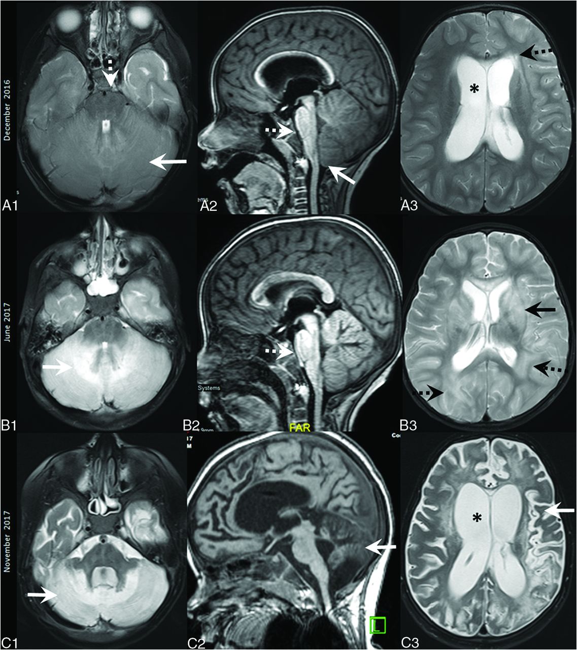
  • FIG 2.
    • Download figure
    • Open in new tab
    • Download powerpoint
    FIG 2.

    Findings of subpattern 1.3 (cerebellitis). Axial T2WI at the level of the fourth ventricle (A1–C1), lateral ventricles (A3–C3), and midline sagittal T1WI (A2–C2). Onset MR imaging (December 2016) shows severe cerebellar edema and expansion (white arrow, A1) with mass effect on the brainstem, effacement of the prepontine cistern (dashed white arrow, A2), and foramen magnum crowding (white arrow, A2). Lateral ventricular dilation (asterisk, A3) and transependymal CSF seepage (dashed black arrow, A3) are also noted. Mild reduction in cerebellar edema and mass effect (dashed arrow, B2) with new cerebellar (white arrow, B1), parieto-occipital (dashed arrow, B3), and deep gray nuclei (black arrow, B3) hyperintensities were found in June 2017. Last MR imaging in November 2017 shows cerebral (white arrow, C3) and cerebellar (white arrows, C1–2) atrophy, diffuse white matter hyperintensities, and ventriculomegaly (asterisk, C3).

Tables

  • Figures
    • View popup
    Table 1:

    Clinical, treatment, and outcome profilesa

    Profiles
    Age
     Age at onset (median) (IQR) (mo)36 (5.5–80.8)
     Age at CNS presentation (median) (IQR) (mo)49.2 (11–96)
    Male/female (ratio)34:23 (1.4:1)
    General symptoms
     Fever45/56 (80)
     Hepato-/splenomegaly46/56 (82)
     Abdominal distension14/56 (25)
    CNS symptoms45/57, 79%
     Seizures28 (62)
     Decreased sensorium22 (49)
     Meningismus13 (29)
     Gait ataxia12 (27)
     Hypotonia11 (24)
    Minimal symptoms (mild irritability) or clinically silent patients (no symptoms with CSF abnormalities)12 (21)
    CSF findings
     CSF analyzed at presentation50/57 (87)
     Abnormal CSF42/50 (84)
     CSF pleocytosis (>10 leucocytes/μL)25/50 (50)
     CSF proteinosis (>45 mg/dL)32/50 (64)
    Treatment
     HLH 1994/200435/57 (61)
     IT methotrexate17 (30)
     HSCT25 (45)
    Outcome profiles
     Death19 (32)
      Death before 8 weeks7
      Death after 8 weeks12
    Alive at last follow-up36 (63)
    Lost to follow-up2
    • Note:—IQR indicates interquartile range; IT, intrathecal.

    • ↵a Total (n = 57). Data collected are both continuous and categorical, and the analysis method used has been referred to under statistical analysis heading.

    • View popup
    Table 2:

    Pattern characteristics and analysisa

    SubpatternsP ValuePattern 1 OverallPattern 2P Value
    1.11.21.3
    Median age at onset (mo)45.46680.5.555.516.004
    Symptoms
     Seizures11/21 (52%)4/5 (80%)4/6 (67%).519/32 (59%)9/25 (36%).08
     Encephalopathy11/21 (52%)1/5 (20%)4/6 (67%).316/32 (50%)6/25 (24%).04
     Gait ataxia5/21 (24%)3/5 (60%)3/6 (50%).0812/32 (34%)0.001
     Limb weakness6/21 (29%)1/5 (20%)1/6 (17%).88/32 (25%)1/25 (4%).03
     Dysarthria02/5 (40%)1/6 (17%).013/32 (9%)1/25 (4%).4
     Diplopia01/21 (5%)2/5 (40%)2/6 (33%).065/32 (16%)0.03
    Abnormal CSF18/20 (90%)4/5 (80%)5/5 (100%).627/30 (90%)15/20(75%).2
    Proteinosis13 (65%)4 (80%)4 (80%).521 (70%)11 (55%).3
    Pleocytosis10 (50%)4 (80%)5 (100%).0819 (63%)6 (30%).04
    Pooled mutation group4/6 (67%)2/3 (67%)3/3 (100%).329/12 (75%)13/13 (100%).09
     Mortality9/20 (45%)3/5 (60%)2/6 (33%).314/31 (45%)5/24 (21%).06
    Deficit-free at follow-up8/20 (40%)1/5 (20%)1/6 (17%).110/31 (32%)13/25(52%).06
    • ↵a Data collected are both continuous and categorical, and the analysis method used has been referred to under statistical analysis heading.

    • View popup
    Table 3:

    Relevant literature review of CNS HLH cases with brainstem or cerebellitis patterns with available MR imaging and genetic data

    Literature ReviewImaging PatternGenetic VariantsAge of Onset, Relation to Systemic HLHImaging Findings
    Benson et al23 casesBrainstem–predominant pattern (CLIPPERS- like)1 disruptive, 2 with missense mutations and absent protein expression
    Pt 1: PRF1 c.452A>T (p.H151L) and c.666C>A (H222Q), Perforin expression 0%
    Pt 2: PRF1 c.443C>G (p.A148G) and c.666C>A (H222Q), Perforin expression 0%
    Pt 3: UNC13D c.2346_2349delGGAG(p.R782fs), c.2588G>A (p.G863D)
    5–7 yr, all 3 CNS-restricted HLHCLIPPERS MR imaging criteria NA
    Taieb et al284 patientsBrainstem–predominant pattern (CLIPPERS- like)4 cases, all with missense mutations and retained-but-decreased protein expression
    Pt 1: PRF1 c.272C>T(p.A91V) homozygous, perforin expression 38%
    Pt 2: UNC13D c.919C>T (p.Q307*) and c.2038C>T (p.R680W), not applicable
    Pt 3: PRF1 c.116C>A (p.P39H) and c.272C>T (p.A91V), perforin expression 25%
    Pt 4: PRF1 c.82C>T (p.R28C) and c.272C>T (p.A91V), perforin expression 38%
    Adults (42–73 yr), all had CNS-restricted HLHThree-fourths had atypical MR imaging CLIPPERS features (confluent contrast-enhancing lesions)
    Bhoopalan et al26 1 patientCerebellitis1 patient with compound heterozygous PRF1 gene mutations with at least 1 disruptive mutation PRF1: c.50delT (p.L17fs) and c.527G>A(p.C176Y))8 yr, CNS-restricted HLHCerebellitis, tonsillar herniation, no multifocal lesions
    Khan et al271 patientCerebellitis1 patient with homozygous missense mutation c. 173T > C (p.L58P) in STX11 (syntaxin 11) gene2 yr 7 months, systemic HLH already presentCerebellitis, tonsillar herniation, diffuse-to-multifocal lesions already present
    Astigarraga et al3 1 patientCerebellitis1 patient with compound heterozygous missense PRF mutations PRF1: c.643C>A (p.L215I) and c.785C>A (p.Ala262Asp) (perforin expression data NA)3 yr, preceded systemic HLHRecurrent cerebellitis, tonsillar herniation, subsequently multifocal lesions
    Taieb et al281 patientCerebellitisPatient 3’s (in CLIPPERS series) brother’s; granddaughter, monoallelic PRF1 mutation (genetic variant NA)Not specified, self-limited CNS-restricted presentationNA
    Chiapparini et al29 1 patientCerebellitis1 patient with homozygous missense PRF1 mutation c.673C>T (p.Arg225Trp) (perforin expression data NA)13 yr, preceded systemic HLHCerebellitis, tonsillar herniation followed by multifocal lesions
    • Note:—NA indicates not available; Pt, patient.

PreviousNext
Back to top
Advertisement
Print
Download PDF
Email Article

Thank you for your interest in spreading the word on American Journal of Neuroradiology.

NOTE: We only request your email address so that the person you are recommending the page to knows that you wanted them to see it, and that it is not junk mail. We do not capture any email address.

Enter multiple addresses on separate lines or separate them with commas.
MRI Patterns in Pediatric CNS Hemophagocytic Lymphohistiocytosis
(Your Name) has sent you a message from American Journal of Neuroradiology
(Your Name) thought you would like to see the American Journal of Neuroradiology web site.
CAPTCHA
This question is for testing whether or not you are a human visitor and to prevent automated spam submissions.
Cite this article
P. Malik, L. Antonini, P. Mannam, F.N. Aboobacker, A. Merve, K. Gilmour, K. Rao, S. Kumar, S.E. Mani, D. Eleftheriou, A. Rao, C. Hemingway, S.V. Sudhakar, J. Bartram, K. Mankad
MRI Patterns in Pediatric CNS Hemophagocytic Lymphohistiocytosis
American Journal of Neuroradiology Oct 2021, DOI: 10.3174/ajnr.A7292

Citation Manager Formats

  • BibTeX
  • Bookends
  • EasyBib
  • EndNote (tagged)
  • EndNote 8 (xml)
  • Medlars
  • Mendeley
  • Papers
  • RefWorks Tagged
  • Ref Manager
  • RIS
  • Zotero
0 Responses
Respond to this article
Share
Bookmark this article
MRI Patterns in Pediatric CNS Hemophagocytic Lymphohistiocytosis
P. Malik, L. Antonini, P. Mannam, F.N. Aboobacker, A. Merve, K. Gilmour, K. Rao, S. Kumar, S.E. Mani, D. Eleftheriou, A. Rao, C. Hemingway, S.V. Sudhakar, J. Bartram, K. Mankad
American Journal of Neuroradiology Oct 2021, DOI: 10.3174/ajnr.A7292
del.icio.us logo Twitter logo Facebook logo Mendeley logo
  • Tweet Widget
  • Facebook Like
  • Google Plus One
Purchase

Jump to section

  • Article
    • Abstract
    • ABBREVIATIONS:
    • MATERIALS AND METHODS
    • RESULTS
    • DISCUSSION
    • CONCLUSIONS
    • Footnotes
    • References
  • Figures & Data
  • Supplemental
  • Info & Metrics
  • Responses
  • References
  • PDF

Related Articles

  • PubMed
  • Google Scholar

Cited By...

  • Isolated central nervous system haemophagocytic lymphohistiocytosis with PRF1 gene mutation presenting as fever of unknown origin
  • Child Neurology: Familial Hemophagocytic Lymphohistiocytosis Underlying Isolated CNS Inflammation
  • Crossref (19)
  • Google Scholar

This article has been cited by the following articles in journals that are participating in Crossref Cited-by Linking.

  • Childhood Cerebral Vasculitis
    Neetika Gupta, Shivaprakash B. Hiremath, Richard I. Aviv, Nagwa Wilson
    Clinical Neuroradiology 2023 33 1
  • Child Neurology: Familial Hemophagocytic Lymphohistiocytosis Underlying Isolated CNS Inflammation
    Giorgia Bucciol, Nele Willemyns, Benjamin Verhaaren, Xavier Bossuyt, Katrien Lagrou, Anniek Corveleyn, Despina Moshous, Katrien Jansen, Liesbeth De Waele, Isabelle Meyts
    Neurology 2022 99 15
  • Imaging Spectrum of Neurological Manifestations of Hemophagocytic Lymphohistiocytosis in Pediatrics: A Case Series
    Geethapriya Sivaramalingam, Afiya VK, Bagyam Raghavan, Jayaraj Govindaraj
    Indian Journal of Radiology and Imaging 2023 33 04
  • Case report: Cerebellar swelling and hydrocephalus in familial hemophagocytic lymphohistiocytosis
    Taro Yoshida, Kunihiko Moriya, Keisuke Oikawa, Shoko Miura, Yoshiko Asakura, Sachiko Tanifuji, Shuji Kusano, Mikiya Endo, Manami Akasaka
    Frontiers in Pediatrics 2022 10
  • Differential Diagnosis of Suspected Multiple Sclerosis in Pediatric and Late-Onset Populations
    Le H. Hua, Andrew J. Solomon, Silvia Tenembaum, Antonio Scalfari, Àlex Rovira, Kevin Rostasy, Scott D. Newsome, Ruth Ann Marrie, Melinda Magyari, Orhun Kantarci, Bernhard Hemmer, Cheryl Hemingway, Mary Pat Harnegie, Jennifer S. Graves, Jeffrey A. Cohen, Riley Bove, Brenda Banwell, John R. Corboy, Emmanuelle Waubant
    JAMA Neurology 2024 81 11
  • Pediatric CNS-isolated hemophagocytic lymphohistiocytosis with brain hemorrhages: a case report
    Mauricio Borda, Helen Tian, Steven Benitez, Ashley Bonheur, Nagma Dalvi, Ellen Fraint
    BMC Neurology 2024 24 1
  • Imaging of Childhood Cerebral Vasculitis
    Sheng-Che Hung, Carolina Guimaraes
    Neuroimaging Clinics of North America 2024 34 1
  • Infection and inflammation: radiological insights into patterns of pediatric immune-mediated CNS injury
    Prateek Malik, Manohar Shroff
    Neuroradiology 2023 65 3
  • Isolated Sixth Nerve Palsies in a Child With Familial Hemophagocytic Lymphohistiocytosis Type 2
    Homer H. Chiang, Sebastian Fernandez-Pol, Gordon H. Bae, Kerri E. Rieger, Hisham M. Dahmoush, Shannon J. Beres
    Journal of Neuro-Ophthalmology 2023 43 1
  • Neurobrucellosis Presenting as Vocal Cord Paralysis in an Infant: Correspondence
    Prateek Kumar Panda, Indar Kumar Sharawat
    Indian Journal of Pediatrics 2024 91 12

More in this TOC Section

  • FRACTURE MR in Congenital Vertebral Anomalies
  • Neuroimaging Delineation and Progression of SLSMD
  • fetal brain development of 10 weeks gestation
Show more PEDIATRIC NEUROIMAGING

Similar Articles

Advertisement

Indexed Content

  • Current Issue
  • Accepted Manuscripts
  • Article Preview
  • Past Issues
  • Editorials
  • Editor's Choice
  • Fellows' Journal Club
  • Letters to the Editor
  • Video Articles

Cases

  • Case Collection
  • Archive - Case of the Week
  • Archive - Case of the Month
  • Archive - Classic Case

More from AJNR

  • Trainee Corner
  • Imaging Protocols
  • MRI Safety Corner
  • Book Reviews

Multimedia

  • AJNR Podcasts
  • AJNR Scantastics

Resources

  • Turnaround Time
  • Submit a Manuscript
  • Submit a Video Article
  • Submit an eLetter to the Editor/Response
  • Manuscript Submission Guidelines
  • Statistical Tips
  • Fast Publishing of Accepted Manuscripts
  • Graphical Abstract Preparation
  • Imaging Protocol Submission
  • Evidence-Based Medicine Level Guide
  • Publishing Checklists
  • Author Policies
  • Become a Reviewer/Academy of Reviewers
  • News and Updates

About Us

  • About AJNR
  • Editorial Board
  • Editorial Board Alumni
  • Alerts
  • Permissions
  • Not an AJNR Subscriber? Join Now
  • Advertise with Us
  • Librarian Resources
  • Feedback
  • Terms and Conditions
  • AJNR Editorial Board Alumni

American Society of Neuroradiology

  • Not an ASNR Member? Join Now

© 2025 by the American Society of Neuroradiology All rights, including for text and data mining, AI training, and similar technologies, are reserved.
Print ISSN: 0195-6108 Online ISSN: 1936-959X

Powered by HighWire