
Article Figures & Data
Figures
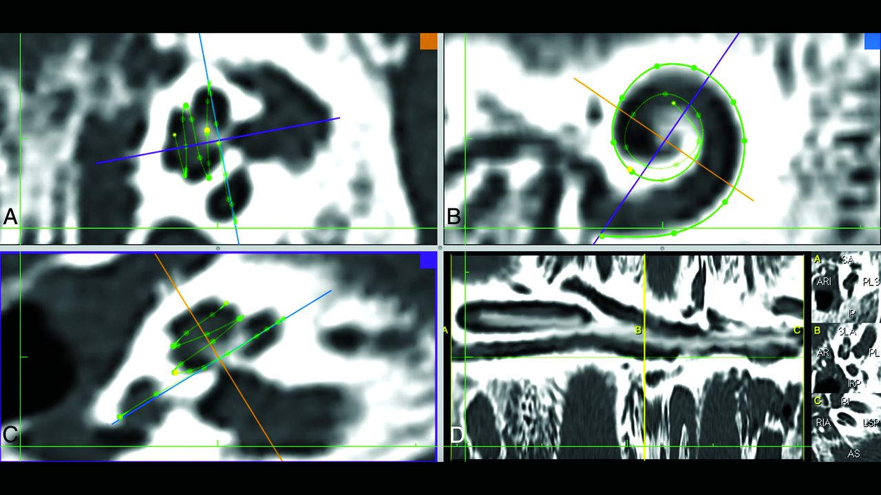
- FIG 1.
Screenshot of the CT postprocessing software used in the study. A and C, Planes perpendicular to the modiolus are formed. B, The cochlear view is shown; in this plane, the basal turn can be fully traced and the bone structure of the round window is visualized as a thin line parallel to the purple line. The purple line also indicates the A-value measured from the niche to the opposite cochlear wall and the diameter of the cochlear basal turn. C, The round window niche is distinguished as a hypodense area below the measuring point. D, The measurement screen.
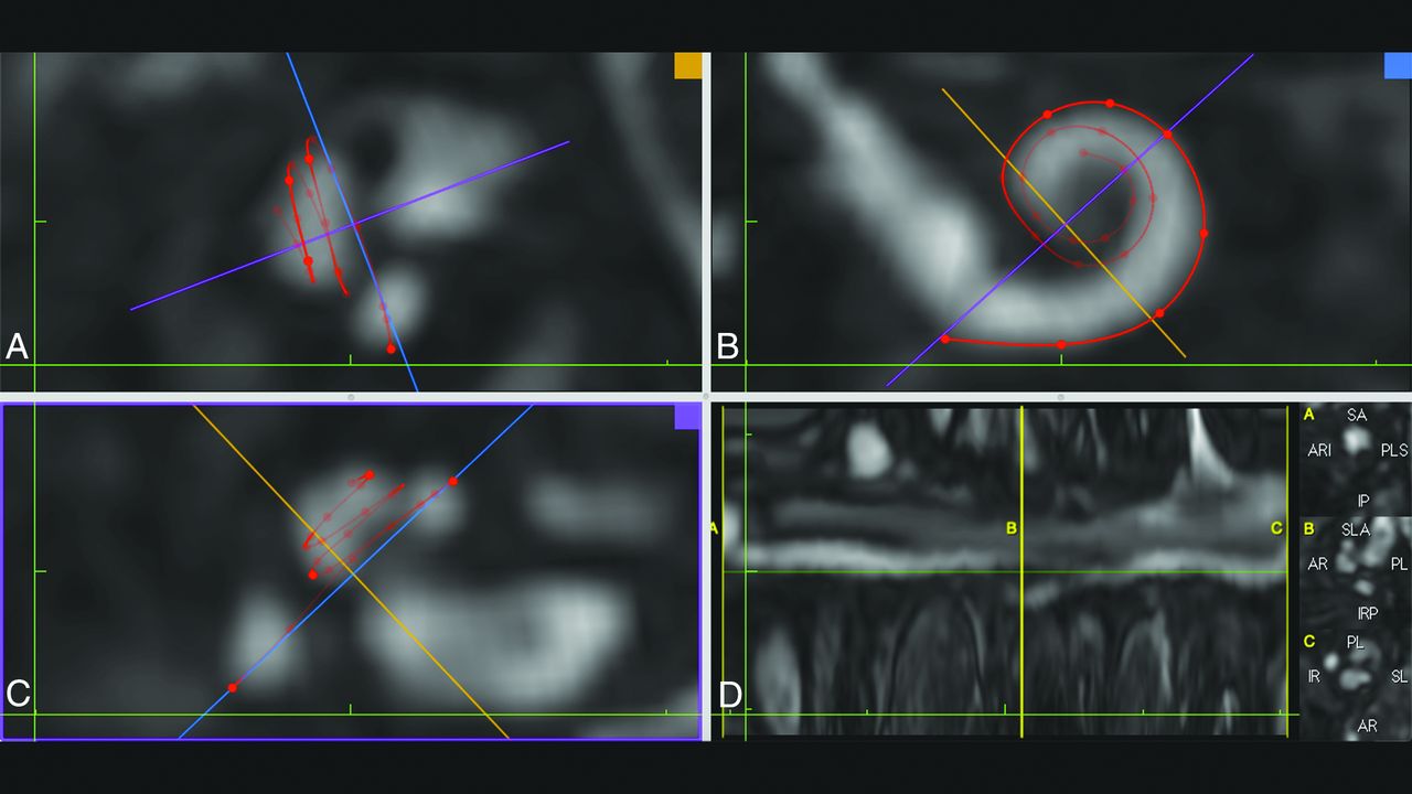
- FIG 2.
Screenshot of the MR imaging postprocessing software. A and C, Planes perpendicular to modiolus are formed. B, The cochlear view is shown; in this plane, the basal turn can be fully traced and the bone structure of the round window is viewed as a thin hypointense line parallel to the purple line. C, The round window niche is not distinguished, and bone and air are shown as hypointense on the FIESTA-C MR imaging sequence. D, The measurement screen.
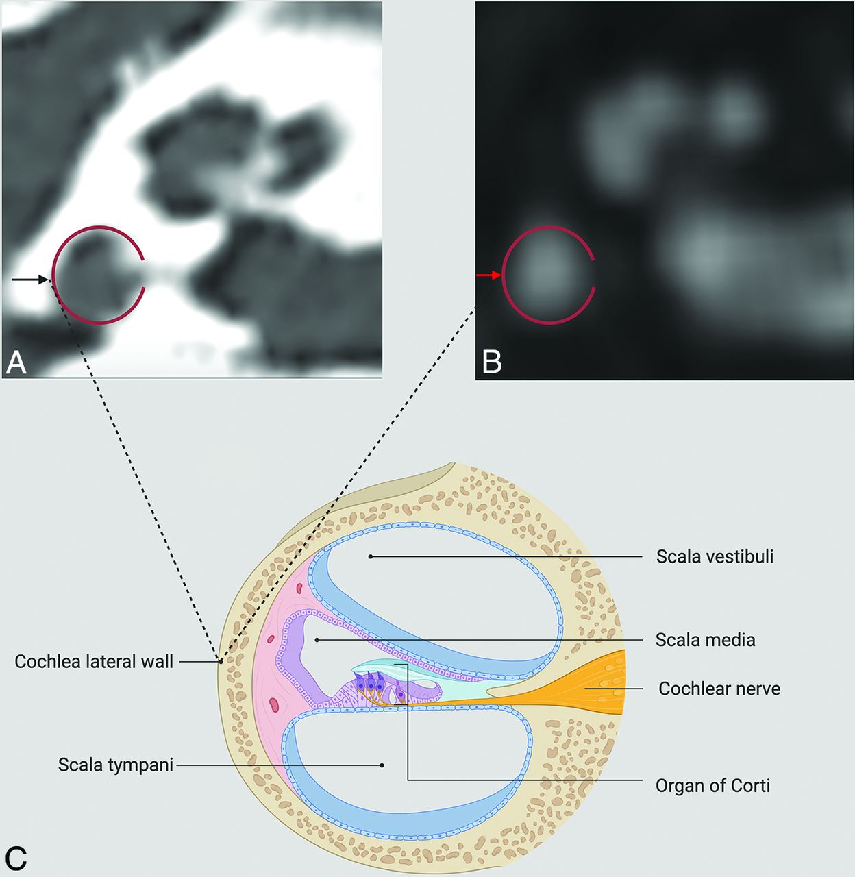
- FIG 3.
CT (A) and MR imaging (B) of the cochlea of the same patient. The red ring seen in these images shows the lateral wall of the cochlea. The black arrow seen on the CT (A) image and the red arrow seen on the MR imaging (B) image indicate the outermost point of the lateral cochlear wall. The lateral cochlear wall, the target point of measurement, and other anatomic structures are summarized in a schematic view (C). Created with BioRender.com.


Tables
3D FIESTA-C Plane Axial + coronal + sagittal oblique Fat suppression + TR (ms) 6.9 TE (ms) Min Flip angle 55° Section thickness (mm) 1 FOV (mm) 320 × 320 Bandwidth (Hz) 90 Matrix (mm × mm) 256 × 192 Note:—Min indicates 2.6–12 ms.
- Table 2:
Intraobserver accuracy and reliability of observer 1’s cochlear LWL measurements between CT and MR imaging
CT (mean) (95% CI) MRI (mean) (95% CI) ICC (95% CI) P Value Cochlea basal turn length (mm) 22.95 (SD, 1.21) (22.65–23.25) 23.37 (SD, 1.12) (23.09–23.65) 0.70 (0.48–0.82) <.001a Cochlea 2 turn length (mm) 35.92 (SD, 1.98) (35.43–36.41) 36.27 (SD, 1.77) (35.83–36.71) 0.85 (0.75–0.91) <.001a Cochlea lateral wall length (mm) 41.52 (SD, 2.25) (40.96–42.08) 41.44 (SD, 2.18) (40.90–41.98) 0.94 (0.90–0.96) <.001a ↵a The P value is statistically significant.
- Table 3:
Intraobserver accuracy and reliability of observer 2’s cochlear LWL measurements between CT and MR imaging
CT (mean) (95% CI) MRI (mean) (95% CI) ICC (95% CI) P Value Cochlea basal turn length (mm) 22.57 (SD, 1.21) (22.28–22.88) 22.91 (SD, 1.18) (22.61–23.20) 0.69 (0.52–0.80) <.001a Cochlea 2 turn length (mm) 35.51 (SD, 2.17) (34.98–36.05) 36.37 (SD, 2.08) (35.85–36.88) 0.79 (0.66–0.87) <.001a Cochlea lateral wall length (mm) 41.74 (SD, 2.69) (41.07–42.40) 42.34 (SD, 2.53) (41.71–42.97) 0.86 (0.73–0.92) <.001a ↵a The P value is statistically significant.


Jump to section
Related Articles
Cited By...
- No citing articles found.