
Article Figures & Data
Figures
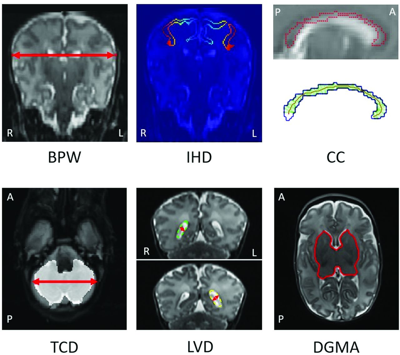
- FIG 1.
Automated MR imaging measures of biparietal width (BPW), interhemispheric distance (IHD), thickness of the corpus callosum (CC), transcerebellar diameter (TCD), left and right LVD, and DGMA. IHD, Blue to orange represents the distance (closest and farthest, respectively) for each voxel to the segmentation in the opposite hemisphere. CC, Upper image, T2WI with the CC segmentation border (red); lower panel, segmentation border (blue), skeleton (red), and distance between the upper and lower borders of the segmentation for each voxel on the skeleton (green). R indicates right; L, left; A, anterior; P, posterior.
- FIG 2.
Associations between the 6 raw conventional MR imaging measures with postmenstrual age (left) and gestational age at birth (right) in the Pearson r, displayed separately for preterm-born infants at early MR imaging (n = 94) and TEA MR imaging (n = 81), as well as term-born controls at TEA MR imaging (PPREMO, n = 22; dHCP, n = 455). Manual measures were available for PPREMO only. Double asterisks indicate P ≤ .001; asterisk, P ≤ .05. BPW indicates biparietal width; CC, callosal thickness (at the genu (CCg), midbody (CCm), and splenium (CCs)); IHD, interhemispheric distance; TCD, transcerebellar diameter; R, right; L, left.


Tables
Measure How to Measure Biparietal width Greatest distance between left and right parietal cortices, measured on a single coronal slice identifying bilateral
cochlea and basilar truncusInterhemispheric distance Distance between crowns of superior frontal gyri, measured on the same coronal slice as biparietal width Callosal thickness Thinning at the genu, midbody, splenium Transcerebellar diameter Single coronal slice at level of ventricular atrium Lateral ventricular diameter Same coronal slice as transcerebellar diameter Deep grey matter area Single axial slice showing caudate heads, lentiform nuclei, and thalami Measure How to Measure Biparietal width Identify most lateral sagittal slices of parietal GM (regionprops3)
For each GM voxel, calculate distance to voxels in opposite hemisphere
Calculate maximum distanceInterhemispheric distance Calculate distance from each voxel of superior frontal gyrus label in left hemisphere to each voxel label in right hemisphere (bwmorph3, bwdist)
Derive minimum distanceCallosal thickness Combine 2D corpus callosum segmentation of the 11 most medial slices
Increase resolution (imresize) and improve mask (bwmorph3, bwmorph)
Derive skeleton (bwskel); for every voxel, calculate normal vector, identify intersection with borders of segmentation (points2contour, polyfit)
Derive distance between upper/lower segmentation borders for every voxel
Apply smoothing (nanfastsmooth), make 3 divisions
Obtain 97th percentile for each division (prctile)Transcerebellar diameter Model cerebellum segmentation as 3D ellipsoid (bwmorph3, regionprops3)
Calculate length of principal axis (regionprops3)Lateral ventricular dilation Identify coronal slice at level of ventricular atrium (regionprops3): find maximum surface area using ventricle label (bwmorph, regionprops)
Model 2D ventricle as ellipse, calculate length of minor axes (regionprops)Deep grey matter area Identify axial slice: centroid of caudate, thalamus, and lentiform nucleus (bwmorph, regionprops)
Combine labels to calculate area (regionprops)Note:—Matlab package used is provided in brackets.
PPREMO (n = 197) dHCP Preterm, Early MR Imaging Preterm, TEA MR Imaging Control, TEA MR Imaging No. 94 of 121 81 of 105 22 of 27 455 of 506 (95 preterm) Sex (female) 61% 63% 50% 45% PMA at MR imaging (wk)a 31.86 ± 1.96 (29–35) 40.71 ± 1.43 (38–47) 41.29 ± 1.25 (39–44) 40.71 ± 2.86 (29–45) GA (wk)a 28.40 ± 2.00 (23–31) 28.50 ± 2.20 (24–31) 40.00 ± 0.98 (38–41) 39.71 ± 2.82 (24–42) Birth weight (g)a 1107 ± 368 (494–1886) 1061 ± 391 (494–1886) 3516 ± 157 (2932–4200) 3250 ± 928 (540–4800) ↵a P < .05 for 2-sided t test comparing preterm infants at 40 weeks with term-born controls (PPREMO). Continuous variables are given as median ± interquartile range (range). Scans with poor quality (PPREMO, n = 39), realignment errors (PPREMO, n = 1; dHCP, n = 39), and severe pathology (PPREMO, n = 16; dHCP, n = 12) were excluded.


Jump to section
Related Articles
Cited By...
- No citing articles found.