
Article Figures & Data
Figures
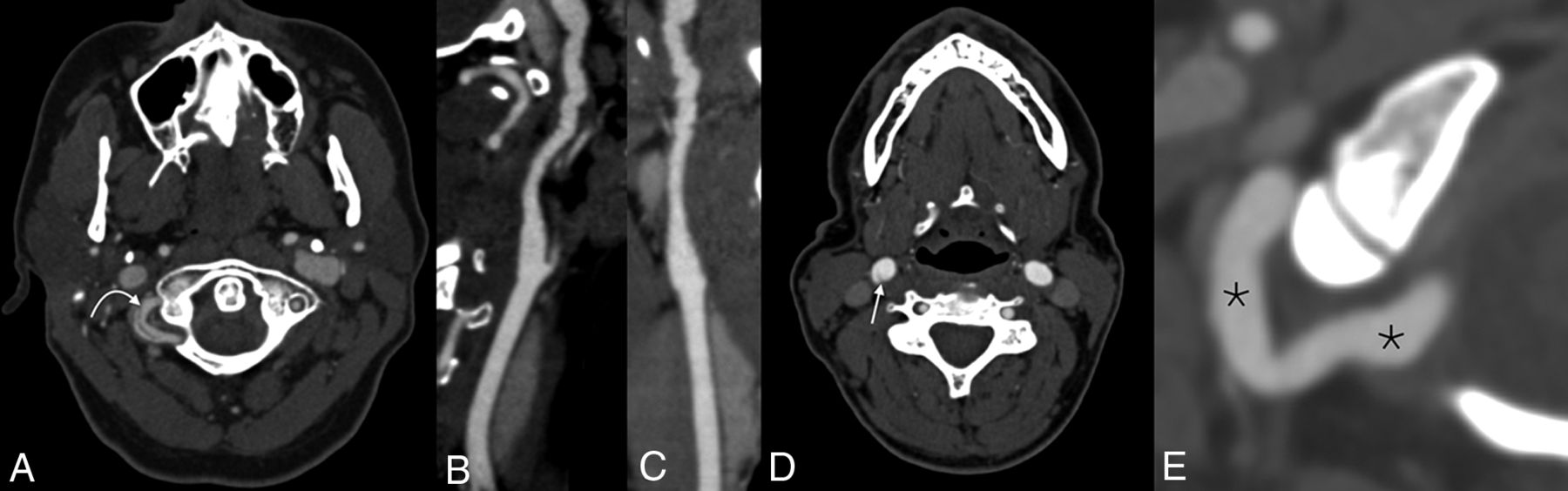
- FIG 1.
Examples of cervical artery abnormalities noted in patients with SCAD. Axial CTA (A) demonstrates a right vertebral artery dissection (curved arrow). Reformatted sagittal (B) and coronal (C) images show multifocal FMD of the right ICA. Axial CTA (D) demonstrates a web in the right ICA bulb (straight arrow). Reformatted axial CTA (E) shows ectasia of the distal right vertebral artery (asterisks).
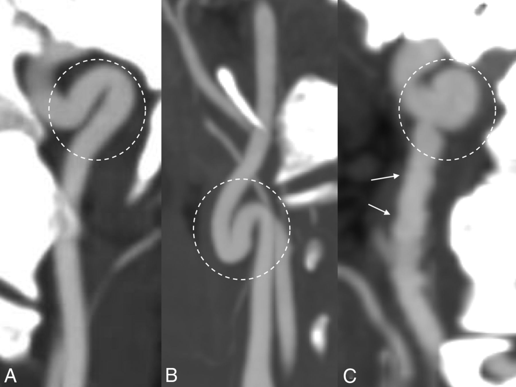
- FIG 2.
Examples of tortuosity subtypes. Arterial kink (A) (a single arterial angulation of ≤90°), loop (B) (2 adjacent acute angulations of the vessel), and coil (C) (complete 360° turn) are all shown. Also note the beaded irregularity of the vessel in C, compatible with FMD (arrows).


Tables
Findings No. (%) FMD 83/214 (38.8%) Dissection and/or pseudoaneurysm 28/214 (13.1%) Aneurysm/ectasia 22/214 (10.3%) Carotid web 10/214 (4.7%) 1+ type of carotid tortuosity 99/214 (46.3%) Atherosclerosis None 185/214 (86.4%) Mild 26/214 (12.2%) Moderate 3/214 (1.4%) Severe 0/214 (0.0%) Vertebral tortuosity None 124/214 (57.9%) Mild 64/214 (29.9%) Moderate 22/214 (10.3%) Severe 4/214 (1.9%) ↵a FMD, dissection, pseudoaneurysm, and aneurysm/ectasia numbers reflect the number of patients with those findings in either the carotid or vertebral artery. Webs were only assessed in the carotid arteries because they are considered to be rarely present in the vertebral arteries. Subtypes of carotid tortuosity include kinks, loops, coils, and retropharyngeal and retrojugular courses. Carotid atherosclerosis and vertebral tortuosity reflect the more severe side, if asymmetry existed.
- Table 2:
Prevalence of FMD, dissection and/or pseudoaneurysm, aneurysm/ectasia, and webs based on arterial involvementa
Right Carotid Artery Left Carotid Artery Both Carotid Arteries Right Vertebral Artery Left Vertebral Artery Both Vertebral Arteries FMD 56 (25.2%) 49 (22.9%) 39 (18.2%) 30 (14.0%) 32 (15.0%) 22 (10.3%) Dissection and/or pseudoaneurysm 5 (2.3%) 8 (3.7%) 0 (0.0%) 8 (3.7%) 12 (5.6%) 1 (0.5%) Aneurysm/ectasia 16 (7.5%) 13 (6.1%) 8 (3.7%) 4 (1.9%) 3 (1.4%) 1 (0.5%) Web 6 (2.8%) 6 (2.8%) 0 (0.0%) NA Note:—NA indicates not applicable.
↵a The presence or absence of webs within the vertebral arteries was not assessed because they are classically found in the carotid bulbs. Data are No. (%).


Jump to section
Related Articles
Cited By...
- No citing articles found.