
Article Figures & Data
Figures
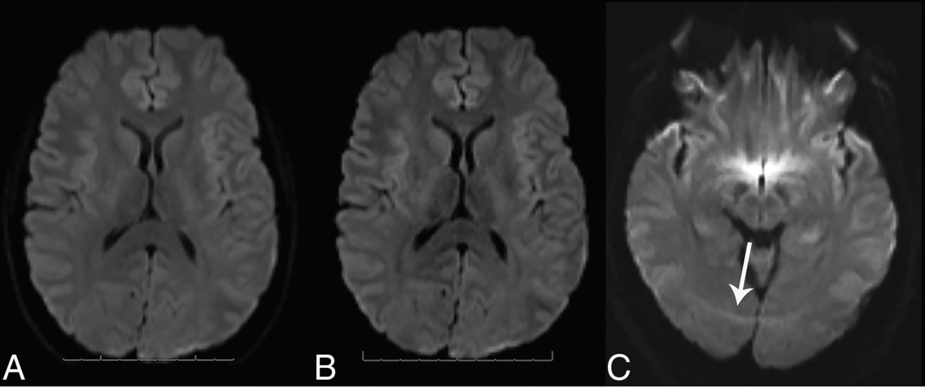
- FIG 1.
Axial diffusion images from standard (A) and SMS diffusion acquisitions (B) in a clinical patient with headaches. There is no appreciable difference in diagnostic utility, even with a reduction in scan time using SMS. An SMS arc-like scalp artifact (C, arrow) is seen in another clinical patient due to poor fat saturation. With SMS, unsaturated fat signal associated with echo-planar acquisitions can alias into all simultaneously acquired slices.
- FIG 2.
Examples of 2-slice SMS-accelerated diffusion trace and ADC maps of 4 different clinical patients. Large acute left-occipital infarct (A) contrasted with a small subacute left-thalamic infarct (B) (arrows). Raters suggested subtle SNR reductions for the posterior fossa using SMS (Table 1); however, small infarcts in the brainstem (C) and cerebellum (D) remain well-visualized.
- FIG 3.
Overlap of diffusion tractography between standard and SMS-acquired data in 2 selected patients. Upper Row: A 56-year-old man with a multifocal right-cerebral hemisphere glioma. The right corticospinal tract volumes are shown in blue (conventional) and red (SMS) at the levels of the cerebral peduncle (A), posterior limb of the internal capsule (B), and precentral gyrus (C). The DSC between standard and SMS tract volumes is 0.76. Lower Row: A 36-year-old man with a left opercular cavernous malformation. The left-arcuate fasciculus volumes are shown in purple (conventional) and yellow (SMS) at the levels of the anterior and posterior frontal projections (D and E) and genu (F). There are slight differences in the edge of the visualized tracts abutting the superior border of the cavernous malformation (E), but these differences would not affect selection of the surgical corridor, and in both situations, the neurosurgeon would be cautious in approaching the superior margin of the lesion. The DSC between standard and SMS tract volumes is 0.75.


Tables
- Table 1:
Quantitative and qualitative comparison of diffusion trace and ADC parameter maps for clinical patients in the emergency department (single single 3T MRI scanner)a
Data Standard (n = 25) SMS (n = 25) 95% CI for Difference P Value Age (yr) 61.5 [SD, 19.5] 63.2 [SD, 15.4] NA .734b Woman (No.) (%) 52% (13/25) 56% (14/25) NA NA ADC, minor forceps (×10–3 mm2/s) 0.833 [SD, 0.077] 0.816 [SD, 0.073] –0.025 to + 0.059 .427b ADC, CSF (×10–3 mm2/s) 3.032 [SD, 0.099] 3.015 [SD, 0.080] –0.033 to + 0.067 .508b SNR, minor forceps 21.4 [SD, 14.6] 22.5 [SD, 9.6] –7.9 to + 5.7 .754b SNR, cerebellum 32.6 [SD, 10.9] 27.3 [SD, 14.8] –1.9 to + 12.5 .156b Artifactsc 3.5 [SD, 0.5] 3.7 [SD, 0.5] –0.5 to + 0.1 .070d Image qualityc 4.0 [SD, 0.7] 3.3 [SD, 0.5] –0.4 to + 1.0 .001d Diagnostic utilityc 3.8 [SD, 0.4] 3.7 [SD, 0.5] –0.2 to + 0.4 .187d - Table 2:
Quantitative comparison of DTI tractography for the corticospinal tract and arcuate fasciculus ipsilateral to the lesion in 13 subjectsa
Diffusion Technique Standard SMS Mean Individual Differenceb 95% CI for Difference P Value Corticospinal tract Volume (mL) 30.7 [SD, 9.1] 32.9 [SD, 9.7] –2.3 [SD, 4.0] –9.4 to +5.0 .062c Length (mm) 125.2 [SD, 13.1] 124.0 [SD, 15.0] 1.2 [SD, 7.0] –9.6 to +12.0 .537c Mean FA 0.45 [SD, 0.04] 0.46 [SD, 0.04] –0.01 [SD, 0.02] –0.04 to +0.02 .076c Arcuate fasciculus Volume (mL) 25.3 [SD, 5.3] 26.0 [SD, 6.7] –0.7 [SD, 3.1] –5.3 to +3.9 .438c Length (mm) 77.4 [SD, 7.1] 76.5 [SD, 6.7] 0.9 [SD, 4.6] –4.4 to +6.2 .482c Mean FA 0.36 [SD, 0.04] 0.36 [SD, 0.04] 0.00 [SD, 0.01] –0.03 to +0.03 1.000c Spatial misregistration (mm)d 3.4 [SD, 1.6] 2.8 [SD, 1.0] 0.6 [SD, 1.3] –0.4 to +1.6 .135c ↵a Data are mean (SD) or 95% CI.
↵b Difference between measurements in the same individual (standard value minus SMS value).
↵c Paired-sample 2-tailed t test.
↵d Difference between the anterior margin of the cervicomedullary junction at the foramen magnum depicted by volumetric T1 MR imaging versus CST diffusion tractography.


Jump to section
Related Articles
Cited By...
- No citing articles found.