Advertisement

Main menu

  • Home
  • Content
    • Current Issue
    • Accepted Manuscripts
    • Article Preview
    • Past Issue Archive
    • Video Articles
    • AJNR Case Collection
    • Case of the Week Archive
    • Case of the Month Archive
    • Classic Case Archive
  • Special Collections
    • AJNR Awards
    • Low-Field MRI
    • Alzheimer Disease
    • ASNR Foundation Special Collection
    • Photon-Counting CT
    • View All
  • Multimedia
    • AJNR Podcasts
    • AJNR SCANtastic
    • Trainee Corner
    • MRI Safety Corner
    • Imaging Protocols
  • For Authors
    • Submit a Manuscript
    • Submit a Video Article
    • Submit an eLetter to the Editor/Response
    • Manuscript Submission Guidelines
    • Statistical Tips
    • Fast Publishing of Accepted Manuscripts
    • Graphical Abstract Preparation
    • Imaging Protocol Submission
    • Author Policies
  • About Us
    • About AJNR
    • Editorial Board
    • Editorial Board Alumni
  • More
    • Become a Reviewer/Academy of Reviewers
    • Subscribers
    • Permissions
    • Alerts
    • Feedback
    • Advertisers
    • ASNR Home

User menu

  • Alerts
  • Log in

Search

  • Advanced search
American Journal of Neuroradiology

American Journal of Neuroradiology

ASHNR American Society of Functional Neuroradiology ASHNR American Society of Pediatric Neuroradiology ASSR
  • Alerts
  • Log in

Advanced Search

  • Home
  • Content
    • Current Issue
    • Accepted Manuscripts
    • Article Preview
    • Past Issue Archive
    • Video Articles
    • AJNR Case Collection
    • Case of the Week Archive
    • Case of the Month Archive
    • Classic Case Archive
  • Special Collections
    • AJNR Awards
    • Low-Field MRI
    • Alzheimer Disease
    • ASNR Foundation Special Collection
    • Photon-Counting CT
    • View All
  • Multimedia
    • AJNR Podcasts
    • AJNR SCANtastic
    • Trainee Corner
    • MRI Safety Corner
    • Imaging Protocols
  • For Authors
    • Submit a Manuscript
    • Submit a Video Article
    • Submit an eLetter to the Editor/Response
    • Manuscript Submission Guidelines
    • Statistical Tips
    • Fast Publishing of Accepted Manuscripts
    • Graphical Abstract Preparation
    • Imaging Protocol Submission
    • Author Policies
  • About Us
    • About AJNR
    • Editorial Board
    • Editorial Board Alumni
  • More
    • Become a Reviewer/Academy of Reviewers
    • Subscribers
    • Permissions
    • Alerts
    • Feedback
    • Advertisers
    • ASNR Home
  • Follow AJNR on Twitter
  • Visit AJNR on Facebook
  • Follow AJNR on Instagram
  • Join AJNR on LinkedIn
  • RSS Feeds

AJNR Awards, New Junior Editors, and more. Read the latest AJNR updates

Research ArticlePediatric Neuroimaging

The Fetus with Ganglionic Eminence Abnormality: Head Size and Extracranial Sonographic Findings Predict Genetic Diagnoses and Postnatal Outcomes

S.K. Goergen, E. Alibrahim, J. Christie, A. Dobrotwir, M. Fahey, L. Fender, K. Frawley, S.A. Manikkam, J.R. Pinner, S. Sinnott, R. Romaniello, S.A. Sandaradura, J. Taylor, A. Vasudevan and A. Righini
American Journal of Neuroradiology May 2021, DOI: https://doi.org/10.3174/ajnr.A7131
S.K. Goergen
aFrom the Monash Imaging (S.K.G.), Monash Health, Victoria, Australia
bDepartments of Imaging and Surgery (S.K.G.), School of Clinical Sciences, Monash University, Clayton, Victoria, Australia
  • Find this author on Google Scholar
  • Find this author on PubMed
  • Search for this author on this site
  • ORCID record for S.K. Goergen
E. Alibrahim
cDepartment of Radiology (E.A., A.D.), Royal Women’s Hospital, Parkville, Victoria, Australia
  • Find this author on Google Scholar
  • Find this author on PubMed
  • Search for this author on this site
  • ORCID record for E. Alibrahim
J. Christie
dPRP Imaging (J.C.), Sydney, New South Wales, Australia
  • Find this author on Google Scholar
  • Find this author on PubMed
  • Search for this author on this site
  • ORCID record for J. Christie
A. Dobrotwir
cDepartment of Radiology (E.A., A.D.), Royal Women’s Hospital, Parkville, Victoria, Australia
  • Find this author on Google Scholar
  • Find this author on PubMed
  • Search for this author on this site
  • ORCID record for A. Dobrotwir
M. Fahey
eDepartment of Paediatrics (M.F.), School of Clinical Sciences, Monash University, Clayton, Victoria, Australia
fNeurogenetics Unit (M.F.), Monash Health, Victoria, Australia
  • Find this author on Google Scholar
  • Find this author on PubMed
  • Search for this author on this site
  • ORCID record for M. Fahey
L. Fender
gDepartment of Radiology (L.F.), King Edward Memorial Hospital, Perth, Western Australia, Australia
  • Find this author on Google Scholar
  • Find this author on PubMed
  • Search for this author on this site
  • ORCID record for L. Fender
K. Frawley
hDepartment of Medical Imaging and Nuclear Medicine (K.F., S.A.M.), Queensland Children’s Hospital, Brisbane, Queensland, Australia
  • Find this author on Google Scholar
  • Find this author on PubMed
  • Search for this author on this site
  • ORCID record for K. Frawley
S.A. Manikkam
hDepartment of Medical Imaging and Nuclear Medicine (K.F., S.A.M.), Queensland Children’s Hospital, Brisbane, Queensland, Australia
  • Find this author on Google Scholar
  • Find this author on PubMed
  • Search for this author on this site
  • ORCID record for S.A. Manikkam
J.R. Pinner
iCentre for Clinical Genetics (J.R.P.), Sydney Children’s Hospital, Sydney, New South Wales, Australia
jUniversity of New South Wales (J.R.P.), Sydney, Australia
  • Find this author on Google Scholar
  • Find this author on PubMed
  • Search for this author on this site
  • ORCID record for J.R. Pinner
S. Sinnott
kSO + GI Scan I-MED Radiology (S.S.), Department of Medical Imaging, Royal Brisbane and Women’s Hospital, Brisbane, Queensland, Australia
  • Find this author on Google Scholar
  • Find this author on PubMed
  • Search for this author on this site
  • ORCID record for S. Sinnott
R. Romaniello
lChild Neuropsychiatry and Neurorehabilitation Department (R.R.), Scientific Institute Eugenio Medea, La Nostra Famiglia, Bosiso Parini, Lecco, Italy
  • Find this author on Google Scholar
  • Find this author on PubMed
  • Search for this author on this site
  • ORCID record for R. Romaniello
S.A. Sandaradura
mDiscipline of Child and Adolescent Health (S.A.S.), Faculty of Medicine and Health, The University of Sydney, Westmead, New South Wales, Australia
nDepartment of Clinical Genetics (S.A.S.), Children’s Hospital at Westmead, Westmead, New South Wales, Australia
  • Find this author on Google Scholar
  • Find this author on PubMed
  • Search for this author on this site
  • ORCID record for S.A. Sandaradura
J. Taylor
oDepartment of Radiology (J.T.), Prince of Wales Hospital, Sydney, New South Wales, Australia
  • Find this author on Google Scholar
  • Find this author on PubMed
  • Search for this author on this site
  • ORCID record for J. Taylor
A. Vasudevan
pDepartment of Clinical Genetics (A.V.), Royal Women’s Hospital, Parkville, Victoria, Australia.
  • Find this author on Google Scholar
  • Find this author on PubMed
  • Search for this author on this site
  • ORCID record for A. Vasudevan
A. Righini
qDepartment of Pediatric Radiology and Neuroradiology (A.R.), Vittore Buzzi Children’s Hospital, Milan, Italy
  • Find this author on Google Scholar
  • Find this author on PubMed
  • Search for this author on this site
  • ORCID record for A. Righini
  • Article
  • Figures & Data
  • Supplemental
  • Info & Metrics
  • Responses
  • References
  • PDF
Loading

Article Figures & Data

Figures

  • FIG 1.
    • Download figure
    • Open in new tab
    • Download powerpoint
    FIG 1.

    PIK3CA pathogenic variant. Case 2 at 31 weeks 3 days’ gestation. Megalencephaly, polymicrogyria, polydactyly hydrocephalus syndrome due to a PIK3CA heterozygous pathogenic variant. The fronto-occipital diameter is =6 SDs above the mean for gestation. Enlarged bilateral ganglionic eminences (arrows, A) are seen on T2-weighted single-shot FSE and DWI (A and B). Diffusion-weighted b = 0 image (C) and T2*-weighted EPI (D) confirm the absence of hemorrhage as the cause for ganglionic eminence enlargement. Abnormal opercularization is present with Sylvian fissures lined by peri-Sylvian polymicrogyria (E). Postaxial polydactyly is seen on sonography with arrow indicating a rudimentary sixth digit medial to the fifth digit of the hand (F).

  • FIG 2.
    • Download figure
    • Open in new tab
    • Download powerpoint
    FIG 2.

    MTOR/PROS pathway mutations. Smith-Kingsmore syndrome (case 5) at 25 weeks’ gestation demonstrates enlarged GEs on DWI and T2-weighted single-shot FSE. There is bilateral underopercularization and left peri-Sylvian polymicrogyria (arrow). The right hemisphere is mildly overgrown.

  • FIG 3.
    • Download figure
    • Open in new tab
    • Download powerpoint
    FIG 3.

    Tuberous sclerosis complex (A and B). Case 1. Dichorionic diamniotic twin at 22 weeks 5 days’ gestation. T2-weighted single-shot FSE image demonstrates hemimegalencephaly and marked enlargement of the ipsilateral GE, which merges with a hypointense masslike lesion in the enlarged right cerebral hemisphere. The fetus had left ventricular cardiac rhabdomyoma on prenatal sonography (arrow, B); the findings likely represent tuberous sclerosis complex with an associated hemispheric malformation or, less likely, coexistent subependymal giant cell astrocytoma. No postmortem data or confirmatory genetic testing was available. Case 8. T2 single-shot FSE at 25 weeks gestation (C) and T2 FSE at 1-week postnatal imaging (D) demonstrate mild enlargement of the right GE, ipsilateral and mild ventriculomegaly on fetal MR imaging, and typical changes of tuberous sclerosis complex postnatally. The fetus’ father also had tuberous sclerosis complex.
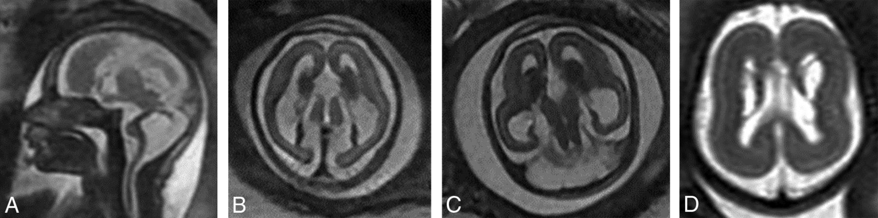
  • FIG 4.
    • Download figure
    • Open in new tab
    • Download powerpoint
    FIG 4.

    TUBA1A mutations (A–C). Case 18. T2-weighted single-shot FSE at 24 weeks 4 days’ gestation. TUBA1A pathogenic heterozygous variant. The bilateral GEs are enlarged. The fronto-occipital diameter is 2.5, standard deviations below the mean. The corpus callosum and cerebellum are severely hypogenetic/hypoplastic. Underopercularization, severe ventriculomegaly, a thin kinked brainstem (A), a ventral pontine cleft suggestive of a Walker-Warburg phenotype, and apparent diencephalic-mesencephalic fusion or dysplasia (C) are also noted. D, Case 19 at 33 weeks. TUBA1A mutation with enlarged, cavitated GEs and abnormal persistence of hemispheric lamination.
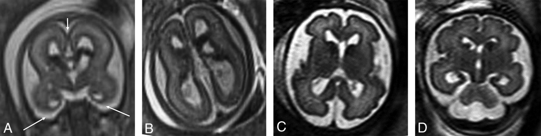
  • FIG 5.
    • Download figure
    • Open in new tab
    • Download powerpoint
    FIG 5.

    PDHA1 and OPHN1 mutations. A and B, PDHA1 mutation. Case 20 at 23 weeks 3 days’ gestation. PDHA1 mutation. Agenesis of the corpus callosum with the anterior commissure present (short arrow), cavitated enlarged GEs, and anterior temporal pole subependymal pseudocysts (germinolytic cysts) (long arrows). C and D, Case 21 at 29 weeks. OPHN1 mutation with mild ventriculomegaly (11 mm) and bilaterally enlarged GEs.

PreviousNext
Back to top
Advertisement
Print
Download PDF
Email Article

Thank you for your interest in spreading the word on American Journal of Neuroradiology.

NOTE: We only request your email address so that the person you are recommending the page to knows that you wanted them to see it, and that it is not junk mail. We do not capture any email address.

Enter multiple addresses on separate lines or separate them with commas.
The Fetus with Ganglionic Eminence Abnormality: Head Size and Extracranial Sonographic Findings Predict Genetic Diagnoses and Postnatal Outcomes
(Your Name) has sent you a message from American Journal of Neuroradiology
(Your Name) thought you would like to see the American Journal of Neuroradiology web site.
CAPTCHA
This question is for testing whether or not you are a human visitor and to prevent automated spam submissions.
Cite this article
S.K. Goergen, E. Alibrahim, J. Christie, A. Dobrotwir, M. Fahey, L. Fender, K. Frawley, S.A. Manikkam, J.R. Pinner, S. Sinnott, R. Romaniello, S.A. Sandaradura, J. Taylor, A. Vasudevan, A. Righini
The Fetus with Ganglionic Eminence Abnormality: Head Size and Extracranial Sonographic Findings Predict Genetic Diagnoses and Postnatal Outcomes
American Journal of Neuroradiology May 2021, DOI: 10.3174/ajnr.A7131

Citation Manager Formats

  • BibTeX
  • Bookends
  • EasyBib
  • EndNote (tagged)
  • EndNote 8 (xml)
  • Medlars
  • Mendeley
  • Papers
  • RefWorks Tagged
  • Ref Manager
  • RIS
  • Zotero
0 Responses
Respond to this article
Share
Bookmark this article
The Fetus with Ganglionic Eminence Abnormality: Head Size and Extracranial Sonographic Findings Predict Genetic Diagnoses and Postnatal Outcomes
S.K. Goergen, E. Alibrahim, J. Christie, A. Dobrotwir, M. Fahey, L. Fender, K. Frawley, S.A. Manikkam, J.R. Pinner, S. Sinnott, R. Romaniello, S.A. Sandaradura, J. Taylor, A. Vasudevan, A. Righini
American Journal of Neuroradiology May 2021, DOI: 10.3174/ajnr.A7131
del.icio.us logo Twitter logo Facebook logo Mendeley logo
  • Tweet Widget
  • Facebook Like
  • Google Plus One
Purchase

Jump to section

  • Article
    • Abstract
    • ABBREVIATIONS:
    • MATERIALS AND METHODS
    • RESULTS
    • DISCUSSION
    • CONCLUSIONS
    • Footnotes
    • References
  • Figures & Data
  • Supplemental
  • Info & Metrics
  • Responses
  • References
  • PDF

Related Articles

  • PubMed
  • Google Scholar

Cited By...

  • Characteristic Fetal Brain MRI Abnormalities in Pyruvate Dehydrogenase Complex Deficiency
  • Prenatal MR Imaging Phenotype of Fetuses with Tuberous Sclerosis: An Institutional Case Series and Literature Review
  • Crossref (13)
  • Google Scholar

This article has been cited by the following articles in journals that are participating in Crossref Cited-by Linking.

  • Prenatal MR Imaging Phenotype of Fetuses with Tuberous Sclerosis: An Institutional Case Series and Literature Review
    S.K. Goergen, M.C. Fahey
    American Journal of Neuroradiology 2022 43 4
  • TUBB3 and KIF21A in neurodevelopment and disease
    Dharmendra Puri, Brenda J. Barry, Elizabeth C. Engle
    Frontiers in Neuroscience 2023 17
  • Normal and abnormal appearance of fetal ganglionic eminence on second‐trimester three‐dimensional ultrasound
    E. Contro, N. Volpe, L. Larcher, A. Dall'Asta, M. A. Penas Da Costa, G. Vairo, E. Di Pasquo, I. Giorgini, T. Ghi
    Ultrasound in Obstetrics & Gynecology 2023 62 3
  • Prenatal assessment of brain malformations on neuroimaging: an expert panel review
    Ivana Pogledic, Kshitij Mankad, Mariasavina Severino, Tally Lerman-Sagie, Andras Jakab, Efrat Hadi, Anna C Jansen, Nadia Bahi-Buisson, Natalya Di Donato, Renske Oegema, Christian Mitter, Ivan Capo, Matthew T Whitehead, Parthiv Haldipur, Grazia Mancini, Thierry A G M Huisman, Andrea Righini, Bill Dobyns, James A Barkovich, Natasa Jovanov Milosevic, Gregor Kasprian, Maarten Lequin
    Brain 2024 147 12
  • Fetal Brain MRI Abnormalities in Pyruvate Dehydrogenase Complex Deficiency
    Olivier Fortin, Kelsey Christoffel, Abdullah B. Shoaib, Charu Venkatesan, Kate Cilli, Jason W. Schroeder, Cesar Alves, Rebecca D. Ganetzky, Jamie L. Fraser
    Neurology 2024 103 4
  • X‐linked pyruvate dehydrogenase complex deficiency due to a novel PDHA1 variant associated with structural brain abnormalities in a fetus
    Laura M. Tanner, Olli Tynninen, Kirsi Piippo, Antti M. Puhakka
    Prenatal Diagnosis 2023 43 6
  • Second trimester fetal MRI of the brain: Through the ground glass
    Nevena Fileva, Mariasavina Severino, Domenico Tortora, Antonia Ramaglia, Dario Paladini, Andrea Rossi
    Journal of Clinical Ultrasound 2023 51 2
  • Posterior subependymal germinal matrix hemorrhage as a mild form of hemorrhage in extremely preterm infants: neurodevelopmental outcomes at corrected ages of 18-24 months
    Jae Mo Koo, So-Young Yoo, Ji Hye Kim, Ji Eun Park, Sun-Young Baek, Tae Yeon Jeon
    Ultrasonography 2025 44 1
  • Fetal Neurosonography
    Ritsuko K. Pooh
    2024
  • Fetal Neurosonography
    Nevena Fileva, Irina Stavarache, Andrea Righini, Andrea Rossi
    2024

More in this TOC Section

  • FRACTURE MR in Congenital Vertebral Anomalies
  • Neuroimaging Delineation and Progression of SLSMD
  • fetal brain development of 10 weeks gestation
Show more PEDIATRIC NEUROIMAGING

Similar Articles

Advertisement

Indexed Content

  • Current Issue
  • Accepted Manuscripts
  • Article Preview
  • Past Issues
  • Editorials
  • Editor's Choice
  • Fellows' Journal Club
  • Letters to the Editor
  • Video Articles

Cases

  • Case Collection
  • Archive - Case of the Week
  • Archive - Case of the Month
  • Archive - Classic Case

More from AJNR

  • Trainee Corner
  • Imaging Protocols
  • MRI Safety Corner
  • Book Reviews

Multimedia

  • AJNR Podcasts
  • AJNR Scantastics

Resources

  • Turnaround Time
  • Submit a Manuscript
  • Submit a Video Article
  • Submit an eLetter to the Editor/Response
  • Manuscript Submission Guidelines
  • Statistical Tips
  • Fast Publishing of Accepted Manuscripts
  • Graphical Abstract Preparation
  • Imaging Protocol Submission
  • Evidence-Based Medicine Level Guide
  • Publishing Checklists
  • Author Policies
  • Become a Reviewer/Academy of Reviewers
  • News and Updates

About Us

  • About AJNR
  • Editorial Board
  • Editorial Board Alumni
  • Alerts
  • Permissions
  • Not an AJNR Subscriber? Join Now
  • Advertise with Us
  • Librarian Resources
  • Feedback
  • Terms and Conditions
  • AJNR Editorial Board Alumni

American Society of Neuroradiology

  • Not an ASNR Member? Join Now

© 2025 by the American Society of Neuroradiology All rights, including for text and data mining, AI training, and similar technologies, are reserved.
Print ISSN: 0195-6108 Online ISSN: 1936-959X

Powered by HighWire