
Article Figures & Data
Figures
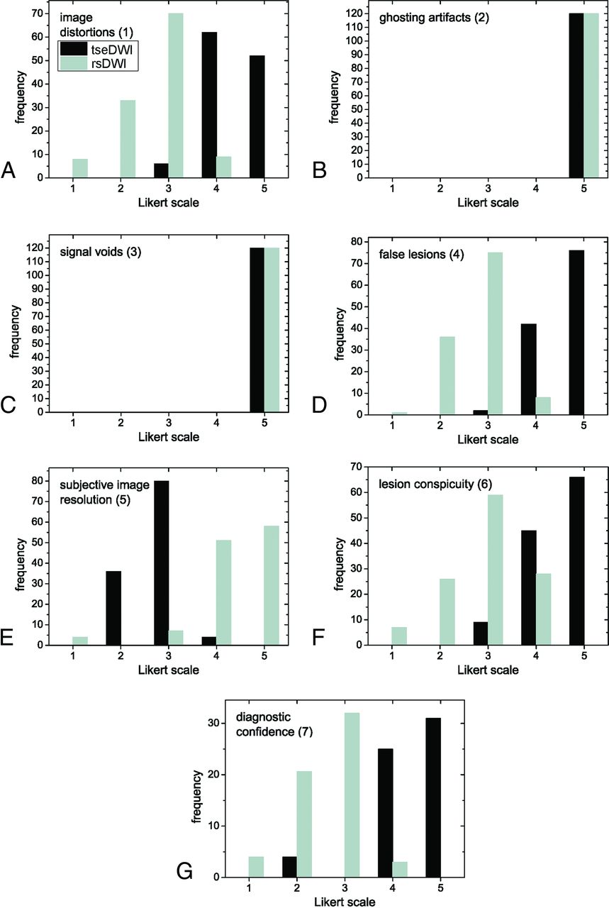
- FIG 1.
Histograms displaying Likert scale scores ranging from 1 (worst) to 5 (best) points. The histograms show the combined frequencies of both readers. The frequencies of axial and coronal images were added.
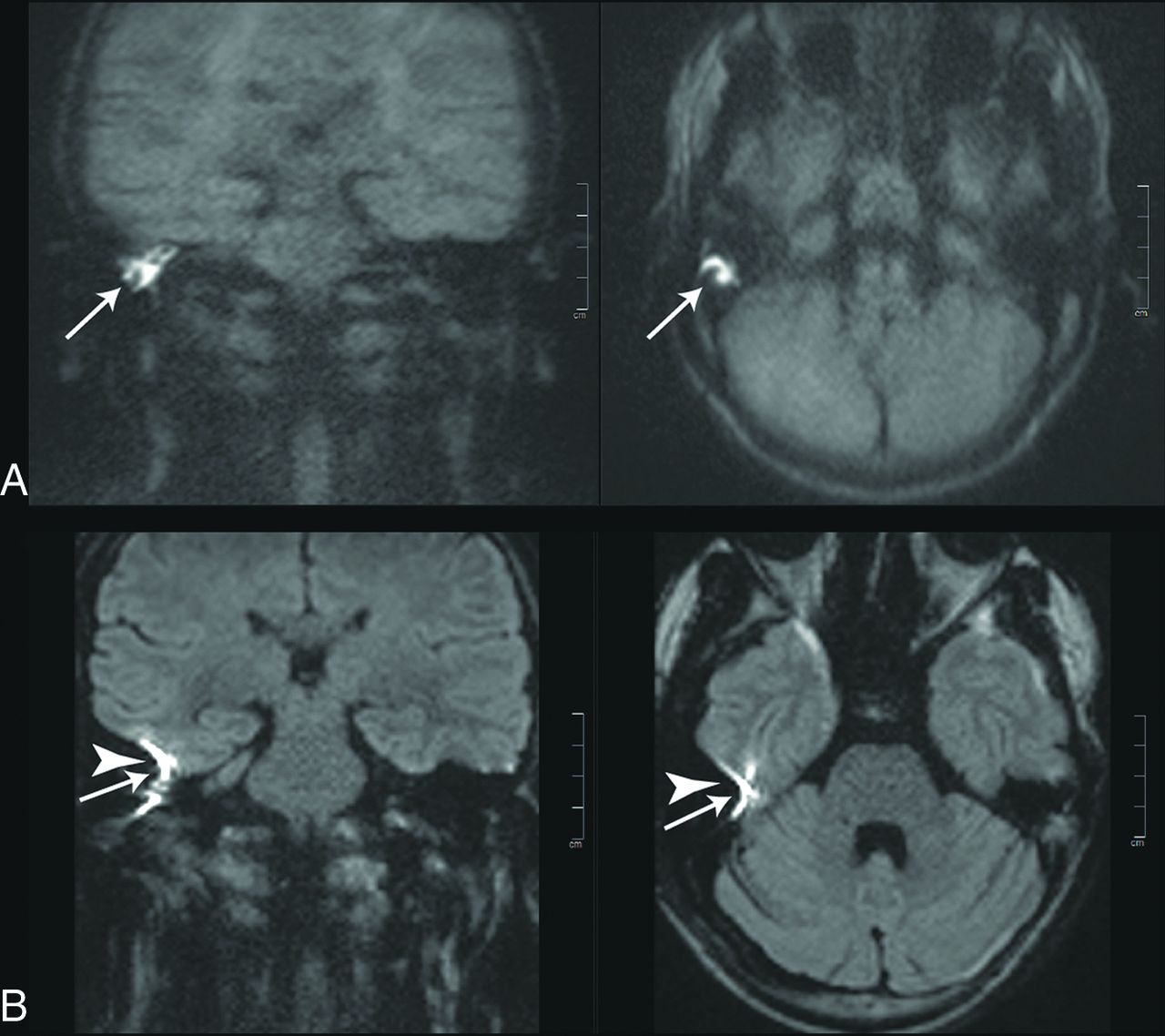
- FIG 2.
Images of a 20-year-old patient with a left-sided cholesteatoma (white arrows). A, tseDWI. B, rsDWI. The lesion can be seen clearly in tseDWI and rsDWI. It is, however, not spheric in the coronal rsDWI but rather is elongated and tilted due to image distortions in phase directions. Field inhomogeneities generate a bright spot in the rsDWI that might be mistaken for a lesion (arrowheads). The tseDWI displayed in A is more blurred than the rsDWI and shows less contrast in the brain. For example, unlike in the rsDWI, both white matter and gray matter are not discernible in the tseDWI.
- FIG 3.
Images of a 55-year-old patient with a left-sided cholesteatoma (white arrows). A, tseDWI. B, rsDWI. The lesion can be seen clearly in the tseDWI. In the rsDWI, however, the lesion is displayed with reduced contrast and is hardly visible; therefore, both readers did not diagnose a cholesteatoma in this case in the rsDWI dataset. Again, a bright spot is present in the coronal rsDWI next to the temporal bone on the right side due to field inhomogeneities, which might be mistaken for a true lesion (arrowhead).
- FIG 4.
Images of a 38-year-old patient with a recurrent cholesteatoma (white arrows). A stapes prosthesis implant caused major image distortions in the rsDWI (arrowheads).
- FIG 5.
Histograms displaying the scores obtained with Likert scores ranging from −2 (rsDWI much better than tseDWI) to 2 points (tseDWI much better than rsDWI). The histograms show the added frequencies of both readers.


Tables
Sequence tseDWI rsDWI TR (ms) 2000 4000 TE (ms) 103 66 (and 91 for phase-correction scan) Voxel size (mm³) 1.1 × 1.5 × 3 1.4 × 1.4 × 3 FOV (mm²) 220 230 FOV in-phase direction 100% 65% Phase direction Anterior-posterior (axial), right to center (coronal) Right to center (axial and coronal) Phase resolution 75% 100% Partial Fourier 50% (phase) 87.5% (readout) Matrix 192 × 144 160 × 104 Section distance 10% 10% No. of slices 13 (axial)11 (coronal) 13 (axial)11 (coronal) Parallel imaging GRAPPA ×2 GRAPPA ×2 Bandwidth (Hz/pixel) 554 977 Echo spacing (ms) 4.48 0.36 Readout segments 1 5 Flip angle 150° 180° b-values (s/mm2) 1000 0, 1000 Averages 10 1 (for b=0 s/mm²), 2 (for b=1000 s/mm²) Diffusion mode 3D diagonal 4-scan trace Diffusion scheme Bipolar Bipolar Acquisition time (minute:second) 4:22 (axial), 3:42 (coronal) 3:06 Note:—GRAPPA indicates generalized autocalibrating partially parallel acquisition.
Category Coronal/Axial tseDWI (Likert Categories 4 and 5) rsDWI (Likert Categories 4 and 5) Mean Difference(tseDWI and rsDWI) Reader 1 Reader 2 Reader 1 Reader 2 Reader 1 Reader 2 1) Geometric image distortion Cor 97% 97% 0% 0% 1.9 2 1) Geometric image distortion Ax 93% 93% 17% 13% 1.5 1.5 2) Ghosting artifacts Cor 100% 100% 100% 100% 0 0 2) Ghosting artifacts Ax 100% 100% 100% 100% 0 0 3) Signal voids or other artifacts Cor 100% 100% 100% 100% 0 0 3) Signal voids or other artifacts Ax 100% 100% 100% 100% 0 0 4) Bright-appearing region Cor 97% 97% 0% 0% 2 2 4) Bright-appearing region Ax 100% 100% 13% 13% 1.7 1.8 5) Subjective image resolution Cor 3% 3% 90% 90% –1.6 –1.7 5) Subjective image resolution Ax 3% 3% 90% 93% –1.5 –1.6 6) Lesion conspicuity Cor 97% 93% 33% 23% 1.6 1.7 6) Lesion conspicuity Ax 90% 90% 20% 17% 1.5 1.5 7) Diagnostic confidence Cor/Ax 93% 93% 7% 3% 1.8 1.8 Note:—Cor indicates coronal; Ax, axial.
κ (tseDWI, Axial) κ (tseDWI, Coronal) κ (rsDWI, Axial) κ (rsDWI, Coronal) 1) Geometric image distortion 0.64 0.62 0.83 0.58 2) Ghosting artifacts 1 1 1 1 3) Signal voids 1 1 1 1 4) Bright-appearing region 0.79 0.78 0.82 0.78 5) Subjective image resolution 0.79 0.78 0.71 0.77 6) Lesion conspicuity 0.71 0.62 0.83 0.53 κ (tseDWI) κ (rsDWI) 7) Diagnostic confidence with κ (tseDWI, axial and coronal combined) and κ (rsDWI, axial and coronal combined) 0.82 0.77 κ (tseDWI vs. rsDWI) 9) Lesion conspicuity with κ (tseDWI vs. rsDWI) 0.80 10) Subjective diagnostic confidence 0.68


